![Immunohistochemistry-Paraffin: FACL4 Antibody [NBP2-16401]](https://resources.rndsystems.com/images/products/FACL4-Antibody-Immunohistochemistry-Paraffin-NBP2-16401-img0012.jpg "Immunohistochemistry-Paraffin: FACL4 Antibody [NBP2-16401]")
Loading...
Key Product Details
Species Reactivity
Validated:
Human, Mouse, Rat
Cited:
Mouse
Predicted:
Porcine (100%), Rhesus Macaque (100%). Backed by our 100% Guarantee.
Applications
Validated:
Immunohistochemistry, Immunohistochemistry-Paraffin, Western Blot, Immunocytochemistry/ Immunofluorescence, Immunoprecipitation
Cited:
Western Blot, IF/IHC
Label
Unconjugated
Antibody Source
Polyclonal Rabbit IgG
Loading...
Product Specifications
Immunogen
Carrier-protein conjugated synthetic peptide encompassing a sequence within the C-terminus region of human FACL4. The exact sequence is proprietary.
Localization
Mitochondrion outer membrane; Single-pass type III membrane protein; Peroxisome membrane; Microsome membrane; Endoplasmic reticulum membrane
Clonality
Polyclonal
Host
Rabbit
Isotype
IgG
Theoretical MW
79 kDa.
Disclaimer note: The observed molecular weight of the protein may vary from the listed predicted molecular weight due to post translational modifications, post translation cleavages, relative charges, and other experimental factors.
Disclaimer note: The observed molecular weight of the protein may vary from the listed predicted molecular weight due to post translational modifications, post translation cleavages, relative charges, and other experimental factors.
Scientific Data Images for FACL4 Antibody
Immunohistochemistry-Paraffin: FACL4 Antibody [NBP2-16401]
Immunohistochemistry-Paraffin: FACL4 Antibody [NBP2-16401] - mouse kidney. FACL4 stained by FACL4 antibody [C3], C-term diluted at 1:500.Antigen Retrieval: Citrate buffer, pH 6.0, 15 min.![Western Blot: FACL4 Antibody [NBP2-16401]](https://resources.rndsystems.com/images/products/FACL4-Antibody-Western-Blot-NBP2-16401-img0014.jpg "Western Blot: FACL4 Antibody [NBP2-16401]")
Western Blot: FACL4 Antibody [NBP2-16401]
Western Blot: FACL4 Antibody [NBP2-16401] - Various whole cell extracts (30 ug) were separated by 7.5% SDS-PAGE, and the membrane was blotted with FACL4 antibody [C3], C-term diluted at 1:1000. The HRP-conjugated anti-rabbit IgG antibody (NBP2-19301) was used to detect the primary antibody.![Immunocytochemistry/ Immunofluorescence: FACL4 Antibody [NBP2-16401]](https://resources.rndsystems.com/images/products/FACL4-Antibody-Immunofluorescence-NBP2-16401-img0005.jpg "Immunocytochemistry/ Immunofluorescence: FACL4 Antibody [NBP2-16401]")
Immunocytochemistry/ Immunofluorescence: FACL4 Antibody [NBP2-16401]
Immunocytochemistry/Immunofluorescence: FACL4 Antibody [NBP2-16401] - analysis of FACL4 in MCF10A cells using anti-FACL4 antibody. Image submitted by a verified customer review.![Immunohistochemistry-Paraffin: FACL4 Antibody [NBP2-16401]](https://resources.rndsystems.com/images/products/FACL4-Antibody-Immunohistochemistry-Paraffin-NBP2-16401-img0008.jpg "Immunohistochemistry-Paraffin: FACL4 Antibody [NBP2-16401]")
Immunohistochemistry-Paraffin: FACL4 Antibody [NBP2-16401]
Immunohistochemistry-Paraffin: FACL4 Antibody [NBP2-16401] - Rat middle brain. FACL4 antibody [C3], C-term dilution: 1:500. Antigen Retrieval: Trilogy™ (EDTA based, pH 8.0) buffer, 15min![Immunohistochemistry-Paraffin: FACL4 Antibody [NBP2-16401]](https://resources.rndsystems.com/images/products/FACL4-Antibody-Immunohistochemistry-Paraffin-NBP2-16401-img0009.jpg "Immunohistochemistry-Paraffin: FACL4 Antibody [NBP2-16401]")
Immunohistochemistry-Paraffin: FACL4 Antibody [NBP2-16401]
Immunohistochemistry-Paraffin: FACL4 Antibody [NBP2-16401] - Mouse lymph node. FACL4 antibody [C3], C-term dilution: 1:500. Antigen Retrieval: Trilogy™ (EDTA based, pH 8.0) buffer, 15min![Immunoprecipitation: FACL4 Antibody [NBP2-16401]](https://resources.rndsystems.com/images/products/FACL4-Antibody-Immunoprecipitation-NBP2-16401-img0007.jpg "Immunoprecipitation: FACL4 Antibody [NBP2-16401]")
Immunoprecipitation: FACL4 Antibody [NBP2-16401]
Immunoprecipitation: FACL4 Antibody [NBP2-16401] - Immunoprecipitation of FACL4 protein from HeLa whole cell extracts using 5 ug of FACL4 antibody [C3], C-term Western blot analysis was performed using FACL4 antibody [C3], C-term. EasyBlot anti-Rabbit IgG was used as a secondary reagent.
Immunohistochemistry-Paraffin: FACL4 Antibody [NBP2-16401] -
FACL4 antibody [C3], C-term detects FACL4 protein by immunohistochemical analysis.Sample: Paraffin-embedded mouse tissues.
FACL4 stained by FACL4 antibody [C3], C-term (NBP2-16401) diluted at 1:500.
Antigen Retrieval: Citrate buffer, pH 6.0, 15 min

Western Blot: FACL4 Antibody [NBP2-16401] -
Various whole cell extracts (30 ug) were separated by 7.5% SDS-PAGE, and the membrane was blotted with FACL4 antibody [C3], C-term (NBP2-16401) diluted at 1:1000. The HRP-conjugated anti-rabbit IgG antibody was used to detect the primary antibody.
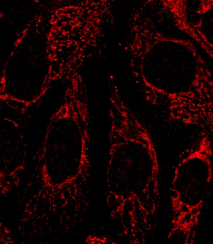
Immunocytochemistry/ Immunofluorescence: FACL4 Antibody [NBP2-16401] -
FACL4 antibody [C3], C-term detects FACL4 protein at cytoplasm by immunofluorescent analysis.Sample: HeLa cells were fixed in 4% paraformaldehyde at RT for 15 min.
Green: FACL4 stained by FACL4 antibody [C3], C-term (NBP2-16401) diluted at 1:500.
Red: alpha Tubulin, a cytoskeleton marker, stained by alpha Tubulin antibody [GT114] diluted at 1:1000.
Blue: Fluoroshield with DAPI.

Western Blot: FACL4 Antibody [NBP2-16401] -
Various whole cell extracts (30 ug) were separated by 7.5% SDS-PAGE, and the membrane was blotted with FACL4 antibody [C3], C-term (NBP2-16401) diluted at 1:1000. The HRP-conjugated anti-rabbit IgG antibody was used to detect the primary antibody.
Immunohistochemistry-Paraffin: FACL4 Antibody [NBP2-16401] -
FACL4 antibody [C3], C-term detects FACL4 protein by immunohistochemical analysis.Sample: Paraffin-embedded rat tissues.
FACL4 stained by FACL4 antibody [C3], C-term (NBP2-16401) diluted at 1:500.
Antigen Retrieval: Citrate buffer, pH 6.0, 15 min

Western Blot: FACL4 Antibody [NBP2-16401] -
Expression and location of ferroptosis-related proteins in diabetic mice retinas. (A) Western blot analysis of ferroptosis-related proteins expression levels in retinas at 1 and 3 months post-diabetes. GAPDH was used as a control. (B) GPX4 and SLC7A11 protein expression was remarkably decreased in diabetic mice retinas compared with the control retinas. Expression of ACSL4, FTH1, and NCOA4 proteins was significantly increased in retinas at 1 and 3 months post-diabetes. (C) Immunofluorescence staining of location of ferroptosis-related proteins (red) and nuclear (blue) in retinas at 1 and 3 months post-diabetes: ganglion cell layer (GCL), inner nuclear layer (INL), outer nuclear layer (ONL), inner segment (IS), outer segment (OS). Data are shown as mean +/- SEM, n = 3 per group for Western blotting. * p < 0.05, ** p < 0.01, *** p < 0.001 versus Ctrl group. Scale bar: 50 μm. Image collected and cropped by CiteAb from the following open publication (https://www.mdpi.com/1422-0067/24/23/16946), licensed under a CC-BY license. Not internally tested by Novus Biologicals.
Western Blot: FACL4 Antibody [NBP2-16401] -
HG causes changes in the expression of ferroptosis-related proteins in 661W cells. (A) Western blot analysis of ferroptosis-related protein expression levels in HG-induced 661W cells. GAPDH was used as a control. (B) Expression of GPX4 and SLC7A11 proteins was significantly downregulated in HG-stimulated 661W cells after 12, 18, and 24 h. HG induced obvious upregulation in the expression of ACSL4, FTH1, and NCOA4 in 661W cells compared with the Ctrl group. (C) Immunofluorescence staining of localization of ferroptosis-related proteins (red) and nuclear (blue) in HG-induced 661W cells after 18 h. Data are shown as mean +/- SEM, n = 3 per group for Western blotting. p = not significant [ns], * p < 0.05, ** p < 0.01, *** p < 0.001 versus Ctrl group. Scale bar: 50 μm. Image collected and cropped by CiteAb from the following open publication (https://www.mdpi.com/1422-0067/24/23/16946), licensed under a CC-BY license. Not internally tested by Novus Biologicals.
Western Blot: FACL4 Antibody [NBP2-16401] -
Fer-1 treatment attenuated changes in ferroptosis-related proteins’ expression in HG-stimulated 661W cells after 18 h. (A) Western blot analysis of ferroptosis-related proteins’ expression levels in HG-induced 661W cells after Fer-1 treatment. GAPDH was used as a control. (B) The downregulation in GPX4 and SLC7A11 protein expression in HG-stimulated 661W cells was significantly attenuated after Fer-1 treatment. The upregulation in ACSL4, FTH1, and NCOA4 protein expression in HG-stimulated 661W cells was effectively abrogated after Fer-1 treatment. (C) Immunofluorescence staining of ferroptosis-related proteins (red) and nuclear (blue) in HG-induced 661W cells after Fer-1 administration. Data are shown as mean +/- SEM, n = 3 per group for Western blotting. p = not significant [ns], * p < 0.05, ** p < 0.01 versus Ctrl group. Scale bar: 50 μm. Image collected and cropped by CiteAb from the following open publication (https://www.mdpi.com/1422-0067/24/23/16946), licensed under a CC-BY license. Not internally tested by Novus Biologicals.
Western Blot: FACL4 Antibody [NBP2-16401] -
Fer-1 administration attenuated changes of ferroptosis-related proteins expression in diabetic mice retinas. (A) Western blot analysis of ferroptosis-related proteins expression levels in diabetic mice retinas after Fer-1 treatment. GAPDH was used as a control. (B) The decrease in GPX4 and SLC7A11 protein expression in diabetic mice retinas was significantly attenuated after Fer-1 treatment. The increase in ACSL4, FTH1, and NCOA4 protein expression in diabetic mice retinas was effectively abrogated after Fer-1 treatment. (C) Immunofluorescence staining of ferroptosis-related proteins (red) and nuclear (blue) in diabetic mice retinas after Fer-1 treatment: ganglion cell layer (GCL), inner nuclear layer (INL), outer nuclear layer (ONL), inner segment (IS), outer segment (OS). Data are shown as mean +/- SEM, n = 3 per group for Western blotting. p = not significant [ns], * p < 0.05, ** p < 0.01 versus Ctrl group. Scale bar: 50 μm. Image collected and cropped by CiteAb from the following open publication (https://www.mdpi.com/1422-0067/24/23/16946), licensed under a CC-BY license. Not internally tested by Novus Biologicals.
Western Blot: FACL4 Antibody [NBP2-16401] -
Extraction ion-containing PMs (PM2.5 EI) cause ferroptosis in macrophages.A Western blot analysis after incubation of RAW264.7 cells with three types of PM (100 μg/ml) for 18 h. B Malondialdehyde (MDA) formation in RAW264.7 cells after incubation with 100 μg/ml PMs for 12 h, investigated using a lipid peroxidation assay kit. C Intracellular ferrous iron levels are detected in J774A.1 cells incubated with 50 μg/ml of PMs for 12 h. D WST-8 assay demonstrating the cell viability analysis in RAW264.7 line after pretreatment with ferrostatin-1 (Fer-1; 2 μM), liproxstatin-1 (Lip-1; 2 μM), or deferiprone (DFP; 100 μM) for 2 h followed by the stimulation with 100 μg/ml of PM2.5 EI for 24 h. E Western blot analysis using RAW264.7 cells after preincubation with Fer-1 (2 μM), Lip-1 (2 μM), or DFP (100 μM) for 2 h followed by stimulation with 100 μg/ml of PM2.5 EI for 24 h. F Detection of lipid peroxidation in terms of MDA in J774A.1 cells after preincubation with Fer-1 (2 μM), Lip-1 (2 μM), or DFP (100 μM) for 2 h followed by treatment using 50 μg/ml of PM2.5 EI for 12 h. All data are presented as the means +/- standard deviations from at least three independent experiments. *P < 0.05, **P < 0.01 and #P < 0.001. All experiments were conducted at least three times. Image collected and cropped by CiteAb from the following open publication (https://www.nature.com/articles/s41420-024-01874-y), licensed under a CC-BY license. Not internally tested by Novus Biologicals.
Western Blot: FACL4 Antibody [NBP2-16401] -
PM2.5 EI induces a correlation between the activation of inflammasomes and the induction of ferroptosis.A and B Western blot analysis in J774A.1 cells after preincubation with CY-09 (10 μM) or YVAD (20 μM) for 2 h and subsequent stimulation with 50 μg/ml of PM2.5 EI for 24 h. C MDA levels in J774A.1 cells after pretreatment with CY-09 (10 μM) or YVAD (20 μM) for 2 h followed by stimulation with 50 μg/ml of PM2.5 EI for 12 h tested using a lipid peroxidation assay kit. D Western blot analysis using J774A.1 cells preincubated with Fer-1 (2 μM), Lip-1 (2 μM), or DFP (100 μM) for 2 h and treated using 50 μg/ml of PM2.5 EI for 24 h. E, F ELISA assay revealed the IL-1 beta and IL-18 levels in the medium of J774A.1 cell culture after pretreatment with CY-09 (10 μM) or YVAD (20 μM) for 2 h and subsequent stimulation with 50 μg/ml of PM2.5 EI for 12 h. All data are presented as the means +/- standard deviations from at least three independent experiments. *P < 0.05, **P < 0.01 and #P < 0.001. All experiments were conducted at least three times. Image collected and cropped by CiteAb from the following open publication (https://www.nature.com/articles/s41420-024-01874-y), licensed under a CC-BY license. Not internally tested by Novus Biologicals.
Immunocytochemistry/ Immunofluorescence: FACL4 Antibody [NBP2-16401] -
HG causes changes in the expression of ferroptosis-related proteins in 661W cells. (A) Western blot analysis of ferroptosis-related protein expression levels in HG-induced 661W cells. GAPDH was used as a control. (B) Expression of GPX4 and SLC7A11 proteins was significantly downregulated in HG-stimulated 661W cells after 12, 18, and 24 h. HG induced obvious upregulation in the expression of ACSL4, FTH1, and NCOA4 in 661W cells compared with the Ctrl group. (C) Immunofluorescence staining of localization of ferroptosis-related proteins (red) and nuclear (blue) in HG-induced 661W cells after 18 h. Data are shown as mean +/- SEM, n = 3 per group for Western blotting. p = not significant [ns], * p < 0.05, ** p < 0.01, *** p < 0.001 versus Ctrl group. Scale bar: 50 μm. Image collected and cropped by CiteAb from the following open publication (https://www.mdpi.com/1422-0067/24/23/16946), licensed under a CC-BY license. Not internally tested by Novus Biologicals.Applications for FACL4 Antibody
Application
Recommended Usage
Immunocytochemistry/ Immunofluorescence
1:100-1:1000
Immunohistochemistry
1:100-1:1000
Immunohistochemistry-Paraffin
1:100-1:1000
Immunoprecipitation
1:1000-1:5000
Western Blot
1:500-1:10000
Reviewed Applications
Read 1 review rated 4 using NBP2-16401 in the following applications:
Formulation, Preparation, and Storage
Purification
Antigen Affinity-purified
Formulation
PBS, 20% Glycerol
Preservative
0.025% Proclin 300
Concentration
Concentrations vary lot to lot. See vial label for concentration. If unlisted please contact technical services.
Shipping
The product is shipped with polar packs. Upon receipt, store it immediately at the temperature recommended below.
Stability & Storage
Aliquot and store at -20C or -80C. Avoid freeze-thaw cycles.
Background: FACL4
Alternate Names
ACS4mental retardation, X-linked 68, acyl-CoA synthetase 4, acyl-CoA synthetase long-chain family member 4, EC 6.2.1.3, FACL4long-chain 4, LACS 4, LACS4MRX68, lignoceroyl-CoA synthase, Long-chain acyl-CoA synthetase 4, long-chain fatty-acid-Coenzyme A ligase 4, long-chain-fatty-acid--CoA ligase 4, mental retardation, X-linked 63, MRX63
Gene Symbol
ACSL4
UniProt
Additional FACL4 Products
Product Documents for FACL4 Antibody
Certificate of Analysis
To download a Certificate of Analysis, please enter a lot or batch number in the search box below.
Product Specific Notices for FACL4 Antibody
This product is for research use only and is not approved for use in humans or in clinical diagnosis. Primary Antibodies are guaranteed for 1 year from date of receipt.
⚠ WARNING: This product can expose you to chemicals including mercury, which is known to the State of California to cause reproductive toxicity with developmental effects. For more information go to www.P65Warnings.ca.gov.Citations for FACL4 Antibody
 Powered by Bioz
Customer Reviews for FACL4 Antibody (1)
4 out of 5
1 Customer Rating
Have you used FACL4 Antibody?
Submit a review and receive an Amazon gift card!
$25/€18/£15/$25CAN/¥2500 Yen for a review with an image
$10/€7/£6/$10CAN/¥1110 Yen for a review without an image
Submit a review
Customer Images
Showing
1
-
1 of
1 review
Showing All
Filter By:
-
Application: ImmunofluorescenceSample Tested: Mcf10A cell lineSpecies: HumanVerified Customer | Posted 04/03/2015MCF10A cell line

There are no reviews that match your criteria.
Protocols
Find general support by application which include: protocols, troubleshooting, illustrated assays, videos and webinars.
- Antigen Retrieval Protocol (PIER)
- Antigen Retrieval for Frozen Sections Protocol
- Appropriate Fixation of IHC/ICC Samples
- Cellular Response to Hypoxia Protocols
- Chromogenic IHC Staining of Formalin-Fixed Paraffin-Embedded (FFPE) Tissue Protocol
- Chromogenic Immunohistochemistry Staining of Frozen Tissue
- ClariTSA™ Fluorophore Kits
- Detection & Visualization of Antibody Binding
- Fluorescent IHC Staining of Frozen Tissue Protocol
- Graphic Protocol for Heat-induced Epitope Retrieval
- Graphic Protocol for the Preparation and Fluorescent IHC Staining of Frozen Tissue Sections
- Graphic Protocol for the Preparation and Fluorescent IHC Staining of Paraffin-embedded Tissue Sections
- Graphic Protocol for the Preparation of Gelatin-coated Slides for Histological Tissue Sections
- ICC Cell Smear Protocol for Suspension Cells
- ICC Immunocytochemistry Protocol Videos
- ICC for Adherent Cells
- IHC Sample Preparation (Frozen sections vs Paraffin)
- Immunocytochemistry (ICC) Protocol
- Immunocytochemistry Troubleshooting
- Immunofluorescence of Organoids Embedded in Cultrex Basement Membrane Extract
- Immunofluorescent IHC Staining of Formalin-Fixed Paraffin-Embedded (FFPE) Tissue Protocol
- Immunohistochemistry (IHC) and Immunocytochemistry (ICC) Protocols
- Immunohistochemistry Frozen Troubleshooting
- Immunohistochemistry Paraffin Troubleshooting
- Immunoprecipitation Protocol
- Preparing Samples for IHC/ICC Experiments
- Preventing Non-Specific Staining (Non-Specific Binding)
- Primary Antibody Selection & Optimization
- Protocol for Heat-Induced Epitope Retrieval (HIER)
- Protocol for Making a 4% Formaldehyde Solution in PBS
- Protocol for VisUCyte™ HRP Polymer Detection Reagent
- Protocol for the Fluorescent ICC Staining of Cell Smears - Graphic
- Protocol for the Fluorescent ICC Staining of Cultured Cells on Coverslips - Graphic
- Protocol for the Preparation & Fixation of Cells on Coverslips
- Protocol for the Preparation and Chromogenic IHC Staining of Frozen Tissue Sections
- Protocol for the Preparation and Chromogenic IHC Staining of Frozen Tissue Sections - Graphic
- Protocol for the Preparation and Chromogenic IHC Staining of Paraffin-embedded Tissue Sections
- Protocol for the Preparation and Chromogenic IHC Staining of Paraffin-embedded Tissue Sections - Graphic
- Protocol for the Preparation and Fluorescent ICC Staining of Cells on Coverslips
- Protocol for the Preparation and Fluorescent ICC Staining of Non-adherent Cells
- Protocol for the Preparation and Fluorescent ICC Staining of Stem Cells on Coverslips
- Protocol for the Preparation and Fluorescent IHC Staining of Frozen Tissue Sections
- Protocol for the Preparation and Fluorescent IHC Staining of Paraffin-embedded Tissue Sections
- Protocol for the Preparation of Gelatin-coated Slides for Histological Tissue Sections
- Protocol for the Preparation of a Cell Smear for Non-adherent Cell ICC - Graphic
- R&D Systems Quality Control Western Blot Protocol
- TUNEL and Active Caspase-3 Detection by IHC/ICC Protocol
- The Importance of IHC/ICC Controls
- Troubleshooting Guide: Immunohistochemistry
- Troubleshooting Guide: Western Blot Figures
- Western Blot Conditions
- Western Blot Protocol
- Western Blot Protocol for Cell Lysates
- Western Blot Troubleshooting
- Western Blot Troubleshooting Guide
- View all Protocols, Troubleshooting, Illustrated assays and Webinars
Loading...