
FAM171B Antibody - BSA Free
Novus Biologicals | Catalog # NBP1-93847

![Western Blot: FAM171B Antibody [NBP1-93847]](https://resources.rndsystems.com/images/products/FAM171B-Antibody-Western-Blot-NBP1-93847-img0005.jpg "Western Blot: FAM171B Antibody [NBP1-93847]")
Loading...
Key Product Details
Species Reactivity
Human, Mouse
Applications
Immunohistochemistry, Immunohistochemistry-Paraffin, Western Blot, Immunocytochemistry/ Immunofluorescence
Label
Unconjugated
Antibody Source
Polyclonal Rabbit IgG
Format
BSA Free
Loading...
Product Specifications
Immunogen
This antibody was developed against Recombinant Protein corresponding to amino acids: FEDTVLITGKLADAKSQPSVQFSKALIKLPDNHHISNVTGYLTVLQQFLKVDNFLHTTGITLNKPGFENIELTPLAAICVKIYSGGKELKVNGSIQVSLPLLRLNDISAGDRIPAWTFDMNTGAWVNHGRGM
Clonality
Polyclonal
Host
Rabbit
Isotype
IgG
Scientific Data Images for FAM171B Antibody - BSA Free
Western Blot: FAM171B Antibody [NBP1-93847]
Western Blot: FAM171B Antibody [NBP1-93847] - Lane 1: Mouse liver tissue lysate. Lane 2: Rat liver tissue lysate.![Immunocytochemistry/ Immunofluorescence: FAM171B Antibody [NBP1-93847]](https://resources.rndsystems.com/images/products/FAM171B-Antibody-Immunocytochemistry-Immunofluorescence-NBP1-93847-img0007.jpg "Immunocytochemistry/ Immunofluorescence: FAM171B Antibody [NBP1-93847]")
Immunocytochemistry/ Immunofluorescence: FAM171B Antibody [NBP1-93847]
Immunocytochemistry/Immunofluorescence: FAM171B Antibody [NBP1-93847] - Staining of human cell line U-2 OS shows localization to mitochondria. Antibody staining is shown in green.![Immunohistochemistry-Paraffin: FAM171B Antibody [NBP1-93847]](https://resources.rndsystems.com/images/products/FAM171B-Antibody-Immunohistochemistry-Paraffin-NBP1-93847-img0006.jpg "Immunohistochemistry-Paraffin: FAM171B Antibody [NBP1-93847]")
Immunohistochemistry-Paraffin: FAM171B Antibody [NBP1-93847]
Immunohistochemistry-Paraffin: FAM171B Antibody [NBP1-93847] - Staining of human cerebral cortex shows moderate cytoplasmic positivity in neuronal cells.![FAM171B Antibody - BSA Free Western Blot: FAM171B Antibody - BSA Free [NBP1-93847]](https://resources.rndsystems.com/images/products/nbp1-93847_rabbit-polyclonal-fam171b-antibody-2952025164149.jpg "Western Blot: FAM171B Antibody - BSA Free [NBP1-93847]")
Western Blot: FAM171B Antibody - BSA Free [NBP1-93847]
Lane 1: Mouse liver tissue lysateLane 2: Rat liver tissue lysate
Western Blot: FAM171B Antibody - BSA Free [NBP1-93847] -
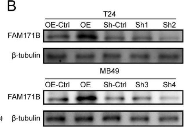
FAM171B promotes the malignant phenotype of bladder cancer cells. A The Chronos dependency score analysis of FAM171B in the 28 human bladder urothelial carcinoma cell lines in the CCLE database. The Chronos dependency score is based on data from a cell depletion assay. A lower Chronos score indicates a higher likelihood that the gene of interest is essential in a given cell line. B Protein levels of FAM171B in the knockdown and overexpression cell lines. C Representative colony formation images in different groups. D Colony formation analysis of T24 and MB49 cells after knockdown and overexpression of FAM171B. E Cell viability of T24 and MB49 after knockdown and overexpression of FAM171B. F Representative transwell images of T24 and MB49 cells after knockdown and overexpression of FAM171B. G Statistical results of the number of invasive cells in each group of T24 and MB49 cells. H Representative images of migration in T24 and MB49 cells after knockdown and overexpression of FAM171B. I Statistical results of the number of migrated cells in each group of T24 and MB49 cells Image collected and cropped by CiteAb from the following open publication (https://pubmed.ncbi.nlm.nih.gov/37915048), licensed under a CC-BY license. Not internally tested by Novus Biologicals.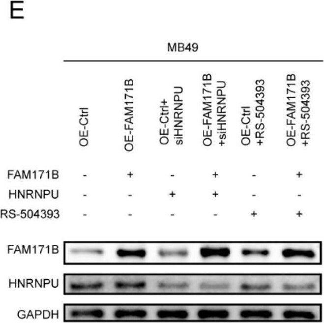
Western Blot: FAM171B Antibody - BSA Free [NBP1-93847] -
FAM171B regulates CCL2 via HNRNPU and promotes TAM migration and infiltration in bladder cancer. A ELISA analysis of CCL2 secretion levels in MB49 cells with FAM171B overexpression or knockdown. B RIP results show that HNRNPU can immunoprecipitate with CCL2 precursor RNA. C qRT-PCR analysis of the relative RNA levels of precursor CCL2 and CCL2 in MB49 cells with HNRNPU knockdown or control. D ELISA analysis of CCL2 secretion levels in MB49 cells with HNRNPU knockdown or control. E Protein levels of FAM171B and HNRNPU in the MB49 cell lines in different groups. F ELISA analysis of CCL2 secretion levels in MB49 cells in different groups. G Analysis of migration of RAW264.7(mouse macrophages) toward MB49 cells in different groups. H Representative images of migration of RAW264.7(mouse macrophages) toward MB49 cells in co-culture chambers. I The pictures of tumors derived from mouse bladder cancer MB49 cell line. J IHC staining pictures of F4/80(macrophage marker) expression in different groups. K Representative images of flow cytometry used to evaluate F4/80 positive subpopulations. L The tumor weight in different groups. M The tumor volume in different groups. N The average body weight of mice in different groups. O Analysis of F4/80 positive population by IHC. P Analysis of F4/80 positive population by flow cytometry Image collected and cropped by CiteAb from the following open publication (https://pubmed.ncbi.nlm.nih.gov/37915048), licensed under a CC-BY license. Not internally tested by Novus Biologicals.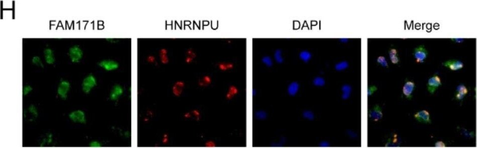
Immunocytochemistry/ Immunofluorescence: FAM171B Antibody - BSA Free [NBP1-93847] -
FAM171B interacts with vimentin and HNRNPU in bladder cancer cells. A Mass spectrometry analysis of interacting proteins enriched by immunoprecipitation. Immunoprecipitation is performed by Anti-DYKDDDDK beads on T24 cells transfected with FLAG-FAM171B or FLAG-control. B Analysis of positive and background proteins based on Mass Spectrometry scores. C The top 16 proteins with the highest scores based on MS results. D Protein interaction network map of proteins bound to FAM171B based on MS results. The protein interactions data is from STRING database. E The predicted protein structure image of FAM171B based on the Alpha Fold. F Immunofluorescence analysis images revealed the localization of FAM171B in T24 cells. G Colocalization of FAM171B and vimentin was visualized by confocal microscope in T24 cells. Cytoplasmic staining of FAM171B and vimentin was mostly merged together. H Colocalization of FAM171B and HNRNPU was visualized by confocal microscope in T24 cells. Nuclear staining of FAM171B and HNRNPU was mostly merged together. I ImageJ was used for colocation analysis of FAM171B and vimentin. Pearson correlation analysis showed that r > 0.5. Manders’ colocation coefficient showed that M1 > 0.5 and M2 > 0.5. J Computational docking model between FAM171B and vimentin. K ImageJ was used for colocation analysis of FAM171B and HNRNPU. Pearson correlation analysis showed that r > 0.5. Manders’ colocation coefficient showed that M1 > 0.5 and M2 > 0.5. L Computational docking model between FAM171B and HNRNPU Image collected and cropped by CiteAb from the following open publication (https://pubmed.ncbi.nlm.nih.gov/37915048), licensed under a CC-BY license. Not internally tested by Novus Biologicals.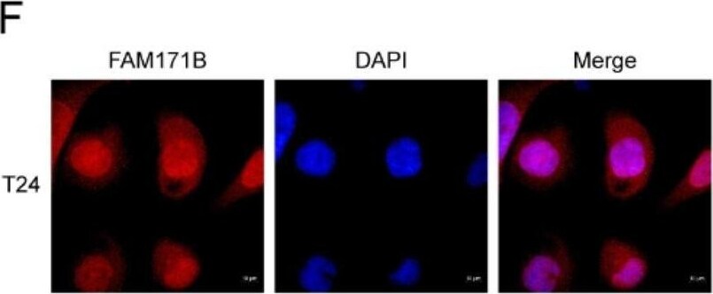
Immunocytochemistry/ Immunofluorescence: FAM171B Antibody - BSA Free [NBP1-93847] -
FAM171B interacts with vimentin and HNRNPU in bladder cancer cells. A Mass spectrometry analysis of interacting proteins enriched by immunoprecipitation. Immunoprecipitation is performed by Anti-DYKDDDDK beads on T24 cells transfected with FLAG-FAM171B or FLAG-control. B Analysis of positive and background proteins based on Mass Spectrometry scores. C The top 16 proteins with the highest scores based on MS results. D Protein interaction network map of proteins bound to FAM171B based on MS results. The protein interactions data is from STRING database. E The predicted protein structure image of FAM171B based on the Alpha Fold. F Immunofluorescence analysis images revealed the localization of FAM171B in T24 cells. G Colocalization of FAM171B and vimentin was visualized by confocal microscope in T24 cells. Cytoplasmic staining of FAM171B and vimentin was mostly merged together. H Colocalization of FAM171B and HNRNPU was visualized by confocal microscope in T24 cells. Nuclear staining of FAM171B and HNRNPU was mostly merged together. I ImageJ was used for colocation analysis of FAM171B and vimentin. Pearson correlation analysis showed that r > 0.5. Manders’ colocation coefficient showed that M1 > 0.5 and M2 > 0.5. J Computational docking model between FAM171B and vimentin. K ImageJ was used for colocation analysis of FAM171B and HNRNPU. Pearson correlation analysis showed that r > 0.5. Manders’ colocation coefficient showed that M1 > 0.5 and M2 > 0.5. L Computational docking model between FAM171B and HNRNPU Image collected and cropped by CiteAb from the following open publication (https://pubmed.ncbi.nlm.nih.gov/37915048), licensed under a CC-BY license. Not internally tested by Novus Biologicals.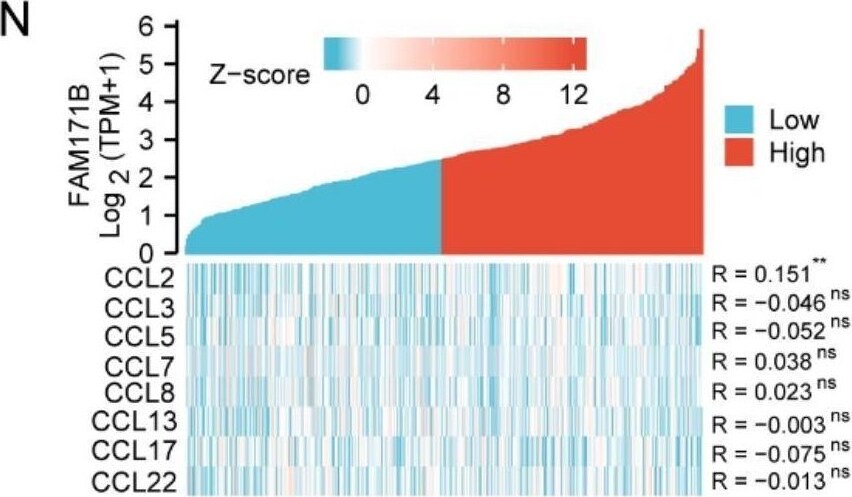
Immunohistochemistry: FAM171B Antibody - BSA Free [NBP1-93847] -
FAM171B expression is positively correlated with tumor progression and M2 TAM infiltration in BLCA patients. A Analysis of FAM171B expression in tumor tissue and paired normal tissue from TCGA dataset. B Representative IHC staining images of FAM171B in BLCA tissue array. C IHC score in different T stages from BLCA tissue array. D IHC score in different N stages from BLCA tissue array. E Relationship between FAM171B expression level and overall survival from TCGA dataset. F Relationship between FAM171B expression level and progression free survival from TCGA dataset. G Relationship between FAM171B expression level and disease specific survival from TCGA dataset. H Relationship between FAM171B expression level and disease-free survival from TCGA dataset. I Relationship between FAM171B expression level and overall survival from GEO dataset (GSE13507/GSE31684/GSE32894/GSE37815). J Analysis of FAM171B expression in different T stages from TCGA dataset. K Analysis of FAM171B expression in different N stages from TCGA dataset. L Analysis of FAM171B expression in different pathologic stages from TCGA dataset. M Lollipop plot demonstrating the correlation between FAM171B expression and different immune cell infiltration. N Heat map of the correlation between FAM171B expression and major monocyte/macrophage chemokines. O Immunoscore analysis of FAM171B and M1 macrophages in bladder cancer using QUANTISEQ algorithm. P Immunoscore analysis of FAM171B and M2 macrophages in bladder cancer using QUANTISEQ algorithm. Q Representative IHC staining images (FAM171B) and IF staining images (CD206, CD8 and alpha -SMA) in BLCA tissue array. R Proportion of immunofluorescence-positive cells (CD206, CD8 and alpha -SMA) in FAM171B high or low expression group from tissue array Image collected and cropped by CiteAb from the following open publication (https://pubmed.ncbi.nlm.nih.gov/37915048), licensed under a CC-BY license. Not internally tested by Novus Biologicals.Applications for FAM171B Antibody - BSA Free
Application
Recommended Usage
Immunocytochemistry/ Immunofluorescence
0.25-2 ug/ml
Immunohistochemistry
1:50 - 1:200
Immunohistochemistry-Paraffin
1:50 - 1:200
Western Blot
0.04-0.4 ug/ml
Application Notes
For IHC-Paraffin, HIER pH 6 retrieval is recommended. ICC/IF Fixation Permeabilization: Use PFA/Triton X-100.
Formulation, Preparation, and Storage
Purification
Affinity purified
Formulation
PBS (pH 7.2) and 40% Glycerol
Format
BSA Free
Preservative
0.02% Sodium Azide
Concentration
Concentrations vary lot to lot. See vial label for concentration. If unlisted please contact technical services.
Shipping
The product is shipped with polar packs. Upon receipt, store it immediately at the temperature recommended below.
Stability & Storage
Store at 4C short term. Aliquot and store at -20C long term. Avoid freeze-thaw cycles.
Background: FAM171B
Alternate Names
family with sequence similarity 171, member B, FLJ34104, KIAA1946, NPD019
Gene Symbol
FAM171B
Additional FAM171B Products
Product Documents for FAM171B Antibody - BSA Free
Certificate of Analysis
To download a Certificate of Analysis, please enter a lot or batch number in the search box below.
Product Specific Notices for FAM171B Antibody - BSA Free
This product is for research use only and is not approved for use in humans or in clinical diagnosis. Primary Antibodies are guaranteed for 1 year from date of receipt.
Citations for FAM171B Antibody - BSA Free
 Powered by Bioz
Customer Reviews for FAM171B Antibody - BSA Free
There are currently no reviews for this product. Be the first to review FAM171B Antibody - BSA Free and earn rewards!
Have you used FAM171B Antibody - BSA Free?
Submit a review and receive an Amazon gift card!
$25/€18/£15/$25CAN/¥2500 for a review with an image
$10/€7/£6/$10CAN/¥1110 for a review without an image
Submit a review
Protocols
Find general support by application which include: protocols, troubleshooting, illustrated assays, videos and webinars.
- Antigen Retrieval Protocol (PIER)
- Antigen Retrieval for Frozen Sections Protocol
- Appropriate Fixation of IHC/ICC Samples
- Cellular Response to Hypoxia Protocols
- Chromogenic IHC Staining of Formalin-Fixed Paraffin-Embedded (FFPE) Tissue Protocol
- Chromogenic Immunohistochemistry Staining of Frozen Tissue
- ClariTSA™ Fluorophore Kits
- Detection & Visualization of Antibody Binding
- Fluorescent IHC Staining of Frozen Tissue Protocol
- Graphic Protocol for Heat-induced Epitope Retrieval
- Graphic Protocol for the Preparation and Fluorescent IHC Staining of Frozen Tissue Sections
- Graphic Protocol for the Preparation and Fluorescent IHC Staining of Paraffin-embedded Tissue Sections
- Graphic Protocol for the Preparation of Gelatin-coated Slides for Histological Tissue Sections
- ICC Cell Smear Protocol for Suspension Cells
- ICC Immunocytochemistry Protocol Videos
- ICC for Adherent Cells
- IHC Sample Preparation (Frozen sections vs Paraffin)
- Immunocytochemistry (ICC) Protocol
- Immunocytochemistry Troubleshooting
- Immunofluorescence of Organoids Embedded in Cultrex Basement Membrane Extract
- Immunofluorescent IHC Staining of Formalin-Fixed Paraffin-Embedded (FFPE) Tissue Protocol
- Immunohistochemistry (IHC) and Immunocytochemistry (ICC) Protocols
- Immunohistochemistry Frozen Troubleshooting
- Immunohistochemistry Paraffin Troubleshooting
- Preparing Samples for IHC/ICC Experiments
- Preventing Non-Specific Staining (Non-Specific Binding)
- Primary Antibody Selection & Optimization
- Protocol for Heat-Induced Epitope Retrieval (HIER)
- Protocol for Making a 4% Formaldehyde Solution in PBS
- Protocol for VisUCyte™ HRP Polymer Detection Reagent
- Protocol for the Fluorescent ICC Staining of Cell Smears - Graphic
- Protocol for the Fluorescent ICC Staining of Cultured Cells on Coverslips - Graphic
- Protocol for the Preparation & Fixation of Cells on Coverslips
- Protocol for the Preparation and Chromogenic IHC Staining of Frozen Tissue Sections
- Protocol for the Preparation and Chromogenic IHC Staining of Frozen Tissue Sections - Graphic
- Protocol for the Preparation and Chromogenic IHC Staining of Paraffin-embedded Tissue Sections
- Protocol for the Preparation and Chromogenic IHC Staining of Paraffin-embedded Tissue Sections - Graphic
- Protocol for the Preparation and Fluorescent ICC Staining of Cells on Coverslips
- Protocol for the Preparation and Fluorescent ICC Staining of Non-adherent Cells
- Protocol for the Preparation and Fluorescent ICC Staining of Stem Cells on Coverslips
- Protocol for the Preparation and Fluorescent IHC Staining of Frozen Tissue Sections
- Protocol for the Preparation and Fluorescent IHC Staining of Paraffin-embedded Tissue Sections
- Protocol for the Preparation of Gelatin-coated Slides for Histological Tissue Sections
- Protocol for the Preparation of a Cell Smear for Non-adherent Cell ICC - Graphic
- R&D Systems Quality Control Western Blot Protocol
- TUNEL and Active Caspase-3 Detection by IHC/ICC Protocol
- The Importance of IHC/ICC Controls
- Troubleshooting Guide: Immunohistochemistry
- Troubleshooting Guide: Western Blot Figures
- Western Blot Conditions
- Western Blot Protocol
- Western Blot Protocol for Cell Lysates
- Western Blot Troubleshooting
- Western Blot Troubleshooting Guide
- View all Protocols, Troubleshooting, Illustrated assays and Webinars
Loading...