FCRN/FCGRT Antibody - BSA Free
Novus Biologicals | Catalog # NBP1-89128
![Knockout Validated: FCRN/FCGRT Antibody [NBP1-89128]](https://resources.rndsystems.com/images/products/FCRN-FCGRT-Antibody-Knockout-Validated-NBP1-89128-img0025.jpg "Western Blot: FCRN/FCGRT Antibody [NBP1-89128]")
Key Product Details
Validated by
Species Reactivity
Validated:
Cited:
Applications
Validated:
Cited:
Label
Antibody Source
Format
Product Specifications
Immunogen
Clonality
Host
Isotype
Theoretical MW
Disclaimer note: The observed molecular weight of the protein may vary from the listed predicted molecular weight due to post translational modifications, post translation cleavages, relative charges, and other experimental factors.
Scientific Data Images for FCRN/FCGRT Antibody - BSA Free
Western Blot: FCRN/FCGRT Antibody [NBP1-89128]
Western Blot: FCRN/FCGRT Antibody [NBP1-89128] - Western blot shows lysates of 293 human embryonic kidney parental cell line and FCGRT knockout (KO) 293 cell line. PVDF membrane was probed with 1:500 of Rabbit Anti-Human FCGRT Polyclonal Antibody (Catalog # NBP1-89128) followed by HRP-conjugated Anti-Rabbit IgG Secondary Antibody (Catalog #HAF008). Specific band was detected for FCGRT at approximately 40 kDa (as indicated) in the parental 293 cell line, but is not detectable in the knockout 293 cell line. This experiment was conducted under reducing conditions.![Western Blot: FCRN/FCGRT Antibody [NBP1-89128]](https://resources.rndsystems.com/images/products/FCRN-FCGRT-Antibody-Western-Blot-NBP1-89128-img0024.jpg "Western Blot: FCRN/FCGRT Antibody [NBP1-89128]")
![Western Blot: FCRN/FCGRT Antibody [NBP1-89128]](https://resources.rndsystems.com/images/products/FCRN-FCGRT-Antibody-Western-Blot-NBP1-89128-img0029.jpg "Western Blot: FCRN/FCGRT Antibody [NBP1-89128]")
Western Blot: FCRN/FCGRT Antibody [NBP1-89128]
FCRN-FCGRT-Antibody-Western-Blot-NBP1-89128-img0029.jpg![Immunohistochemistry-Paraffin: FCRN/FCGRT Antibody [NBP1-89128]](https://resources.rndsystems.com/images/products/FCRN-FCGRT-Antibody-Immunohistochemistry-Paraffin-NBP1-89128-img0028.jpg "Immunohistochemistry-Paraffin: FCRN/FCGRT Antibody [NBP1-89128]")
Immunohistochemistry-Paraffin: FCRN/FCGRT Antibody [NBP1-89128]
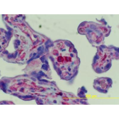
Immunohistochemistry-Paraffin: FCRN/FCGRT Antibody [NBP1-89128] - Staining in human placental Hoffbauer cells at a 1:65 dilution. Image from a verified customer review.![Immunohistochemistry-Paraffin: FCRN/FCGRT Antibody [NBP1-89128]](https://resources.rndsystems.com/images/products/FCRN-FCGRT-Antibody-Immunohistochemistry-Paraffin-NBP1-89128-img0011.jpg "Immunohistochemistry-Paraffin: FCRN/FCGRT Antibody [NBP1-89128]")
Immunohistochemistry-Paraffin: FCRN/FCGRT Antibody [NBP1-89128]
Immunohistochemistry-Paraffin: FCRN/FCGRT Antibody [NBP1-89128] - Immunohistochemical staining of human liver shows moderate to strong cytoplasmic positivity in Kupffer cells.![Immunohistochemistry-Paraffin: FCRN/FCGRT Antibody [NBP1-89128]](https://resources.rndsystems.com/images/products/FCRN-FCGRT-Antibody-Immunohistochemistry-Paraffin-NBP1-89128-img0012.jpg "Immunohistochemistry-Paraffin: FCRN/FCGRT Antibody [NBP1-89128]")
Immunohistochemistry-Paraffin: FCRN/FCGRT Antibody [NBP1-89128]
Immunohistochemistry-Paraffin: FCRN/FCGRT Antibody [NBP1-89128] - Immunohistochemical staining of human cerebellum shows no positivity in neuronal cells as expected.![Immunohistochemistry-Paraffin: FCRN/FCGRT Antibody [NBP1-89128]](https://resources.rndsystems.com/images/products/FCRN-FCGRT-Antibody-Immunohistochemistry-Paraffin-NBP1-89128-img0015.jpg "Immunohistochemistry-Paraffin: FCRN/FCGRT Antibody [NBP1-89128]")
Immunohistochemistry-Paraffin: FCRN/FCGRT Antibody [NBP1-89128]
Immunohistochemistry-Paraffin: FCRN/FCGRT Antibody [NBP1-89128] - Staining of human placenta shows moderate to strong cytoplasmic positivity in Hofbauer cells.![Immunohistochemistry-Paraffin: FCRN/FCGRT Antibody [NBP1-89128]](https://resources.rndsystems.com/images/products/FCRN-FCGRT-Antibody-Immunohistochemistry-Paraffin-NBP1-89128-img0027.jpg "Immunohistochemistry-Paraffin: FCRN/FCGRT Antibody [NBP1-89128]")
Immunohistochemistry-Paraffin: FCRN/FCGRT Antibody [NBP1-89128]
Immunohistochemistry-Paraffin: FCRN/FCGRT Antibody [NBP1-89128] - Staining of human pancreas shows no positivity in exocrine glandular cells as expected.![Simple Western: FCRN/FCGRT Antibody [NBP1-89128]](https://resources.rndsystems.com/images/products/FCRN-FCGRT-Antibody-Simple-Western-NBP1-89128-img0008.jpg "Simple Western: FCRN/FCGRT Antibody [NBP1-89128]")
Simple Western: FCRN/FCGRT Antibody [NBP1-89128]
Simple Western: FCRN/FCGRT Antibody [NBP1-89128] - Simple Western lane view shows a specific band for FCGRT in 0.2 mg/ml of h. Liver (left), HepG2 (middle) and THP-1 (right) lysate. This experiment was performed under reducing conditions using the 12-230 kDa separation system.![Simple Western: FCRN/FCGRT Antibody [NBP1-89128]](https://resources.rndsystems.com/images/products/FCRN-FCGRT-Antibody-Simple-Western-NBP1-89128-img0010.jpg "Simple Western: FCRN/FCGRT Antibody [NBP1-89128]")
Simple Western: FCRN/FCGRT Antibody [NBP1-89128]
Simple Western: FCRN/FCGRT Antibody [NBP1-89128] - Electropherogram image(s) of corresponding Simple Western lane view. FCRN/FCGRT antibody was used at 1:20 dilution on h. Liver, HepG2, and THP-1 lysate(s).
Immunohistochemistry: FCRN/FCGRT Antibody - BSA Free [NBP1-89128] -
Expression of FcRn in non-cancerous A. and cancerous B. serial lung sections from a small set of patients (n=8)(A) FcRn expression was very week in bronchial epithelial cells (left panel) and marked in alveolar macrophages (right panel) in the non-cancerous lung. (B) In cancerous tissue, the staining revealed that FcRn is expressed in interstitial stromal cells including DCs (PS100), macrophages (CD163) (arrowheads indicate areas of colocalization) but no CD8+ T cells (CD8). A very weak staining was also observed in carcinomatous cells. Pictures from 3 patients were selected as they are representative of the different staining. Image collected and cropped by CiteAb from the following open publication (https://pubmed.ncbi.nlm.nih.gov/27384673), licensed under a CC-BY license. Not internally tested by Novus Biologicals.
Immunohistochemistry: FCRN/FCGRT Antibody - BSA Free [NBP1-89128] -
Expression of FcRn in non-cancerous A. and cancerous B. serial lung sections from a small set of patients (n=8)(A) FcRn expression was very week in bronchial epithelial cells (left panel) and marked in alveolar macrophages (right panel) in the non-cancerous lung. (B) In cancerous tissue, the staining revealed that FcRn is expressed in interstitial stromal cells including DCs (PS100), macrophages (CD163) (arrowheads indicate areas of colocalization) but no CD8+ T cells (CD8). A very weak staining was also observed in carcinomatous cells. Pictures from 3 patients were selected as they are representative of the different staining. Image collected and cropped by CiteAb from the following open publication (https://pubmed.ncbi.nlm.nih.gov/27384673), licensed under a CC-BY license. Not internally tested by Novus Biologicals.Applications for FCRN/FCGRT Antibody - BSA Free
Immunohistochemistry
Immunohistochemistry-Paraffin
Simple Western
Western Blot
See Simple Western Antibody Database for Simple Western validation: Tested in h. Liver, HepG2 (middle) and THP-1, separated by Size, antibody dilution of 1:20, apparent MW was 51, 44 kDa. Separated by Size-Wes, Sally Sue/Peggy Sue
Reviewed Applications
Read 2 reviews rated 4.5 using NBP1-89128 in the following applications:
Formulation, Preparation, and Storage
Purification
Formulation
Format
Preservative
Concentration
Shipping
Stability & Storage
Background: FCRN
Long Name
Alternate Names
Entrez Gene IDs
Gene Symbol
UniProt
Additional FCRN Products
Product Documents for FCRN/FCGRT Antibody - BSA Free
Certificate of Analysis
To download a Certificate of Analysis, please enter a lot or batch number in the search box below.
Product Specific Notices for FCRN/FCGRT Antibody - BSA Free
This product is for research use only and is not approved for use in humans or in clinical diagnosis. Primary Antibodies are guaranteed for 1 year from date of receipt.
Related Research Areas
Citations for FCRN/FCGRT Antibody - BSA Free
 Powered by Bioz
Customer Reviews for FCRN/FCGRT Antibody - BSA Free (2)
Have you used FCRN/FCGRT Antibody - BSA Free?
Submit a review and receive an Amazon gift card!
$25/€18/£15/$25CAN/¥2500 Yen for a review with an image
$10/€7/£6/$10CAN/¥1110 Yen for a review without an image
Submit a review
Customer Images
-
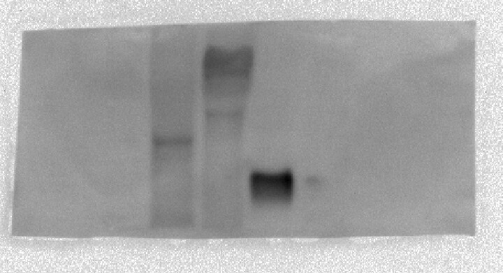
Application: Western BlotSample Tested: HepG2Species: HumanVerified Customer | Posted 10/16/2019Antibody was able to visualize endogenous FcRn (first lane), an FcRn-GFP fusion protein (middle lane), and a recombinant, shortened version of FcRn (third lane), with some background.

-
Application: Immunohistochemistry-ParaffinSample Tested: Penile urethra and placental tissueSpecies: HumanVerified Customer | Posted 12/04/2012Human Placental Hoffbauer cells at a 1:65 dilution.
There are no reviews that match your criteria.
Protocols
Find general support by application which include: protocols, troubleshooting, illustrated assays, videos and webinars.
- Antigen Retrieval Protocol (PIER)
- Antigen Retrieval for Frozen Sections Protocol
- Appropriate Fixation of IHC/ICC Samples
- Cellular Response to Hypoxia Protocols
- Chromogenic IHC Staining of Formalin-Fixed Paraffin-Embedded (FFPE) Tissue Protocol
- Chromogenic Immunohistochemistry Staining of Frozen Tissue
- ClariTSA™ Fluorophore Kits
- Detection & Visualization of Antibody Binding
- Fluorescent IHC Staining of Frozen Tissue Protocol
- Graphic Protocol for Heat-induced Epitope Retrieval
- Graphic Protocol for the Preparation and Fluorescent IHC Staining of Frozen Tissue Sections
- Graphic Protocol for the Preparation and Fluorescent IHC Staining of Paraffin-embedded Tissue Sections
- Graphic Protocol for the Preparation of Gelatin-coated Slides for Histological Tissue Sections
- IHC Sample Preparation (Frozen sections vs Paraffin)
- Immunofluorescent IHC Staining of Formalin-Fixed Paraffin-Embedded (FFPE) Tissue Protocol
- Immunohistochemistry (IHC) and Immunocytochemistry (ICC) Protocols
- Immunohistochemistry Frozen Troubleshooting
- Immunohistochemistry Paraffin Troubleshooting
- Preparing Samples for IHC/ICC Experiments
- Preventing Non-Specific Staining (Non-Specific Binding)
- Primary Antibody Selection & Optimization
- Protocol for Heat-Induced Epitope Retrieval (HIER)
- Protocol for Making a 4% Formaldehyde Solution in PBS
- Protocol for VisUCyte™ HRP Polymer Detection Reagent
- Protocol for the Preparation & Fixation of Cells on Coverslips
- Protocol for the Preparation and Chromogenic IHC Staining of Frozen Tissue Sections
- Protocol for the Preparation and Chromogenic IHC Staining of Frozen Tissue Sections - Graphic
- Protocol for the Preparation and Chromogenic IHC Staining of Paraffin-embedded Tissue Sections
- Protocol for the Preparation and Chromogenic IHC Staining of Paraffin-embedded Tissue Sections - Graphic
- Protocol for the Preparation and Fluorescent IHC Staining of Frozen Tissue Sections
- Protocol for the Preparation and Fluorescent IHC Staining of Paraffin-embedded Tissue Sections
- Protocol for the Preparation of Gelatin-coated Slides for Histological Tissue Sections
- R&D Systems Quality Control Western Blot Protocol
- TUNEL and Active Caspase-3 Detection by IHC/ICC Protocol
- The Importance of IHC/ICC Controls
- Troubleshooting Guide: Immunohistochemistry
- Troubleshooting Guide: Western Blot Figures
- Western Blot Conditions
- Western Blot Protocol
- Western Blot Protocol for Cell Lysates
- Western Blot Troubleshooting
- Western Blot Troubleshooting Guide
- View all Protocols, Troubleshooting, Illustrated assays and Webinars
FAQs for FCRN/FCGRT Antibody - BSA Free
-
Q: I use the anti-FcRn (Ref #NBP1-89128) and I was wondering if you have any clue about the nature of the higher slight band that we can see on the datasheet. Is it an isoform ?
A: The theoretical molecular weight of FCGRT is ~39.7kDa. We typically see this antibody run true to size, as although there is a signal peptide lost, there is a glycoslation site that will make up for the lost weight. In our image on our datasheet, lane 3 is the FCGRT protein co-expressed with a C- terminal myc-DDK tag, which is why we are seeing the faint signal ~3kDa above the protein of interest.