Ferritin Heavy Chain Antibody
Novus Biologicals | Catalog # NBP1-06985
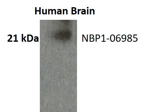
![Western Blot: Ferritin Heavy Chain Antibody [NBP1-06985]](https://resources.rndsystems.com/images/products/Ferritin-Heavy-Chain-Antibody-Western-Blot-NBP1-06985-img0001.jpg "Western Blot: Ferritin Heavy Chain Antibody [NBP1-06985]")
Loading...
Key Product Details
Species Reactivity
Validated:
Human
Cited:
Human
Applications
Validated:
Immunohistochemistry, Immunohistochemistry-Paraffin, Western Blot, Peptide ELISA, Immunocytochemistry/ Immunofluorescence
Cited:
Western Blot
Label
Unconjugated
Antibody Source
Polyclonal Goat IgG
Loading...
Product Specifications
Immunogen
Peptide with sequence C-DKHTLGDSDNES corresponding to C-Terminus according to NP_002023.2.
Clonality
Polyclonal
Host
Goat
Isotype
IgG
Scientific Data Images for Ferritin Heavy Chain Antibody
Western Blot: Ferritin Heavy Chain Antibody [NBP1-06985]
Western Blot: Ferritin Heavy Chain Antibody [NBP1-06985] - Staining of Human Placenta lysate (35 ug protein in RIPA buffer). Antibody at 1 ug/mL. Detected by chemiluminescence.![Immunocytochemistry/ Immunofluorescence: Ferritin Heavy Chain Antibody [NBP1-06985]](https://resources.rndsystems.com/images/products/Ferritin-Heavy-Chain-Antibody-Immunocytochemistry-Immunofluorescence-NBP1-06985-img0003.jpg "Immunocytochemistry/ Immunofluorescence: Ferritin Heavy Chain Antibody [NBP1-06985]")
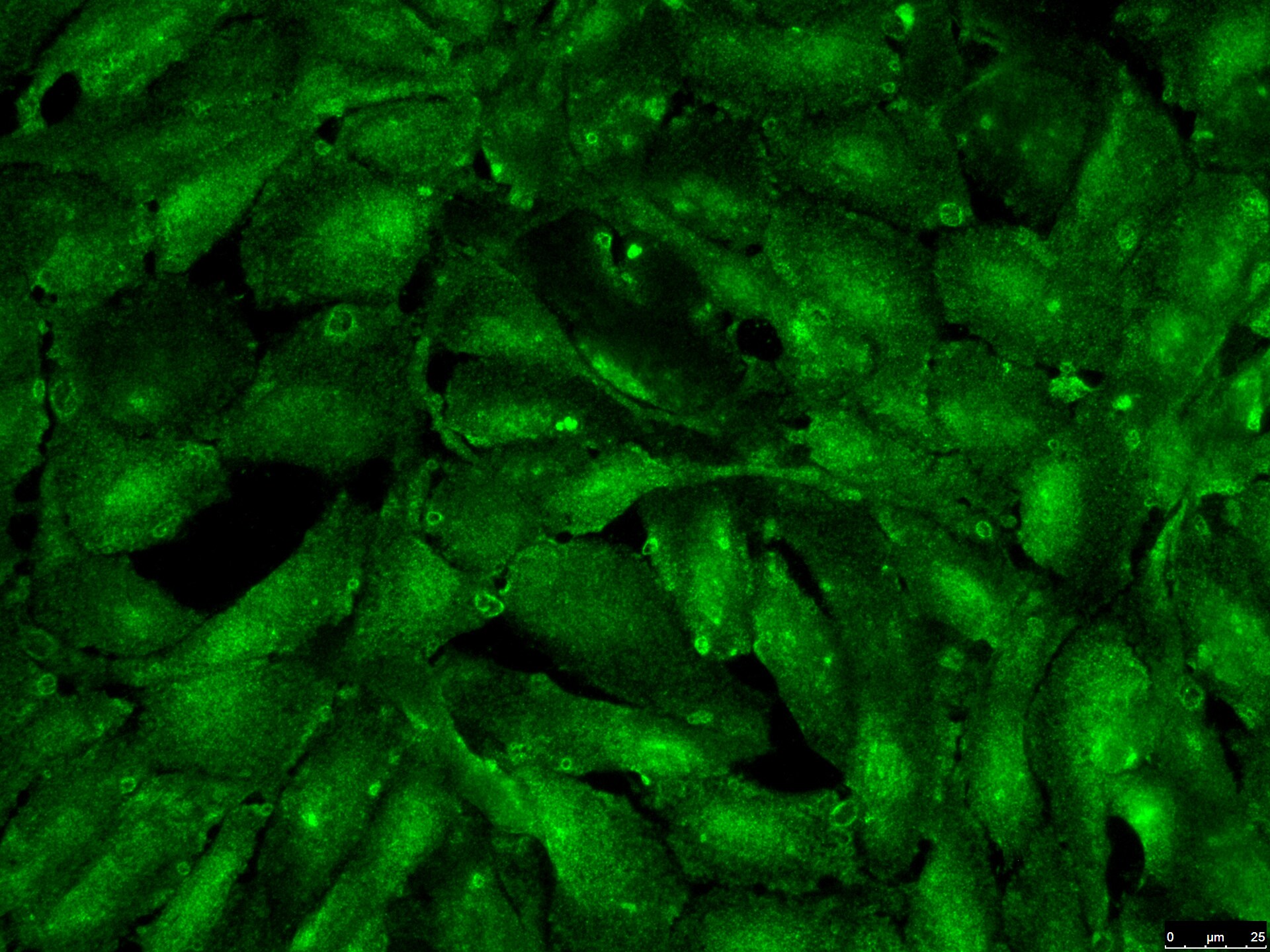
Immunocytochemistry/ Immunofluorescence: Ferritin Heavy Chain Antibody [NBP1-06985]
Immunocytochemistry/Immunofluorescence: Ferritin Heavy Chain Antibody [NBP1-06985] - Human ARPE-19 cells. Antibody at 1:100 overnight. ICC/IF image submitted by a verified customer review.![Immunohistochemistry-Paraffin: Ferritin Heavy Chain Antibody [NBP1-06985]](https://resources.rndsystems.com/images/products/Ferritin-Heavy-Chain-Antibody-Immunohistochemistry-Paraffin-NBP1-06985-img0002.jpg "Immunohistochemistry-Paraffin: Ferritin Heavy Chain Antibody [NBP1-06985]")
Immunohistochemistry-Paraffin: Ferritin Heavy Chain Antibody [NBP1-06985]
Immunohistochemistry-Paraffin: Ferritin Heavy Chain Antibody [NBP1-06985] - Staining of paraffin embedded Human Liver. Antibody at 2 ug/mL. Steamed antigen retrieval with citrate buffer pH 6, HRP-staining.Applications for Ferritin Heavy Chain Antibody
Application
Recommended Usage
Immunohistochemistry-Paraffin
2 - 4 ug/mL
Peptide ELISA
Detection limit 1:4000
Western Blot
1 - 3 ug/mL
Application Notes
This Ferritin Heavy Chain Antibody is validated for ICC/IF from a verified customer review.
Reviewed Applications
Read 2 reviews rated 5 using NBP1-06985 in the following applications:
Formulation, Preparation, and Storage
Purification
Immunogen affinity purified
Formulation
Tris saline (20 mM Tris pH 7.3, 150 mM NaCl), 0.5% BSA
Preservative
0.02% Sodium Azide
Concentration
0.5 mg/ml
Shipping
The product is shipped with polar packs. Upon receipt, store it immediately at the temperature recommended below.
Stability & Storage
Store at -20C. Avoid freeze-thaw cycles.
Background: Ferritin Heavy Chain
Alternate Names
apoferritin, Cell proliferation-inducing gene 15 protein, Ferritin H subunit, ferritin heavy chain, ferritin, heavy polypeptide 1, FHC, FTHEC 1.16.3.1, FTHL6MGC104426, PIG15, placenta immunoregulatory factor, PLIF, proliferation-inducing protein 15
Entrez Gene IDs
2495 (Human)
Gene Symbol
FTH1
UniProt
Additional Ferritin Heavy Chain Products
Product Documents for Ferritin Heavy Chain Antibody
Certificate of Analysis
To download a Certificate of Analysis, please enter a lot or batch number in the search box below.
Product Specific Notices for Ferritin Heavy Chain Antibody
This product is for research use only and is not approved for use in humans or in clinical diagnosis. Primary Antibodies are guaranteed for 1 year from date of receipt.
Citations for Ferritin Heavy Chain Antibody
 Powered by Bioz
Customer Reviews for Ferritin Heavy Chain Antibody (2)
5 out of 5
2 Customer Ratings
Have you used Ferritin Heavy Chain Antibody?
Submit a review and receive an Amazon gift card!
$25/€18/£15/$25CAN/¥2500 Yen for a review with an image
$10/€7/£6/$10CAN/¥1110 Yen for a review without an image
Submit a review
Customer Images
Showing
1
-
2 of
2 reviews
Showing All
Filter By:
-
Application: ImmunocytochemistrySample Tested: ARPE-19 cellsSpecies: HumanVerified Customer | Posted 01/27/2021Works really Well !!! Use at 1:100 (at 4 degrees overnight)Use at a concentration of 1:100 overnight

-
Application: Western BlotSample Tested: BrainSpecies: HumanVerified Customer | Posted 06/16/2017
There are no reviews that match your criteria.
Protocols
Find general support by application which include: protocols, troubleshooting, illustrated assays, videos and webinars.
- Antigen Retrieval Protocol (PIER)
- Antigen Retrieval for Frozen Sections Protocol
- Appropriate Fixation of IHC/ICC Samples
- Cellular Response to Hypoxia Protocols
- Chromogenic IHC Staining of Formalin-Fixed Paraffin-Embedded (FFPE) Tissue Protocol
- Chromogenic Immunohistochemistry Staining of Frozen Tissue
- ClariTSA™ Fluorophore Kits
- Detection & Visualization of Antibody Binding
- ELISA Sample Preparation & Collection Guide
- ELISA Troubleshooting Guide
- Fluorescent IHC Staining of Frozen Tissue Protocol
- Graphic Protocol for Heat-induced Epitope Retrieval
- Graphic Protocol for the Preparation and Fluorescent IHC Staining of Frozen Tissue Sections
- Graphic Protocol for the Preparation and Fluorescent IHC Staining of Paraffin-embedded Tissue Sections
- Graphic Protocol for the Preparation of Gelatin-coated Slides for Histological Tissue Sections
- How to Run an R&D Systems DuoSet ELISA
- How to Run an R&D Systems Quantikine ELISA
- How to Run an R&D Systems Quantikine™ QuicKit™ ELISA
- ICC Cell Smear Protocol for Suspension Cells
- ICC Immunocytochemistry Protocol Videos
- ICC for Adherent Cells
- IHC Sample Preparation (Frozen sections vs Paraffin)
- Immunocytochemistry (ICC) Protocol
- Immunocytochemistry Troubleshooting
- Immunofluorescence of Organoids Embedded in Cultrex Basement Membrane Extract
- Immunofluorescent IHC Staining of Formalin-Fixed Paraffin-Embedded (FFPE) Tissue Protocol
- Immunohistochemistry (IHC) and Immunocytochemistry (ICC) Protocols
- Immunohistochemistry Frozen Troubleshooting
- Immunohistochemistry Paraffin Troubleshooting
- Preparing Samples for IHC/ICC Experiments
- Preventing Non-Specific Staining (Non-Specific Binding)
- Primary Antibody Selection & Optimization
- Protocol for Heat-Induced Epitope Retrieval (HIER)
- Protocol for Making a 4% Formaldehyde Solution in PBS
- Protocol for VisUCyte™ HRP Polymer Detection Reagent
- Protocol for the Fluorescent ICC Staining of Cell Smears - Graphic
- Protocol for the Fluorescent ICC Staining of Cultured Cells on Coverslips - Graphic
- Protocol for the Preparation & Fixation of Cells on Coverslips
- Protocol for the Preparation and Chromogenic IHC Staining of Frozen Tissue Sections
- Protocol for the Preparation and Chromogenic IHC Staining of Frozen Tissue Sections - Graphic
- Protocol for the Preparation and Chromogenic IHC Staining of Paraffin-embedded Tissue Sections
- Protocol for the Preparation and Chromogenic IHC Staining of Paraffin-embedded Tissue Sections - Graphic
- Protocol for the Preparation and Fluorescent ICC Staining of Cells on Coverslips
- Protocol for the Preparation and Fluorescent ICC Staining of Non-adherent Cells
- Protocol for the Preparation and Fluorescent ICC Staining of Stem Cells on Coverslips
- Protocol for the Preparation and Fluorescent IHC Staining of Frozen Tissue Sections
- Protocol for the Preparation and Fluorescent IHC Staining of Paraffin-embedded Tissue Sections
- Protocol for the Preparation of Gelatin-coated Slides for Histological Tissue Sections
- Protocol for the Preparation of a Cell Smear for Non-adherent Cell ICC - Graphic
- Quantikine HS ELISA Kit Assay Principle, Alkaline Phosphatase
- Quantikine HS ELISA Kit Principle, Streptavidin-HRP Polymer
- R&D Systems Quality Control Western Blot Protocol
- Sandwich ELISA (Colorimetric) – Biotin/Streptavidin Detection Protocol
- Sandwich ELISA (Colorimetric) – Direct Detection Protocol
- TUNEL and Active Caspase-3 Detection by IHC/ICC Protocol
- The Importance of IHC/ICC Controls
- Troubleshooting Guide: ELISA
- Troubleshooting Guide: Immunohistochemistry
- Troubleshooting Guide: Western Blot Figures
- Western Blot Conditions
- Western Blot Protocol
- Western Blot Protocol for Cell Lysates
- Western Blot Troubleshooting
- Western Blot Troubleshooting Guide
- View all Protocols, Troubleshooting, Illustrated assays and Webinars
Loading...