Fibrillarin Antibody (38F3)
Novus Biologicals | Catalog # NB300-269
![Immunocytochemistry/ Immunofluorescence: Fibrillarin Antibody (38F3) [NB300-269]](https://resources.rndsystems.com/images/products/Fibrillarin-Antibody-38F3-Immunocytochemistry-Immunofluorescence-NB300-269-img0008.jpg "Immunocytochemistry/ Immunofluorescence: Fibrillarin Antibody (38F3) [NB300-269]")
Loading...
Key Product Details
Species Reactivity
Validated:
Human, Mouse, Rat, Porcine, Bovine, C. elegans, Chicken, Drosophila, Equine, Plant, S. pombe, Yeast
Cited:
Human, Mouse, Insect - Drosophila, Nematode - Caenorhabditis elegans, Plant, S. pombe
Applications
Validated:
Immunohistochemistry, Immunohistochemistry-Paraffin, Immunohistochemistry-Frozen, Immunohistochemistry Whole-Mount, Western Blot, Flow Cytometry, Immunocytochemistry/ Immunofluorescence, Knockdown Validated
Cited:
Immunohistochemistry-Paraffin, Immunohistochemistry-Frozen, Immunohistochemistry Whole-Mount, Western Blot, Immunoblotting, Immunocytochemistry/ Immunofluorescence, Immunoprecipitation, IF/IHC
Label
Unconjugated
Antibody Source
Monoclonal Mouse IgG1 Clone # 38F3
Loading...
Product Specifications
Immunogen
Yeast nuclear preparation (S. cerevisiae).
Reactivity Notes
C. elegans and plant reactivity reported in scientific literature (PMID: 24722283 and 27588463 respectively). Use in S. pombe reported in scientific literature (PMID:31855181).
Localization
Nucleus > nucleolus. Note: Fibrillar region of the nucleolus.
Marker
Nucleolar Marker
Clonality
Monoclonal
Host
Mouse
Isotype
IgG1
Theoretical MW
34.5 kDa.
Disclaimer note: The observed molecular weight of the protein may vary from the listed predicted molecular weight due to post translational modifications, post translation cleavages, relative charges, and other experimental factors.
Disclaimer note: The observed molecular weight of the protein may vary from the listed predicted molecular weight due to post translational modifications, post translation cleavages, relative charges, and other experimental factors.
Scientific Data Images for Fibrillarin Antibody (38F3)
Immunocytochemistry/ Immunofluorescence: Fibrillarin Antibody (38F3) [NB300-269]
Immunocytochemistry/Immunofluorescence: Fibrillarin Antibody (38F3) [NB300-269] - High magnification confocal image of HeLa cells stained with fibrillarin antibody, dilution 1:100 from concentrated tissue culture media (Green), and costained with chicken vimentin pAb (Red), dilution 1:1,000. Nuclear DNA is revealed with the DAPI stain (Blue). The fibrillarin antibody shows strong staining of nucleoli in the nucleus, while the vimentin antibody reveals cytoplasmic intermediate filaments.![Western Blot: Fibrillarin Antibody (38F3) [NB300-269]](https://resources.rndsystems.com/images/products/Fibrillarin-Antibody-38F3-Western-Blot-NB300-269-img0007.jpg "Western Blot: Fibrillarin Antibody (38F3) [NB300-269]")
Western Blot: Fibrillarin Antibody (38F3) [NB300-269]
Western Blot: Fibrillarin Antibody (38F3) [NB300-269] - Analysis of lysates of cell fractions probed with Fibrillarin antibody, dilution 1:500 (Green): [1] protein standard, [2] C6 cytosol, [3] C6 nuclear, [4] HEK293 cytosol, [5] HEK293 nuclear, [6] NIH-3T3 cytosol and [7] NIH-3T3 nuclear fractions. The band at 37kDa corresponds to the fibrillarin protein detected exclusively in the nuclear fractions.![Immunocytochemistry/ Immunofluorescence: Fibrillarin Antibody (38F3) [NB300-269]](https://resources.rndsystems.com/images/products/Fibrillarin-Antibody-38F3-Immunocytochemistry-Immunofluorescence-NB300-269-img0012.jpg "Immunocytochemistry/ Immunofluorescence: Fibrillarin Antibody (38F3) [NB300-269]")
Immunocytochemistry/ Immunofluorescence: Fibrillarin Antibody (38F3) [NB300-269]
Fibrillarin-Antibody-38F3-Immunocytochemistry-Immunofluorescence-NB300-269-img0012.jpg![Immunohistochemistry-Paraffin: Fibrillarin Antibody (38F3) [NB300-269]](https://resources.rndsystems.com/images/products/Fibrillarin-Antibody-38F3-Immunohistochemistry-Paraffin-NB300-269-img0003.jpg "Immunohistochemistry-Paraffin: Fibrillarin Antibody (38F3) [NB300-269]")
Immunohistochemistry-Paraffin: Fibrillarin Antibody (38F3) [NB300-269]
Immunohistochemistry-Paraffin: Fibrillarin Antibody (38F3) [NB300-269] - IHC staining of Fibrillarin in human ovarian cancer using DAB with hematoxylin counterstain.![Immunocytochemistry/ Immunofluorescence: Fibrillarin Antibody (38F3) [NB300-269]](https://resources.rndsystems.com/images/products/Fibrillarin-Antibody-38F3-Immunocytochemistry-Immunofluorescence-NB300-269-img0013.jpg "Immunocytochemistry/ Immunofluorescence: Fibrillarin Antibody (38F3) [NB300-269]")
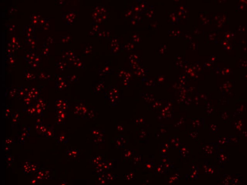
Immunocytochemistry/ Immunofluorescence: Fibrillarin Antibody (38F3) [NB300-269]
Immunocytochemistry/Immunofluorescence: Fibrillarin Antibody (38F3) [NB300-269] - Mouse embryonic fibroblast fibrillarin stained with fibrillarin antibody and Alexa-594 secondary antibody. ICC/IF image submitted by a verified customer review.![Flow Cytometry: Fibrillarin Antibody (38F3) [NB300-269]](https://resources.rndsystems.com/images/products/Fibrillarin-Antibody-38F3-Flow-Cytometry-NB300-269-img0004.jpg "Flow Cytometry: Fibrillarin Antibody (38F3) [NB300-269]")
Flow Cytometry: Fibrillarin Antibody (38F3) [NB300-269]
Flow Cytometry: Fibrillarin Antibody (38F3) [NB300-269] - Fibrillarin antibody was tested at 1:400 in HEK293 cells using an Alexa Fluor 488 secondary (shown in green) alongside unstained cells (shown in red).![Western Blot: Fibrillarin Antibody (38F3) [NB300-269]](https://resources.rndsystems.com/images/products/Fibrillarin-Antibody-38F3-Western-Blot-NB300-269-img0002.jpg "Western Blot: Fibrillarin Antibody (38F3) [NB300-269]")
Western Blot: Fibrillarin Antibody (38F3) [NB300-269]
Western Blot: Fibrillarin Antibody (38F3) [NB300-269] - Western blot analysis of Fibrillarin expression on yeast protein extracts using NB300-269.![Immunocytochemistry/ Immunofluorescence: Fibrillarin Antibody (38F3) [NB300-269]](https://resources.rndsystems.com/images/products/Fibrillarin-Antibody-38F3-Immunocytochemistry-Immunofluorescence-NB300-269-img0005.jpg "Immunocytochemistry/ Immunofluorescence: Fibrillarin Antibody (38F3) [NB300-269]")
Immunocytochemistry/ Immunofluorescence: Fibrillarin Antibody (38F3) [NB300-269]
Immunocytochemistry/Immunofluorescence: Fibrillarin Antibody (38F3) [NB300-269] - Fibrillarin immunofluorescence in fibroblasts. ICC/IF image submitted by a verified customer review.![Immunocytochemistry/ Immunofluorescence: Fibrillarin Antibody (38F3) [NB300-269]](https://resources.rndsystems.com/images/products/Fibrillarin-Antibody-38F3-Immunocytochemistry-Immunofluorescence-NB300-269-img0006.jpg "Immunocytochemistry/ Immunofluorescence: Fibrillarin Antibody (38F3) [NB300-269]")
Immunocytochemistry/ Immunofluorescence: Fibrillarin Antibody (38F3) [NB300-269]
Immunocytochemistry/Immunofluorescence: Fibrillarin Antibody (38F3) [NB300-269] - IF Confocal analysis of MCF7 cells using Fibrillarin antibody (NB300-269, 1:5). An Alexa Fluor 488-conjugated Goat to mouse IgG was used as secondary antibody (green). Actin filaments were labeled with Alexa Fluor 568 phalloidin (red). DAPI was used to stain the cell nuclei (blue). [NB300-269] -")
Immunocytochemistry/ Immunofluorescence: Fibrillarin Antibody (38F3) [NB300-269] -
Immunocytochemistry/ Immunofluorescence: Fibrillarin Antibody (38F3) [NB300-269] - The nuclei of hth mutants have dispersed rDNA foci & ectopic nucleoli & produce more rRNA.A) Wild type nuclei stained with anti-Fibrillarin (red) & a specific probe that recognizes the rDNA genomic region (green). B) hth mutant nuclei stained as in A. The mutant nuclei exhibit an increased number of nucleoli with different sizes. C) Quantification of nuclear dots marked with both the rDNA probe & anti-Fibrillarin in wild type & mutant nuclei. 45 nuclei were quantified for each genotype. (p-value: 0,00010132, calculated using T-Test). D) qRT-PCR comparing the levels of 18S RNA transcripts in wild type & Dfhth mutant embryos. The experiment was performed three independent times to be significant (p-value: 0,01046, calculated using T-Test). Error bars in the graph show the standard deviation & have being calculated as in Fig. 1E. The experiment was normalized using the bicoid (bcd) mRNA. Image collected & cropped by CiteAb from the following publication (https://pubmed.ncbi.nlm.nih.gov/25794008), licensed under a CC-BY license. Not internally tested by Novus Biologicals. [NB300-269] -")
Western Blot: Fibrillarin Antibody (38F3) [NB300-269] -
In vitro VM triggers nuclear YAP1 expression in U87 and U251 glioblastoma cells and is inhibited by Hippo pathway pharmacological targeting. (A) Human U87 and U251 glioblastoma cells were seeded either as monolayers (2D) or on top of Cultrex (3D) to generate capillary‐like structures. Subcellular fractionation was performed to isolate the cytosol and nuclear fractions from 2D or 3D cells. Representative blots for YAP1, Fibrillarin and GAPDH are presented from three independent fractionations. (B) Cytosolic (Cyt) and nuclear (Nuc) fractions were isolated from U87 glioblastoma cells seeded on Cultrex and treated with either vehicle or 10 μM of the indicated Hippo pathway inhibitors for 24 h. Representative blots for YAP1 and Fibrillarin are presented from two independent fractionations. (C) Total RNA was extracted from U87 cells cultured as in (A). RT‐qPCR was performed and relative gene expression of CTGF, Cyr61 and PPIA normalised over GAPDH. Data presented are representative triplicates from two independent experiments (*p < 0.05). Image collected and cropped by CiteAb from the following open publication (https://pubmed.ncbi.nlm.nih.gov/39718433), licensed under a CC-BY license. Not internally tested by Novus Biologicals. [NB300-269] -")
Western Blot: Fibrillarin Antibody (38F3) [NB300-269] -
In vitro VM triggers nuclear YAP1 expression in U87 and U251 glioblastoma cells and is inhibited by Hippo pathway pharmacological targeting. (A) Human U87 and U251 glioblastoma cells were seeded either as monolayers (2D) or on top of Cultrex (3D) to generate capillary‐like structures. Subcellular fractionation was performed to isolate the cytosol and nuclear fractions from 2D or 3D cells. Representative blots for YAP1, Fibrillarin and GAPDH are presented from three independent fractionations. (B) Cytosolic (Cyt) and nuclear (Nuc) fractions were isolated from U87 glioblastoma cells seeded on Cultrex and treated with either vehicle or 10 μM of the indicated Hippo pathway inhibitors for 24 h. Representative blots for YAP1 and Fibrillarin are presented from two independent fractionations. (C) Total RNA was extracted from U87 cells cultured as in (A). RT‐qPCR was performed and relative gene expression of CTGF, Cyr61 and PPIA normalised over GAPDH. Data presented are representative triplicates from two independent experiments (*p < 0.05). Image collected and cropped by CiteAb from the following open publication (https://pubmed.ncbi.nlm.nih.gov/39718433), licensed under a CC-BY license. Not internally tested by Novus Biologicals.Applications for Fibrillarin Antibody (38F3)
Application
Recommended Usage
Flow Cytometry
1:20-1:400
Immunocytochemistry/ Immunofluorescence
1:500-1:1000
Immunohistochemistry
1:500-1:1000
Immunohistochemistry-Frozen
1:500-1:1000
Immunohistochemistry-Paraffin
1:500-1:1000
Western Blot
1:1000-1:5000
Application Notes
This Fibrillarin Antibody (38F3) is useful for Western blot, Immunocytochemistry/Immunofluorescence, Flow Cytometry and Immunohistochemistry on both paraffin-embedded and frozen sections. In WB a band can be seen at approximately 34.5 kDa. In ICC/IF, this antibody shows prominent specular nucleolar staining.
Use in IHC-WhMt and IHC-Fr reported in scientific literature (PMID: 24722283 and 21539824 respectively). Use in Immunohistochemistry-paraffin reported in scientific literature (PMID: 23542174).
Use in IHC-WhMt and IHC-Fr reported in scientific literature (PMID: 24722283 and 21539824 respectively). Use in Immunohistochemistry-paraffin reported in scientific literature (PMID: 23542174).
Reviewed Applications
Read 4 reviews rated 5 using NB300-269 in the following applications:
Flow Cytometry Panel Builder
Bio-Techne Knows Flow Cytometry
Save time and reduce costly mistakes by quickly finding compatible reagents using the Panel Builder Tool.
Advanced Features
- Spectra Viewer - Custom analysis of spectra from multiple fluorochromes
- Spillover Popups - Visualize the spectra of individual fluorochromes
- Antigen Density Selector - Match fluorochrome brightness with antigen density

Formulation, Preparation, and Storage
Purification
Unpurified
Formulation
Supplied as concentrated hybridoma cell culture media.
Preservative
0.035% Sodium Azide
Concentration
This product is unpurified. The exact concentration of antibody is not quantifiable.
Shipping
The product is shipped with polar packs. Upon receipt, store it immediately at the temperature recommended below.
Stability & Storage
Store at 4C short term. Aliquot and store at -20C long term. Avoid freeze-thaw cycles.
Background: Fibrillarin
Alternate Names
EC 2.1.1,34-kD nucleolar scleroderma antigen, EC 2.1.1.-, EC 2.1.1.37, FIB, FIB1,34 kDa nucleolar scleroderma antigen, fibrillarin, FLRNrRNA 2'-O-methyltransferase fibrillarin, RNA, U3 small nucleolar interacting protein 1, RNU3IP1
Gene Symbol
FBL
Additional Fibrillarin Products
Product Documents for Fibrillarin Antibody (38F3)
Certificate of Analysis
To download a Certificate of Analysis, please enter a lot or batch number in the search box below.
Product Specific Notices for Fibrillarin Antibody (38F3)
This product is for research use only and is not approved for use in humans or in clinical diagnosis. Primary Antibodies are guaranteed for 1 year from date of receipt.
Citations for Fibrillarin Antibody (38F3)
 Powered by Bioz
Customer Reviews for Fibrillarin Antibody (38F3) (4)
5 out of 5
4 Customer Ratings
Have you used Fibrillarin Antibody (38F3)?
Submit a review and receive an Amazon gift card!
$25/€18/£15/$25CAN/¥2500 Yen for a review with an image
$10/€7/£6/$10CAN/¥1110 Yen for a review without an image
Submit a review
Customer Images
-(025-ml)_NB300-269_8066.bmp)
Showing
1
-
4 of
4 reviews
Showing All
Filter By:
-
Application: ImmunofluorescenceSample Tested: Mouse embryonic fibroblasts and fibroblastsSpecies: MouseVerified Customer | Posted 06/14/2021immunofluorescence of fibrillarin in mouse embryonic fibroblast using Alexa-594 secondary antibody

-
Application: Western BlotSample Tested: Tissue Lysates and 4% PFA-PBS fixed frozen sections from murine brainSpecies: C. elegansVerified Customer | Posted 01/09/2019
-
Application: ImmunocytochemistrySample Tested:Species: HumanVerified Customer | Posted 06/09/2014IF Confocal analysis of MCF7 cells using Fibrillarin antibody (NB300-269, 1:5).
-
Application: Western BlotSample Tested: fibroblastsSpecies: OtherVerified Customer | Posted 04/24/2014Fibrillarin immunofluorescence in fibroblasts
There are no reviews that match your criteria.
Protocols
View specific protocols for Fibrillarin Antibody (38F3) (NB300-269):
Materials
1) 1 Phosphate buffered saline (pH 7.6): NaCl 137mmol/L, KCl 2.7mmol/L, Na2HPO4 4.3mmol/L, KH2PO4 1.4 mmol/L
2) Citrate buffer, 0.01 M, pH6.0, Sodium Citrate 3g, Citric acid 0.4g
3) 3% Hydrogen peroxide
4) Primary antibody
5) Blocking serum (normal serum)
6) Biotinylated secondary antibody
7) DAB staining kit
Methods
1. Dewax and hydration of slides using xylene and EtOH:
Dry slides for 20 min in a 60 C oven
Add Xylene, 2 x 10 min
100%, 95%, 80%, and 70% EtOH, 5 min each EtOH concentration
Rinse in PBS, 5'
2 Antigen retrieval method (only for paraffin slides)
1a. High-pressure antigen retrieval procedure (recommended method)
Place slides in a glass slide holder (ensure that the slide holder is completely filled with slides, slides without sections if necessary, to ensure even heating. The entire slide holder is immersed in 1000 ml of Citrate buffer (0.01M, pH6.0) within a pressure cooker
Once steam is produced, and ONLY when steam is visible, from the pressure cooker (usually 15-20 min), the required high-pressure will have been reached, and slides will be incubated for 2 min.
Turn off heat, and allow buffer and slides to cool to room temperature
Slides are then rinsed in PBS for 5 minutes
2. Add 3% hydrogen peroxide solution, 10'at RT, then PBS, 3X5'
3. Normal blocking serum, 20'at RT
4. Incubate with Primary Ab, 4C overnight or 1.5 hours at 37C
5. Rinse with PBS, 3 X 5' each rinse
6. Add Biotin-conjugated second antibody, 10'at RT
7. Rinse with PBS, 3 X 5' each rinse
8. Add Streptavidin-Peroxidase, 10'at RT
9. Rinse with PBS, 3 X 5' each rinse
10. Staining with DAB solution, 2-5'under microscope
11. Stop the reaction by washing in tap water
12. Counterstain in Haematoxylin for 3-5 minutes
13. 75%, 80%, 95% and 100% ethanol, 5x2', xylene 2 x 10'
Find general support by application which include: protocols, troubleshooting, illustrated assays, videos and webinars.
- 7-Amino Actinomycin D (7-AAD) Cell Viability Flow Cytometry Protocol
- Antigen Retrieval Protocol (PIER)
- Antigen Retrieval for Frozen Sections Protocol
- Appropriate Fixation of IHC/ICC Samples
- Cellular Response to Hypoxia Protocols
- Chromogenic IHC Staining of Formalin-Fixed Paraffin-Embedded (FFPE) Tissue Protocol
- Chromogenic Immunohistochemistry Staining of Frozen Tissue
- ClariTSA™ Fluorophore Kits
- Detection & Visualization of Antibody Binding
- Extracellular Membrane Flow Cytometry Protocol
- Flow Cytometry Protocol for Cell Surface Markers
- Flow Cytometry Protocol for Staining Membrane Associated Proteins
- Flow Cytometry Staining Protocols
- Flow Cytometry Troubleshooting Guide
- Fluorescent IHC Staining of Frozen Tissue Protocol
- Graphic Protocol for Heat-induced Epitope Retrieval
- Graphic Protocol for the Preparation and Fluorescent IHC Staining of Frozen Tissue Sections
- Graphic Protocol for the Preparation and Fluorescent IHC Staining of Paraffin-embedded Tissue Sections
- Graphic Protocol for the Preparation of Gelatin-coated Slides for Histological Tissue Sections
- ICC Cell Smear Protocol for Suspension Cells
- ICC Immunocytochemistry Protocol Videos
- ICC for Adherent Cells
- IHC Sample Preparation (Frozen sections vs Paraffin)
- Immunocytochemistry (ICC) Protocol
- Immunocytochemistry Troubleshooting
- Immunofluorescence of Organoids Embedded in Cultrex Basement Membrane Extract
- Immunofluorescent IHC Staining of Formalin-Fixed Paraffin-Embedded (FFPE) Tissue Protocol
- Immunohistochemistry (IHC) and Immunocytochemistry (ICC) Protocols
- Immunohistochemistry Frozen Troubleshooting
- Immunohistochemistry Paraffin Troubleshooting
- Intracellular Flow Cytometry Protocol Using Alcohol (Methanol)
- Intracellular Flow Cytometry Protocol Using Detergents
- Intracellular Nuclear Staining Flow Cytometry Protocol Using Detergents
- Intracellular Staining Flow Cytometry Protocol Using Alcohol Permeabilization
- Intracellular Staining Flow Cytometry Protocol Using Detergents to Permeabilize Cells
- Preparing Samples for IHC/ICC Experiments
- Preventing Non-Specific Staining (Non-Specific Binding)
- Primary Antibody Selection & Optimization
- Propidium Iodide Cell Viability Flow Cytometry Protocol
- Protocol for Heat-Induced Epitope Retrieval (HIER)
- Protocol for Liperfluo
- Protocol for Making a 4% Formaldehyde Solution in PBS
- Protocol for VisUCyte™ HRP Polymer Detection Reagent
- Protocol for the Characterization of Human Th22 Cells
- Protocol for the Characterization of Human Th9 Cells
- Protocol for the Fluorescent ICC Staining of Cell Smears - Graphic
- Protocol for the Fluorescent ICC Staining of Cultured Cells on Coverslips - Graphic
- Protocol for the Preparation & Fixation of Cells on Coverslips
- Protocol for the Preparation and Chromogenic IHC Staining of Frozen Tissue Sections
- Protocol for the Preparation and Chromogenic IHC Staining of Frozen Tissue Sections - Graphic
- Protocol for the Preparation and Chromogenic IHC Staining of Paraffin-embedded Tissue Sections
- Protocol for the Preparation and Chromogenic IHC Staining of Paraffin-embedded Tissue Sections - Graphic
- Protocol for the Preparation and Fluorescent ICC Staining of Cells on Coverslips
- Protocol for the Preparation and Fluorescent ICC Staining of Non-adherent Cells
- Protocol for the Preparation and Fluorescent ICC Staining of Stem Cells on Coverslips
- Protocol for the Preparation and Fluorescent IHC Staining of Frozen Tissue Sections
- Protocol for the Preparation and Fluorescent IHC Staining of Paraffin-embedded Tissue Sections
- Protocol for the Preparation of Gelatin-coated Slides for Histological Tissue Sections
- Protocol for the Preparation of a Cell Smear for Non-adherent Cell ICC - Graphic
- Protocol: Annexin V and PI Staining by Flow Cytometry
- Protocol: Annexin V and PI Staining for Apoptosis by Flow Cytometry
- R&D Systems Quality Control Western Blot Protocol
- TUNEL and Active Caspase-3 Detection by IHC/ICC Protocol
- The Importance of IHC/ICC Controls
- Troubleshooting Guide: Fluorokine Flow Cytometry Kits
- Troubleshooting Guide: Immunohistochemistry
- Troubleshooting Guide: Western Blot Figures
- Western Blot Conditions
- Western Blot Protocol
- Western Blot Protocol for Cell Lysates
- Western Blot Troubleshooting
- Western Blot Troubleshooting Guide
- View all Protocols, Troubleshooting, Illustrated assays and Webinars
Loading...