![Fibulin 1 Antibody - BSA Free Western Blot: Fibulin 1 Antibody - BSA Free [NBP1-84725]](https://resources.rndsystems.com/images/products/nbp1-84725_rabbit-polyclonal-fibulin-1-antibody-104202515321323.jpg "Western Blot: Fibulin 1 Antibody - BSA Free [NBP1-84725]")
Fibulin 1 is a matricellular glycoprotein component of the elastic fiber core in connective tissues. It self-aggregates and additionally binds to several extracellular matrix and cellular proteins including Aggrecan, Versican, Fibronectin, ECM1, NOV/CCN3, and pro-HB-EGF. Fibulin 1c enhances ADAMTS-1 mediated cleavage of Aggrecan and Versican and can itself be cleaved by neutrophil elastase or MMP-13. It functions as a bridge between Fibrinogen and Fibrin, enabling Fibrin attachment to the platelet cell surface via Integrin alpha 2 beta 3. Fibulin 1 inhibits cardiomyocyte proliferation and Fibronectin-mediated cell adhesion and can promote bone formation.
Fibulin 1 Antibody - BSA Free
Novus Biologicals | Catalog # NBP1-84725
![Immunohistochemistry-Paraffin: Fibulin 1 Antibody [NBP1-84725]](https://resources.rndsystems.com/images/products/Fibulin-1-Antibody-Immunohistochemistry-Paraffin-NBP1-84725-img0012.jpg "Immunohistochemistry-Paraffin: Fibulin 1 Antibody [NBP1-84725]")
Loading...
Key Product Details
Validated by
Orthogonal Validation
Species Reactivity
Validated:
Human
Cited:
Human
Predicted:
Mouse (94%), Rat (94%). Backed by our 100% Guarantee.
Applications
Validated:
Immunohistochemistry, Immunohistochemistry-Paraffin, Western Blot
Cited:
Immunohistochemistry-Paraffin, Western Blot, Block/Neutralize
Label
Unconjugated
Antibody Source
Polyclonal Rabbit IgG
Format
BSA Free
Loading...
Product Specifications
Immunogen
This antibody was developed against Recombinant Protein corresponding to amino acids: NTLGSYLCSCSVGFRLSVDGRSCEDINECSSSPCSQECANVYGSYQCYCRRGYQLSDVDGVTCEDIDECALPTGGHICSYRCINIPGSFQCSCPSSGYRLAPNGRNCQDIDECVTGIHNCSINETCFNIQGGFRCLAFECPEN
Clonality
Polyclonal
Host
Rabbit
Isotype
IgG
Scientific Data Images for Fibulin 1 Antibody - BSA Free
![Immunohistochemistry-Paraffin: Fibulin 1 Antibody [NBP1-84725]](https://resources.rndsystems.com/images/products/Fibulin-1-Antibody-Immunohistochemistry-Paraffin-NBP1-84725-img0016.jpg "Immunohistochemistry-Paraffin: Fibulin 1 Antibody [NBP1-84725]")
Immunohistochemistry-Paraffin: Fibulin 1 Antibody [NBP1-84725]
Immunohistochemistry-Paraffin: Fibulin 1 Antibody [NBP1-84725] - Staining of human skeletal muscle shows no positivity in myocytes as expected.![Immunohistochemistry-Paraffin: Fibulin 1 Antibody [NBP1-84725]](https://resources.rndsystems.com/images/products/Fibulin-1-Antibody-Immunohistochemistry-Paraffin-NBP1-84725-img0013.jpg "Immunohistochemistry-Paraffin: Fibulin 1 Antibody [NBP1-84725]")
Immunohistochemistry-Paraffin: Fibulin 1 Antibody [NBP1-84725]
Immunohistochemistry-Paraffin: Fibulin 1 Antibody [NBP1-84725] - Staining of human placenta shows strong cytoplasmic positivity in trophoblastic cells.![Immunohistochemistry-Paraffin: Fibulin 1 Antibody [NBP1-84725]](https://resources.rndsystems.com/images/products/Fibulin-1-Antibody-Immunohistochemistry-Paraffin-NBP1-84725-img0014.jpg "Immunohistochemistry-Paraffin: Fibulin 1 Antibody [NBP1-84725]")
Immunohistochemistry-Paraffin: Fibulin 1 Antibody [NBP1-84725]
Immunohistochemistry-Paraffin: Fibulin 1 Antibody [NBP1-84725] - Staining of human endometrium shows moderate positivity in extracellular matrix.![Immunohistochemistry-Paraffin: Fibulin 1 Antibody [NBP1-84725]](https://resources.rndsystems.com/images/products/Fibulin-1-Antibody-Immunohistochemistry-Paraffin-NBP1-84725-img0015.jpg "Immunohistochemistry-Paraffin: Fibulin 1 Antibody [NBP1-84725]")
Immunohistochemistry-Paraffin: Fibulin 1 Antibody [NBP1-84725]
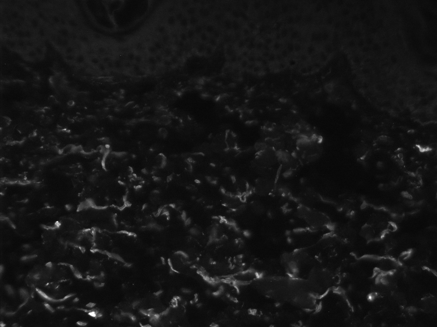
Immunohistochemistry-Paraffin: Fibulin 1 Antibody [NBP1-84725] - Staining of human skin shows strong positivity in extracellular matrix.Applications for Fibulin 1 Antibody - BSA Free
Application
Recommended Usage
Immunohistochemistry
1:50 - 1:200
Immunohistochemistry-Paraffin
1:50 - 1:200
Western Blot
0.04 - 0.4 ug/ml
Application Notes
IHC,HIER pH6 retrieval is recommended.
Reviewed Applications
Read 1 review rated 3 using NBP1-84725 in the following applications:
Formulation, Preparation, and Storage
Purification
Affinity purified
Formulation
PBS (pH 7.2) and 40% Glycerol
Format
BSA Free
Preservative
0.02% Sodium Azide
Concentration
Concentrations vary lot to lot. See vial label for concentration. If unlisted please contact technical services.
Shipping
The product is shipped with polar packs. Upon receipt, store it immediately at the temperature recommended below.
Stability & Storage
Store at 4C short term. Aliquot and store at -20C long term. Avoid freeze-thaw cycles.
Background: Fibulin 1
Alternate Names
FBLN1, FIBL1
Gene Symbol
FBLN1
Additional Fibulin 1 Products
Product Documents for Fibulin 1 Antibody - BSA Free
Certificate of Analysis
To download a Certificate of Analysis, please enter a lot or batch number in the search box below.
Product Specific Notices for Fibulin 1 Antibody - BSA Free
This product is for research use only and is not approved for use in humans or in clinical diagnosis. Primary Antibodies are guaranteed for 1 year from date of receipt.
Related Research Areas
Citations for Fibulin 1 Antibody - BSA Free
 Powered by Bioz
Customer Reviews for Fibulin 1 Antibody - BSA Free (1)
3 out of 5
1 Customer Rating
Have you used Fibulin 1 Antibody - BSA Free?
Submit a review and receive an Amazon gift card!
$25/€18/£15/$25CAN/¥2500 Yen for a review with an image
$10/€7/£6/$10CAN/¥1110 Yen for a review without an image
Submit a review
Customer Images
Showing
1
-
1 of
1 review
Showing All
Filter By:
-
Application: Immunohistochemistry-ParaffinSample Tested: human skinSpecies: HumanVerified Customer | Posted 04/11/2019

There are no reviews that match your criteria.
Protocols
Find general support by application which include: protocols, troubleshooting, illustrated assays, videos and webinars.
- Antigen Retrieval Protocol (PIER)
- Antigen Retrieval for Frozen Sections Protocol
- Appropriate Fixation of IHC/ICC Samples
- Cellular Response to Hypoxia Protocols
- Chromogenic IHC Staining of Formalin-Fixed Paraffin-Embedded (FFPE) Tissue Protocol
- Chromogenic Immunohistochemistry Staining of Frozen Tissue
- ClariTSA™ Fluorophore Kits
- Detection & Visualization of Antibody Binding
- Fluorescent IHC Staining of Frozen Tissue Protocol
- Graphic Protocol for Heat-induced Epitope Retrieval
- Graphic Protocol for the Preparation and Fluorescent IHC Staining of Frozen Tissue Sections
- Graphic Protocol for the Preparation and Fluorescent IHC Staining of Paraffin-embedded Tissue Sections
- Graphic Protocol for the Preparation of Gelatin-coated Slides for Histological Tissue Sections
- IHC Sample Preparation (Frozen sections vs Paraffin)
- Immunofluorescent IHC Staining of Formalin-Fixed Paraffin-Embedded (FFPE) Tissue Protocol
- Immunohistochemistry (IHC) and Immunocytochemistry (ICC) Protocols
- Immunohistochemistry Frozen Troubleshooting
- Immunohistochemistry Paraffin Troubleshooting
- Preparing Samples for IHC/ICC Experiments
- Preventing Non-Specific Staining (Non-Specific Binding)
- Primary Antibody Selection & Optimization
- Protocol for Heat-Induced Epitope Retrieval (HIER)
- Protocol for Making a 4% Formaldehyde Solution in PBS
- Protocol for VisUCyte™ HRP Polymer Detection Reagent
- Protocol for the Preparation & Fixation of Cells on Coverslips
- Protocol for the Preparation and Chromogenic IHC Staining of Frozen Tissue Sections
- Protocol for the Preparation and Chromogenic IHC Staining of Frozen Tissue Sections - Graphic
- Protocol for the Preparation and Chromogenic IHC Staining of Paraffin-embedded Tissue Sections
- Protocol for the Preparation and Chromogenic IHC Staining of Paraffin-embedded Tissue Sections - Graphic
- Protocol for the Preparation and Fluorescent IHC Staining of Frozen Tissue Sections
- Protocol for the Preparation and Fluorescent IHC Staining of Paraffin-embedded Tissue Sections
- Protocol for the Preparation of Gelatin-coated Slides for Histological Tissue Sections
- R&D Systems Quality Control Western Blot Protocol
- TUNEL and Active Caspase-3 Detection by IHC/ICC Protocol
- The Importance of IHC/ICC Controls
- Troubleshooting Guide: Immunohistochemistry
- Troubleshooting Guide: Western Blot Figures
- Western Blot Conditions
- Western Blot Protocol
- Western Blot Protocol for Cell Lysates
- Western Blot Troubleshooting
- Western Blot Troubleshooting Guide
- View all Protocols, Troubleshooting, Illustrated assays and Webinars
Loading...