FPRL1/FPR2 Antibody - BSA Free
Novus Biologicals | Catalog # NLS1878
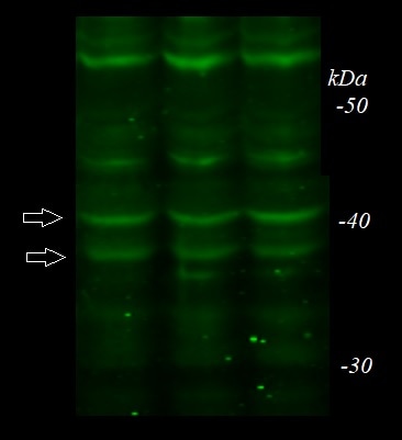
![Western Blot: FPRL1/FPR2 AntibodyBSA Free [NLS1878]](https://resources.rndsystems.com/images/products/FPRL1-FPR2-Antibody---BSA-Free-Western-Blot-NLS1878-img0012.jpg "Western Blot: FPRL1/FPR2 AntibodyBSA Free [NLS1878]")
Key Product Details
Validated by
Biological Validation
Species Reactivity
Validated:
Human, Mouse, Rat, Bacteria
Cited:
Human, Mouse, Rat, Bacteria
Applications
Validated:
Immunohistochemistry, Immunohistochemistry-Paraffin, Immunohistochemistry-Frozen, Western Blot, Flow Cytometry, Immunocytochemistry/ Immunofluorescence, Electron Microscopy
Cited:
Immunohistochemistry, Immunohistochemistry-Paraffin, Immunohistochemistry-Frozen, Western Blot, Flow Cytometry, Immunofluorescence, Immunocytochemistry/ Immunofluorescence, IF/IHC
Label
Unconjugated
Antibody Source
Polyclonal Rabbit IgG
Format
BSA Free
Loading...
Product Specifications
Immunogen
This FPRL1/FPR2 antibody is made to a synthetic peptide from the human FPRL1 protein (between residues 300-350) [UniProt P25090]
Reactivity Notes
Rat reactivity reported in scientific literature (PMID: 24086560). Bacteria reactivity reported in scientific literature (PMID: 31234710).
Localization
Cell membrane; Multi-pass membrane protein.
Clonality
Polyclonal
Host
Rabbit
Isotype
IgG
Theoretical MW
38 kDa.
Disclaimer note: The observed molecular weight of the protein may vary from the listed predicted molecular weight due to post translational modifications, post translation cleavages, relative charges, and other experimental factors.
Disclaimer note: The observed molecular weight of the protein may vary from the listed predicted molecular weight due to post translational modifications, post translation cleavages, relative charges, and other experimental factors.
Scientific Data Images for FPRL1/FPR2 Antibody - BSA Free
Western Blot: FPRL1/FPR2 AntibodyBSA Free [NLS1878]
FPRL1-FPR2-Antibody---BSA-Free-Western-Blot-NLS1878-img0012.jpg![Immunocytochemistry/ Immunofluorescence: FPRL1/FPR2 Antibody - BSA Free [NLS1878]](https://resources.rndsystems.com/images/products/FPRL1-FPR2-Antibody---BSA-Free-Immunocytochemistry-Immunofluorescence-NLS1878-img0008.jpg "Immunocytochemistry/ Immunofluorescence: FPRL1/FPR2 Antibody - BSA Free [NLS1878]")
Immunocytochemistry/ Immunofluorescence: FPRL1/FPR2 Antibody - BSA Free [NLS1878]
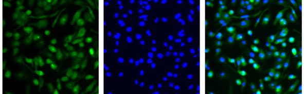
Immunocytochemistry/Immunofluorescence: FPRL1/FPR2 Antibody - BSA Free [NLS1878] - Mouse J774A.1, mouse reticulum cell sarcoma macrophage cell line. Left panel is + Anti-Rabbit FITC; middle panel indicates the cell nuclei stained with Hoechst; the right panel is a merged image. ICC/IF image submitted by a verified customer review.![Immunohistochemistry-Paraffin: FPRL1/FPR2 Antibody - BSA Free [NLS1878]](https://resources.rndsystems.com/images/products/FPRL1-FPR2-Antibody---BSA-Free-Immunohistochemistry-Paraffin-NLS1878-img0011.jpg "Immunohistochemistry-Paraffin: FPRL1/FPR2 Antibody - BSA Free [NLS1878]")
Immunohistochemistry-Paraffin: FPRL1/FPR2 Antibody - BSA Free [NLS1878]
Immunohistochemistry-Paraffin: FPRL1/FPR2 Antibody - BSA Free [NLS1878] - SPM receptors differentially expressed on endothelium and infiltrating leukocytes AE" ALX/FPR2 and ChemR23. Acute inflammation triggered by ventral aspect of forearm of healthy volunteers by intradermal injection of 1. 5 A-107 UV-killed E.coli (UVkEc) suspended in 100 i1/4l of saline. 4hrs after injection a 3-mm skin punch biopsy taken from inflamed site under local anesthesia. Naive skin treated as baseline. IHC-P on skin sections for receptor identification. Low mag(A-5) and high-mag (A-40) images at baseline and the 4 hr time point shown for ALX/FPR2. Red arrows highlight endothelium. Black arrow highlights infiltrating leukocytes. Image collected and cropped by Citeab from the (Pro-resolving mediators promote resolution in human skin model of UV-killed Escherichia coli-driven acute inflammation JCI Insight (2018)) licensed under, CC-BY license.![Flow Cytometry: FPRL1/FPR2 Antibody - BSA Free [NLS1878]](https://resources.rndsystems.com/images/products/FPRL1-FPR2-Antibody---BSA-Free-Flow-Cytometry-NLS1878-img0007.jpg "Flow Cytometry: FPRL1/FPR2 Antibody - BSA Free [NLS1878]")
Flow Cytometry: FPRL1/FPR2 Antibody - BSA Free [NLS1878]
Flow Cytometry: FPRL1/FPR2 Antibody - BSA Free [NLS1878] - Flow Cytometry: [Alexa Fluor® 700] [NLS1878AF700] - Mouse splenocytes. Cells were pre-gated with live/dead and FSC-A/W to exclude dead cells and cell doublets. Image from verified customer review. Image using the Alexa Fluor 700 form of this antibody.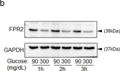
![Western Blot: FPRL1/FPR2 AntibodyBSA Free [NLS1878]](https://resources.rndsystems.com/images/products/FPRL1-FPR2-Antibody---BSA-Free-Western-Blot-NLS1878-img0004.jpg "Western Blot: FPRL1/FPR2 AntibodyBSA Free [NLS1878]")
Western Blot: FPRL1/FPR2 AntibodyBSA Free [NLS1878]
Western Blot: FPRL1/FPR2 Antibody - BSA Free [NLS1878] - Analysis in HL-60 cell lysate.![Immunocytochemistry/ Immunofluorescence: FPRL1/FPR2 Antibody - BSA Free [NLS1878]](https://resources.rndsystems.com/images/products/FPRL1-FPR2-Antibody---BSA-Free-Immunocytochemistry-Immunofluorescence-NLS1878-img0005.jpg "Immunocytochemistry/ Immunofluorescence: FPRL1/FPR2 Antibody - BSA Free [NLS1878]")
Immunocytochemistry/ Immunofluorescence: FPRL1/FPR2 Antibody - BSA Free [NLS1878]
Immunocytochemistry/Immunofluorescence: FPRL1/FPR2 Antibody - BSA Free [NLS1878] - Antibody was tested in Raw264.7 cells with DyLight 488 (green). Nuclei were counterstained with DAPI (blue).![Immunohistochemistry: FPRL1/FPR2 Antibody - BSA Free [NLS1878]](https://resources.rndsystems.com/images/products/FPRL1-FPR2-Antibody---BSA-Free-Immunohistochemistry-NLS1878-img0006.jpg "Immunohistochemistry: FPRL1/FPR2 Antibody - BSA Free [NLS1878]")
Immunohistochemistry: FPRL1/FPR2 Antibody - BSA Free [NLS1878]
Immunohistochemistry: FPRL1/FPR2 Antibody - BSA Free [NLS1878] - Analysis in human kidney cancer using DAB with hematoxylin counterstain.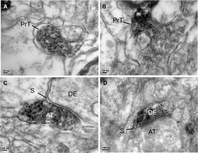
FPRL1-FPR2-Antibody---BSA-Free-Electron-Microscopy-NLS1878-img0010.jpg

Immunohistochemistry: FPRL1/FPR2 Antibody - BSA Free [NLS1878] -
Immunohistochemistry: FPRL1/FPR2 Antibody - BSA Free [NLS1878] - Electron micrographs of FPR2 immunostained sections from the prefrontal cortex. a, b Immunostaining is mostly present in axon pre-terminals (PrT) that did not form synapses with postsynaptic structures. c Occasional axon terminals (AT) are observed to form asymmetrical, putatively glutamatergic synapses (S) with unlabelled dendrites (DE). d Occasional labelled dendrites (DE) are also found, that formed asymmetrical synapses (S) with unlabelled axon terminals (AT). Scale: a, b, c = 50 nm, e = 100 nm Image collected & cropped by CiteAb from the following publication (https://pubmed.ncbi.nlm.nih.gov/29948727), licensed under a CC-BY license. Not internally tested by Novus Biologicals.
Immunocytochemistry/ Immunofluorescence: FPRL1/FPR2 Antibody - BSA Free [NLS1878] -
Immunocytochemistry/ Immunofluorescence: FPRL1/FPR2 Antibody - BSA Free [NLS1878] - Lipoxin A4, along with its precursors & ligand, were altered by hAECs. (A): Expression of the lipoxin A4 precursor genes ALOX‐5, ‐12, & ‐15 was increased at days 5 & 7 compared with saline controls (ALOX‐5: 0.393 ± 0.114 vs. 3.777 ± 1.98; ALOX‐12: 0.484 ± 0.248 vs. 1.520 ± 0.203; ALOX‐15: 0.020 ± 0.007 vs. 1.998 ± 0.983). ∗, p <.05. (B): Lipoxin A4 protein levels were elevated in lung lysates at day 7 in animals treated with hAECs compared with controls (0.103 ± 0.021 ng/ml vs. 0.249 ± 0.072 ng/ml, respectively). ∗, p <.05. (C): At day 7, bleomycin challenge resulted in positively stained F4/80/FPR2 cells, which were was elevated in mice treated with hAECs compared with control mice (5.720% ± 0.587% vs. 8.795% ± 0.687%, respectively). ∗, p <.05. (D): Representative images of F4/80‐ & FPR2‐positive‐stained lung sections from hAEC‐treated animals. Magnification: ×200. Scale bar =100 μm. Abbreviations: DAPI, 4′,6‐diamidino‐2‐phenylindole; FPR2, N‐formyl peptide receptor 2; hAEC, human amnion epithelial cell. Image collected & cropped by CiteAb from the following publication (https://pubmed.ncbi.nlm.nih.gov/28371562), licensed under a CC-BY license. Not internally tested by Novus Biologicals.
Western Blot: FPRL1/FPR2 Antibody - BSA Free [NLS1878] -
Western Blot: FPRL1/FPR2 Antibody - BSA Free [NLS1878] - Higher expression of Fpr2 in the livers of female mice is related with hepatocyte protection.a qRT-PCP analysis for Fpr2 expression in primary hepatocytes (pHEPs) from WT (male n = 2, female n = 2) & KO mice (male n = 2, female n = 2). Total eight mice were employed in each hepatocyte isolation, & the experiments were replicated at least three times & the mean ± S.E.M. results are graphed (*p < 0.05, **p < 0.005 vs WT male-pHEPs). b Western blot analysis & c double immunofluorescent images of Fpr2 (red) with albumin (green) in these cells. Gapdh was used as internal control. DAPI (blue) was used as nuclear counterstaining. Data shown represent one of three experiments with similar results (Scale bar, 20 μm). d qRT-PCR analysis for Fpr2 & glucose-6-phosphatase (G6pc) in, e cell viability of, & f western blot analysis of cleaved Caspase-3 & pro Caspase-3 in WT & KO female mice-isolated pHEPs treated with vehicle (Veh) or 250 μM of palmitate (PA). The data shown represent one of three experiments with similar results. The mean ± S.E.M. results obtained from three repetitive experiments are graphed (*p < 0.05, **p < 0.005 vs WT-Veh). Gray circles represent individual data points. See Supplementary Data for statistical details. Image collected & cropped by CiteAb from the following publication (https://pubmed.ncbi.nlm.nih.gov/35102146), licensed under a CC-BY license. Not internally tested by Novus Biologicals.Applications for FPRL1/FPR2 Antibody - BSA Free
Application
Recommended Usage
Immunocytochemistry/ Immunofluorescence
1:20-1:75
Immunohistochemistry
1:300
Immunohistochemistry-Frozen
reported in scientific literature (PMID 24086560)
Immunohistochemistry-Paraffin
1:300
Western Blot
1:1000
Application Notes
In Western Blot, a band is seen ~38 kDa representing FPRL1. In ICC/IF, plasma membrane staining was observed in Raw264.7 cells. In IHC-P, staining was also observed in the plasma membrane of human kidney cancer tissue. Prior to immunostaining paraffin tissues, antigen retrieval with sodium citrate buffer (pH 6.0) is recommended. Customers have reported success in IF on FFPE mouse kidney tissue, following microwave antigen retrieval.
Reviewed Applications
Read 4 reviews rated 4.5 using NLS1878 in the following applications:
Flow Cytometry Panel Builder
Bio-Techne Knows Flow Cytometry
Save time and reduce costly mistakes by quickly finding compatible reagents using the Panel Builder Tool.
Advanced Features
- Spectra Viewer - Custom analysis of spectra from multiple fluorochromes
- Spillover Popups - Visualize the spectra of individual fluorochromes
- Antigen Density Selector - Match fluorochrome brightness with antigen density

Formulation, Preparation, and Storage
Purification
Immunogen affinity purified
Formulation
PBS, 30% Glycerol
Format
BSA Free
Preservative
0.01% Sodium Azide
Concentration
1.1 mg/ml
Shipping
The product is shipped with polar packs. Upon receipt, store it immediately at the temperature recommended below.
Stability & Storage
Store at -20 degrees C. Avoid freeze/thaw cycles.
Background: FPRL1/FPR2
FPRL1 binding to its agonists triggers the activation of intracellular signaling molecules including calcium, PKC isoforms, phospholipases A2 and D, and MAPKs/p38MAPK. FPRL1 activation may result in an anti-inflammatory outcome counteracting the actions of proinflammatory signals such as leukotriene B4.
Expression of FPR1, FPRL1, and FPRL2 has recently been documented to enhance invasive potential of melanoma cells. Moreover, FPRL1 interacts with amyloid beta peptides is implicated in phagocyte attraction to sites of amyloid plaques in Alzheimer's disease.
Long Name
Formyl Peptide Receptor-like 1
Alternate Names
ALXR, FMLP-R-I, FPR2, HM63, LXA4 Receptor, RFP
Gene Symbol
FPR2
UniProt
Additional FPRL1/FPR2 Products
Product Documents for FPRL1/FPR2 Antibody - BSA Free
Certificate of Analysis
To download a Certificate of Analysis, please enter a lot or batch number in the search box below.
Product Specific Notices for FPRL1/FPR2 Antibody - BSA Free
This product is for research use only and is not approved for use in humans or in clinical diagnosis. Primary Antibodies are guaranteed for 1 year from date of receipt.
Citations for FPRL1/FPR2 Antibody - BSA Free
 Powered by Bioz
Customer Reviews for FPRL1/FPR2 Antibody - BSA Free (4)
4.5 out of 5
4 Customer Ratings
Have you used FPRL1/FPR2 Antibody - BSA Free?
Submit a review and receive an Amazon gift card!
$25/€18/£15/$25CAN/¥2500 Yen for a review with an image
$10/€7/£6/$10CAN/¥1110 Yen for a review without an image
Submit a review
Customer Images
Showing
1
-
4 of
4 reviews
Showing All
Filter By:
-
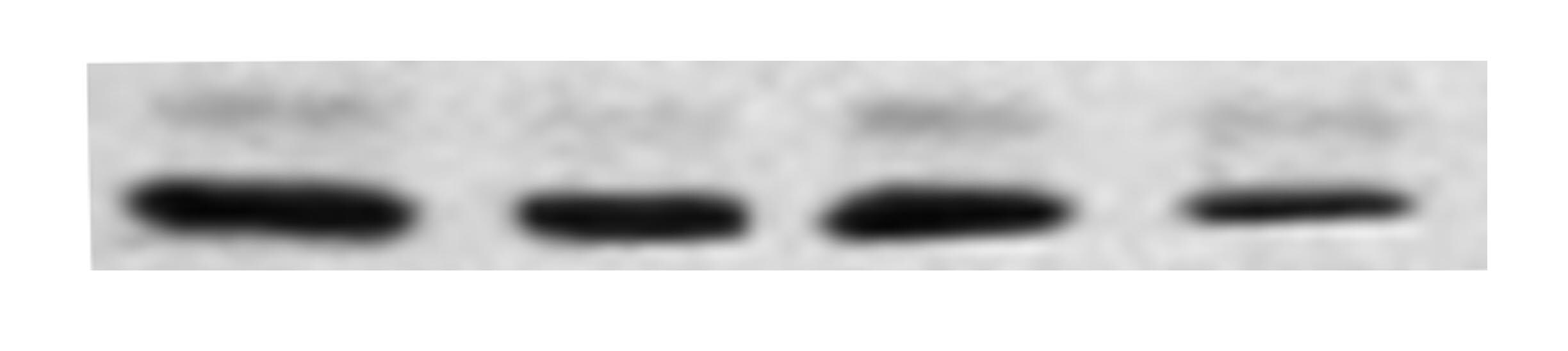
Application: Western BlotSample Tested: mouse primary cell and macrophage cell lysateSpecies: MouseVerified Customer | Posted 09/14/2019Mouse bone marrow cells. Lane 1: DMSO; Lane 2: chemical agonist #1; Lane 3: chemical agonist #2; Lane 4: chemical agonist #3.

-
Application: ImmunocytochemistrySample Tested: J774A.1 mouse reticulum cell sarcoma macrophage cell lineSpecies: MouseVerified Customer | Posted 08/10/2019The left panel is FPRL1/FPR2 Antibody + Anti-Rabbit FITC; the middle panel indicates the cell nuclei stained with Hoechst; the right panel is merged one.
-
Application: Western BlotSample Tested: Mouse brainSpecies: MouseVerified Customer | Posted 08/16/2018Western blot of FRPL1 (or LxA4R)Homogenates of Mouse Brain in RIPA Buffer
-
Application: Immunohistochemistry-FrozenSample Tested: See PMID 23603910Species: MouseVerified Customer | Posted 12/29/2014
There are no reviews that match your criteria.
Protocols
View specific protocols for FPRL1/FPR2 Antibody - BSA Free (NLS1878):
Immunocytochemistry Protocol
Culture cells to appropriate density in 35 mm culture dishes or 6-well plates.
1. Remove culture medium and wash the cells briefly in PBS. Add 10% formalin to the dish and fix at room temperature for 10 minutes.
2. Remove the formalin and wash the cells in PBS.
3. Permeablize the cells with 0.1% Triton X100 or other suitable detergent for 10 min.
4. Remove the permeablization buffer and wash three times for 10 minutes each in PBS. Be sure to not let the specimen dry out.
5. To block nonspecific antibody binding, incubate in 10% normal goat serum from 1 hour to overnight at room temperature.
6. Add primary antibody at appropriate dilution and incubate overnight at 4C.
7. Remove primary antibody and replace with PBS. Wash three times for 10 minutes each.
8. Add secondary antibody at appropriate dilution. Incubate for 1 hour at room temperature.
9. Remove secondary antibody and replace with PBS. Wash three times for 10 minutes each.
10. Counterstain DNA with DAPi if required.
Immunohistochemistry-Paraffin Embedded Sections
Antigen Unmasking:
Bring slides to a boil in 10 mM sodium citrate buffer (pH 6.0) then maintain at a sub-boiling temperature for 10 minutes. Cool slides on bench-top for 30 minutes.
Staining:
1. Wash sections in deionized water three times for 5 minutes each.
2. Wash sections in wash buffer for 5 minutes.
3. Block each section with 100-400 ul blocking solution for 1 hour at room temperature.
4. Remove blocking solution and add 100-400 ul diluted primary antibody. Incubate overnight at 4 C.
5. Remove antibody solution and wash sections in wash buffer three times for 5 minutes each.
6. Add 100-400 ul biotinylated diluted secondary antibody. Incubate 30 minutes at room temperature.
7. Remove secondary antibody solution and wash sections three times with wash buffer for 5 minutes each.
8. Add 100-400 ul Streptavidin-HRP reagent to each section and incubate for 30 minutes at room temperature.
9. Wash sections three times in wash buffer for 5 minutes each.
10. Add 100-400 ul DAB substrate to each section and monitor staining closely.
11. As soon as the sections develop, immerse slides in deionized water.
12. Counterstain sections in hematoxylin.
13. Wash sections in deionized water two times for 5 minutes each.
14. Dehydrate sections.
15. Mount coverslips.
*The above information is only intended as a guide. The researcher should determine what protocol best meets their needs. Please follow safe laboratory procedures.
Western Blot Protocol
1. Perform SDS-PAGE on samples to be analyzed, loading 40 ug of total protein per lane.
2. Transfer proteins to membrane according to the instructions provided by the manufacturer of the membrane and transfer apparatus.
3. Stain according to standard Ponceau S procedure (or similar product) to assess transfer success, and mark molecular weight standards where appropriate.
4. Rinse the blot.
5. Block the membrane using standard blocking buffer for at least 1 hour.
6. Wash the membrane in wash buffer three times for 10 minutes each.
7. Dilute primary antibody in blocking buffer and incubate 1 hour at room temperature.
8. Wash the membrane in wash buffer three times for 10 minutes each.
9. Apply the diluted HRP conjugated secondary antibody in blocking buffer (as per manufacturers instructions) and incubate 1 hour at room temperature.
10. Wash the blot in wash buffer three times for 10 minutes each (this step can be repeated as required to reduce background).
11. Apply the detection reagent of choice in accordance with the manufacturers instructions.
Note: Tween-20 can be added to the blocking or antibody dilution buffer at a final concentration of 0.05-0.2%.
*The above information is only intended as a guide. The researcher should determine what protocol best meets their needs. Please follow safe laboratory procedures.
Find general support by application which include: protocols, troubleshooting, illustrated assays, videos and webinars.
- 7-Amino Actinomycin D (7-AAD) Cell Viability Flow Cytometry Protocol
- Antigen Retrieval Protocol (PIER)
- Antigen Retrieval for Frozen Sections Protocol
- Appropriate Fixation of IHC/ICC Samples
- Cellular Response to Hypoxia Protocols
- Chromogenic IHC Staining of Formalin-Fixed Paraffin-Embedded (FFPE) Tissue Protocol
- Chromogenic Immunohistochemistry Staining of Frozen Tissue
- ClariTSA™ Fluorophore Kits
- Detection & Visualization of Antibody Binding
- Extracellular Membrane Flow Cytometry Protocol
- Flow Cytometry Protocol for Cell Surface Markers
- Flow Cytometry Protocol for Staining Membrane Associated Proteins
- Flow Cytometry Staining Protocols
- Flow Cytometry Troubleshooting Guide
- Fluorescent IHC Staining of Frozen Tissue Protocol
- Graphic Protocol for Heat-induced Epitope Retrieval
- Graphic Protocol for the Preparation and Fluorescent IHC Staining of Frozen Tissue Sections
- Graphic Protocol for the Preparation and Fluorescent IHC Staining of Paraffin-embedded Tissue Sections
- Graphic Protocol for the Preparation of Gelatin-coated Slides for Histological Tissue Sections
- ICC Cell Smear Protocol for Suspension Cells
- ICC Immunocytochemistry Protocol Videos
- ICC for Adherent Cells
- IHC Sample Preparation (Frozen sections vs Paraffin)
- Immunocytochemistry (ICC) Protocol
- Immunocytochemistry Troubleshooting
- Immunofluorescence of Organoids Embedded in Cultrex Basement Membrane Extract
- Immunofluorescent IHC Staining of Formalin-Fixed Paraffin-Embedded (FFPE) Tissue Protocol
- Immunohistochemistry (IHC) and Immunocytochemistry (ICC) Protocols
- Immunohistochemistry Frozen Troubleshooting
- Immunohistochemistry Paraffin Troubleshooting
- Intracellular Flow Cytometry Protocol Using Alcohol (Methanol)
- Intracellular Flow Cytometry Protocol Using Detergents
- Intracellular Nuclear Staining Flow Cytometry Protocol Using Detergents
- Intracellular Staining Flow Cytometry Protocol Using Alcohol Permeabilization
- Intracellular Staining Flow Cytometry Protocol Using Detergents to Permeabilize Cells
- Preparing Samples for IHC/ICC Experiments
- Preventing Non-Specific Staining (Non-Specific Binding)
- Primary Antibody Selection & Optimization
- Propidium Iodide Cell Viability Flow Cytometry Protocol
- Protocol for Heat-Induced Epitope Retrieval (HIER)
- Protocol for Liperfluo
- Protocol for Making a 4% Formaldehyde Solution in PBS
- Protocol for VisUCyte™ HRP Polymer Detection Reagent
- Protocol for the Characterization of Human Th22 Cells
- Protocol for the Characterization of Human Th9 Cells
- Protocol for the Fluorescent ICC Staining of Cell Smears - Graphic
- Protocol for the Fluorescent ICC Staining of Cultured Cells on Coverslips - Graphic
- Protocol for the Preparation & Fixation of Cells on Coverslips
- Protocol for the Preparation and Chromogenic IHC Staining of Frozen Tissue Sections
- Protocol for the Preparation and Chromogenic IHC Staining of Frozen Tissue Sections - Graphic
- Protocol for the Preparation and Chromogenic IHC Staining of Paraffin-embedded Tissue Sections
- Protocol for the Preparation and Chromogenic IHC Staining of Paraffin-embedded Tissue Sections - Graphic
- Protocol for the Preparation and Fluorescent ICC Staining of Cells on Coverslips
- Protocol for the Preparation and Fluorescent ICC Staining of Non-adherent Cells
- Protocol for the Preparation and Fluorescent ICC Staining of Stem Cells on Coverslips
- Protocol for the Preparation and Fluorescent IHC Staining of Frozen Tissue Sections
- Protocol for the Preparation and Fluorescent IHC Staining of Paraffin-embedded Tissue Sections
- Protocol for the Preparation of Gelatin-coated Slides for Histological Tissue Sections
- Protocol for the Preparation of a Cell Smear for Non-adherent Cell ICC - Graphic
- Protocol: Annexin V and PI Staining by Flow Cytometry
- Protocol: Annexin V and PI Staining for Apoptosis by Flow Cytometry
- R&D Systems Quality Control Western Blot Protocol
- TUNEL and Active Caspase-3 Detection by IHC/ICC Protocol
- The Importance of IHC/ICC Controls
- Troubleshooting Guide: Fluorokine Flow Cytometry Kits
- Troubleshooting Guide: Immunohistochemistry
- Troubleshooting Guide: Western Blot Figures
- Western Blot Conditions
- Western Blot Protocol
- Western Blot Protocol for Cell Lysates
- Western Blot Troubleshooting
- Western Blot Troubleshooting Guide
- View all Protocols, Troubleshooting, Illustrated assays and Webinars
FAQs for FPRL1/FPR2 Antibody - BSA Free
Showing
1
-
3 of
3 FAQs
Showing All
-
Q: I am interested in using this antibody for immunohistochemical staining of formalin-fixed rat tissues. I would like to know if this antibody is labeled with any substrate that may aid immunohistochemical staining, or would the HRP Labeled (NLS1878H) or Biotin Labeled (NLS1878B) form be more suitable for my application of interest?
A: Our product NLS1878 is provided as an unlabeled primary antibody. In order to use this antibody for staining in FFPE tissues, you will need to use a directly conjugated anti-rabbit IgG secondary. If you would like to use a directly conjugated primary antibody like NLS1878H or NLS1878B, you will not have to use a secondary antibody for detection. Depending on which detection reagents you are using, that will determine if you need the HRP conjugated or Biotin conjugated antibodies.
-
Q: I am looking for an FPRL1 antibody that is reactive to mouse only. Does Novus have the antibody in question?
A: To answer your question, unfortunately all our FPRL1 antibodies are polyclonal and would detect both mouse and human formyl peptide receptor-like-1 and we do not have any mouse monoclonal antibody for it.
-
Q: May I know if there is a blocking peptide available for this antibody?
A:
We do have the blocking peptide available for this item. It is sold under product #NLS1878PEP: Blocking Peptide.
-
Q: I am interested in using this antibody for immunohistochemical staining of formalin-fixed rat tissues. I would like to know if this antibody is labeled with any substrate that may aid immunohistochemical staining, or would the HRP Labeled (NLS1878H) or Biotin Labeled (NLS1878B) form be more suitable for my application of interest?
A: Our product NLS1878 is provided as an unlabeled primary antibody. In order to use this antibody for staining in FFPE tissues, you will need to use a directly conjugated anti-rabbit IgG secondary. If you would like to use a directly conjugated primary antibody like NLS1878H or NLS1878B, you will not have to use a secondary antibody for detection. Depending on which detection reagents you are using, that will determine if you need the HRP conjugated or Biotin conjugated antibodies.
-
Q: I am looking for an FPRL1 antibody that is reactive to mouse only. Does Novus have the antibody in question?
A: To answer your question, unfortunately all our FPRL1 antibodies are polyclonal and would detect both mouse and human formyl peptide receptor-like-1 and we do not have any mouse monoclonal antibody for it.
-
Q: May I know if there is a blocking peptide available for this antibody?
A:
We do have the blocking peptide available for this item. It is sold under product #NLS1878PEP: Blocking Peptide.
Loading...
Associated Pathways
