Fumarase Antibody (OTI1F10)
Novus Biologicals | Catalog # NBP1-47754
Key Product Details
Species Reactivity
Validated:
Cited:
Applications
Validated:
Cited:
Label
Antibody Source
Product Specifications
Immunogen
Reactivity Notes
Specificity
Clonality
Host
Isotype
Theoretical MW
Disclaimer note: The observed molecular weight of the protein may vary from the listed predicted molecular weight due to post translational modifications, post translation cleavages, relative charges, and other experimental factors.
Scientific Data Images for Fumarase Antibody (OTI1F10)
![Immunocytochemistry/ Immunofluorescence: Fumarase Antibody (OTI1F10) [NBP1-47754]](https://resources.rndsystems.com/images/products/Fumarase-Antibody-1F10-Immunocytochemistry-Immunofluorescence-NBP1-47754-img0006.jpg "Immunocytochemistry/ Immunofluorescence: Fumarase Antibody (OTI1F10) [NBP1-47754]")
Immunocytochemistry/ Immunofluorescence: Fumarase Antibody (OTI1F10) [NBP1-47754]
Immunocytochemistry/Immunofluorescence: Fumarase Antibody (1F10) [NBP1-47754] - Staining of COS7 cells transiently transfected by pCMV6-ENTRY Fumarase.![Immunohistochemistry: Fumarase Antibody (OTI1F10) [NBP1-47754]](https://resources.rndsystems.com/images/products/Fumarase-Antibody-OTI1F10-Immunohistochemistry-NBP1-47754-img0008.jpg "Immunohistochemistry: Fumarase Antibody (OTI1F10) [NBP1-47754]")
Immunohistochemistry: Fumarase Antibody (OTI1F10) [NBP1-47754]
Immunohistochemistry: Fumarase Antibody (OTI1F10) [NBP1-47754] - Fumarase Antibody (1F10) [NBP1-47754] - Staining of paraffin-embedded Human colon tissue using anti-Fumarase mouse monoclonal antibody.![Flow Cytometry: Fumarase Antibody (OTI1F10) [NBP1-47754]](https://resources.rndsystems.com/images/products/Fumarase-Antibody-1F10-Flow-Cytometry-NBP1-47754-img0001.jpg "Flow Cytometry: Fumarase Antibody (OTI1F10) [NBP1-47754]")
Flow Cytometry: Fumarase Antibody (OTI1F10) [NBP1-47754]
Flow Cytometry: Fumarase Antibody (1F10) [NBP1-47754] - HEK293T cells transfected with either overexpression plasmid (Red) or empty vector control plasmid (Blue) were immunostained by anti-Fumarase antibody, and then analyzed by flow cytometry.![Western Blot: Fumarase Antibody (OTI1F10) [NBP1-47754]](https://resources.rndsystems.com/images/products/Fumarase-Antibody-1F10-Western-Blot-NBP1-47754-img0002.jpg "Western Blot: Fumarase Antibody (OTI1F10) [NBP1-47754]")
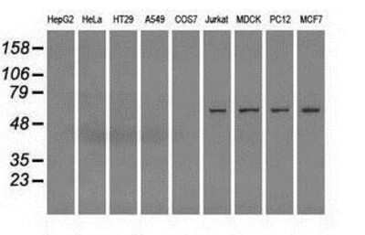
Western Blot: Fumarase Antibody (OTI1F10) [NBP1-47754]
Western Blot: Fumarase Antibody (1F10) [NBP1-47754] - HEK293T cells were transfected with the pCMV6-ENTRY control (Left lane) or pCMV6-ENTRY Fumarase (Right lane) cDNA for 48 hrs and lysed. Equivalent amounts of cell lysates (5 ug per lane) were separated by SDS-PAGE and immunoblotted with anti-Fumarase.![Immunohistochemistry-Paraffin: Fumarase Antibody (OTI1F10) [NBP1-47754]](https://resources.rndsystems.com/images/products/Fumarase-Antibody-1F10-Immunohistochemistry-Paraffin-NBP1-47754-img0004.jpg "Immunohistochemistry-Paraffin: Fumarase Antibody (OTI1F10) [NBP1-47754]")
Immunohistochemistry-Paraffin: Fumarase Antibody (OTI1F10) [NBP1-47754]
Immunohistochemistry-Paraffin: Fumarase Antibody (1F10) [NBP1-47754] - Staining of paraffin-embedded Human endometrium tissue using anti-Fumarase mouse monoclonal antibody.![Immunohistochemistry-Paraffin: Fumarase Antibody (OTI1F10) [NBP1-47754]](https://resources.rndsystems.com/images/products/Fumarase-Antibody-1F10-Immunohistochemistry-Paraffin-NBP1-47754-img0005.jpg "Immunohistochemistry-Paraffin: Fumarase Antibody (OTI1F10) [NBP1-47754]")
Immunohistochemistry-Paraffin: Fumarase Antibody (OTI1F10) [NBP1-47754]
Immunohistochemistry-Paraffin: Fumarase Antibody (1F10) [NBP1-47754] - Staining of paraffin-embedded Human Kidney tissue using anti-Fumarase mouse monoclonal antibody.Applications for Fumarase Antibody (OTI1F10)
Flow Cytometry
Immunocytochemistry/ Immunofluorescence
Immunohistochemistry
Immunohistochemistry-Paraffin
Western Blot
Flow Cytometry Panel Builder
Bio-Techne Knows Flow Cytometry
Save time and reduce costly mistakes by quickly finding compatible reagents using the Panel Builder Tool.
Advanced Features
- Spectra Viewer - Custom analysis of spectra from multiple fluorochromes
- Spillover Popups - Visualize the spectra of individual fluorochromes
- Antigen Density Selector - Match fluorochrome brightness with antigen density

Formulation, Preparation, and Storage
Purification
Formulation
Preservative
Concentration
Shipping
Stability & Storage
Background: Fumarase
Alternate Names
Gene Symbol
UniProt
Additional Fumarase Products
Product Documents for Fumarase Antibody (OTI1F10)
Certificate of Analysis
To download a Certificate of Analysis, please enter a lot or batch number in the search box below.
Product Specific Notices for Fumarase Antibody (OTI1F10)
This product is for research use only and is not approved for use in humans or in clinical diagnosis. Primary Antibodies are guaranteed for 1 year from date of receipt.
Citations for Fumarase Antibody (OTI1F10)
 Powered by Bioz
Customer Reviews for Fumarase Antibody (OTI1F10)
There are currently no reviews for this product. Be the first to review Fumarase Antibody (OTI1F10) and earn rewards!
Have you used Fumarase Antibody (OTI1F10)?
Submit a review and receive an Amazon gift card!
$25/€18/£15/$25CAN/¥2500 Yen for a review with an image
$10/€7/£6/$10CAN/¥1110 Yen for a review without an image
Submit a review
Protocols
Find general support by application which include: protocols, troubleshooting, illustrated assays, videos and webinars.
- 7-Amino Actinomycin D (7-AAD) Cell Viability Flow Cytometry Protocol
- Antigen Retrieval Protocol (PIER)
- Antigen Retrieval for Frozen Sections Protocol
- Appropriate Fixation of IHC/ICC Samples
- Cellular Response to Hypoxia Protocols
- Chromogenic IHC Staining of Formalin-Fixed Paraffin-Embedded (FFPE) Tissue Protocol
- Chromogenic Immunohistochemistry Staining of Frozen Tissue
- ClariTSA™ Fluorophore Kits
- Detection & Visualization of Antibody Binding
- Extracellular Membrane Flow Cytometry Protocol
- Flow Cytometry Protocol for Cell Surface Markers
- Flow Cytometry Protocol for Staining Membrane Associated Proteins
- Flow Cytometry Staining Protocols
- Flow Cytometry Troubleshooting Guide
- Fluorescent IHC Staining of Frozen Tissue Protocol
- Graphic Protocol for Heat-induced Epitope Retrieval
- Graphic Protocol for the Preparation and Fluorescent IHC Staining of Frozen Tissue Sections
- Graphic Protocol for the Preparation and Fluorescent IHC Staining of Paraffin-embedded Tissue Sections
- Graphic Protocol for the Preparation of Gelatin-coated Slides for Histological Tissue Sections
- ICC Cell Smear Protocol for Suspension Cells
- ICC Immunocytochemistry Protocol Videos
- ICC for Adherent Cells
- IHC Sample Preparation (Frozen sections vs Paraffin)
- Immunocytochemistry (ICC) Protocol
- Immunocytochemistry Troubleshooting
- Immunofluorescence of Organoids Embedded in Cultrex Basement Membrane Extract
- Immunofluorescent IHC Staining of Formalin-Fixed Paraffin-Embedded (FFPE) Tissue Protocol
- Immunohistochemistry (IHC) and Immunocytochemistry (ICC) Protocols
- Immunohistochemistry Frozen Troubleshooting
- Immunohistochemistry Paraffin Troubleshooting
- Intracellular Flow Cytometry Protocol Using Alcohol (Methanol)
- Intracellular Flow Cytometry Protocol Using Detergents
- Intracellular Nuclear Staining Flow Cytometry Protocol Using Detergents
- Intracellular Staining Flow Cytometry Protocol Using Alcohol Permeabilization
- Intracellular Staining Flow Cytometry Protocol Using Detergents to Permeabilize Cells
- Preparing Samples for IHC/ICC Experiments
- Preventing Non-Specific Staining (Non-Specific Binding)
- Primary Antibody Selection & Optimization
- Propidium Iodide Cell Viability Flow Cytometry Protocol
- Protocol for Heat-Induced Epitope Retrieval (HIER)
- Protocol for Liperfluo
- Protocol for Making a 4% Formaldehyde Solution in PBS
- Protocol for VisUCyte™ HRP Polymer Detection Reagent
- Protocol for the Characterization of Human Th22 Cells
- Protocol for the Characterization of Human Th9 Cells
- Protocol for the Fluorescent ICC Staining of Cell Smears - Graphic
- Protocol for the Fluorescent ICC Staining of Cultured Cells on Coverslips - Graphic
- Protocol for the Preparation & Fixation of Cells on Coverslips
- Protocol for the Preparation and Chromogenic IHC Staining of Frozen Tissue Sections
- Protocol for the Preparation and Chromogenic IHC Staining of Frozen Tissue Sections - Graphic
- Protocol for the Preparation and Chromogenic IHC Staining of Paraffin-embedded Tissue Sections
- Protocol for the Preparation and Chromogenic IHC Staining of Paraffin-embedded Tissue Sections - Graphic
- Protocol for the Preparation and Fluorescent ICC Staining of Cells on Coverslips
- Protocol for the Preparation and Fluorescent ICC Staining of Non-adherent Cells
- Protocol for the Preparation and Fluorescent ICC Staining of Stem Cells on Coverslips
- Protocol for the Preparation and Fluorescent IHC Staining of Frozen Tissue Sections
- Protocol for the Preparation and Fluorescent IHC Staining of Paraffin-embedded Tissue Sections
- Protocol for the Preparation of Gelatin-coated Slides for Histological Tissue Sections
- Protocol for the Preparation of a Cell Smear for Non-adherent Cell ICC - Graphic
- Protocol: Annexin V and PI Staining by Flow Cytometry
- Protocol: Annexin V and PI Staining for Apoptosis by Flow Cytometry
- R&D Systems Quality Control Western Blot Protocol
- TUNEL and Active Caspase-3 Detection by IHC/ICC Protocol
- The Importance of IHC/ICC Controls
- Troubleshooting Guide: Fluorokine Flow Cytometry Kits
- Troubleshooting Guide: Immunohistochemistry
- Troubleshooting Guide: Western Blot Figures
- Western Blot Conditions
- Western Blot Protocol
- Western Blot Protocol for Cell Lysates
- Western Blot Troubleshooting
- Western Blot Troubleshooting Guide
- View all Protocols, Troubleshooting, Illustrated assays and Webinars
FAQs for Fumarase Antibody (OTI1F10)
-
Q: I'm going to be using this antibody for Immunohistochemistry. Can I use a secondary antibody directly conjugated with the HRP, without the biotin/avidin system? Would the sensitivity be drastically affected? The PBS working concentration is 1X, right (PBS 1X + 0.1% Triton X-100, pH 7.4)? Which will be the same used for primary antibody? Which is the most adequate HIER protocol for this antibody? Can you provide a list of references where they show satisfactory results using this antibody?
A: You may use NBP1-47754 with a secondary that is directly conjugated to HRP, but the sensitivity will be reduced. Using a biotin/strepavidin system allows greater staining essentially because multiple enzymes associate with each primary antibody. If you already have an HRP-conjugated secondary, I would start with that but keep in mind that you may need to change your detection system if you get no signal or weak staining. I'm sorry but we don't have any peer-reviewed references for this product. Here is the recommended protocol for antigen retrieval: Antigen Retrieval Solution: 0.01M Sodium Citrate Buffer, pH 6.0 To prepare stock solutions: Solution A. 0.1 M citric acid solution: dissolve 21.0 g of citric acid, monohydrate (C6H8O7.H2O) in 100 ml of dH2O. Solution B. 0.1M sodium citrate solution: dissolve 29.4 g trisodium citrate dihydrate (C6H5Na3O7.2H2O) in 100 ml of dH2O. Working solution: Add 9 ml of Stock solution A and 41 ml of stock solution B to 450 ml of dH2O. Adjust pH to 6.0.