![Immunocytochemistry/ Immunofluorescence: GABPA Antibody [NBP1-32105]](https://resources.rndsystems.com/images/products/GABPA-Antibody-Immunocytochemistry-Immunofluorescence-NBP1-32105-img0006.jpg "Immunocytochemistry/ Immunofluorescence: GABPA Antibody [NBP1-32105]")
Loading...
Key Product Details
Validated by
Knockout/Knockdown
Species Reactivity
Validated:
Human, Mouse, Rat
Predicted:
Bovine (99%), Rhesus Macaque (100%). Backed by our 100% Guarantee.
Applications
Immunohistochemistry, Immunohistochemistry-Paraffin, Western Blot, Immunocytochemistry/ Immunofluorescence, Immunoprecipitation, Knockdown Validated
Label
Unconjugated
Antibody Source
Polyclonal Rabbit IgG
Format
BSA Free
Loading...
Product Specifications
Immunogen
Recombinant protein encompassing a sequence within the center region of human GABPA. The exact sequence is proprietary.
Reactivity Notes
Xenopus laevis (80%), Chicken (88%).
Localization
Nucleus
Clonality
Polyclonal
Host
Rabbit
Isotype
IgG
Theoretical MW
51 kDa.
Disclaimer note: The observed molecular weight of the protein may vary from the listed predicted molecular weight due to post translational modifications, post translation cleavages, relative charges, and other experimental factors.
Disclaimer note: The observed molecular weight of the protein may vary from the listed predicted molecular weight due to post translational modifications, post translation cleavages, relative charges, and other experimental factors.
Scientific Data Images for GABPA Antibody - BSA Free
Immunocytochemistry/ Immunofluorescence: GABPA Antibody [NBP1-32105]
Immunocytochemistry/Immunofluorescence: GABPA Antibody [NBP1-32105] - HeLa cells were fixed in 4% paraformaldehyde at RT for 15 min. Green: GABPA protein stained by GABPA antibody [N2C2], Internal diluted at 1:1000. Red: phalloidin, a cytoskeleton marker, diluted at 1:200. Blue: Hoechst 33342 staining.Scale bar = 10 um.
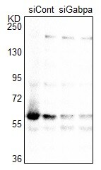
Western Blot: Rabbit Polyclonal GABPA Antibody [NBP1-32105] -
Western Blot: Rabbit Polyclonal GABPA Antibody [NBP1-32105] - Primary rat neonatal ventricular cardiomyocytes lysates with siRNA targeting Gabpa (siGabpa) and control (siCont). After 5 days of transfection with siRNA targeting rat Gabpa, the cells were lysed and subject to immunoblot analysis. Primary antibody dilution: 1/5000. Image from verified customer review.![Immunohistochemistry-Paraffin: GABPA Antibody [NBP1-32105]](https://resources.rndsystems.com/images/products/GABPA-Antibody-Immunohistochemistry-Paraffin-NBP1-32105-img0009.jpg "Immunohistochemistry-Paraffin: GABPA Antibody [NBP1-32105]")
Immunohistochemistry-Paraffin: GABPA Antibody [NBP1-32105]
Immunohistochemistry-Paraffin: GABPA Antibody [NBP1-32105] - Rat spleen. GABPA stained by GABPA antibody [N2C2], Internal diluted at 1:500.Antigen Retrieval: Citrate buffer, pH 6.0, 15 min.![Immunoprecipitation: GABPA Antibody [NBP1-32105]](https://resources.rndsystems.com/images/products/GABPA-Antibody-Immunoprecipitation-NBP1-32105-img0008.jpg "Immunoprecipitation: GABPA Antibody [NBP1-32105]")
Immunoprecipitation: GABPA Antibody [NBP1-32105]
Immunoprecipitation: GABPA Antibody [NBP1-32105] - Immunoprecipitation of GABPA protein from 293T whole cell extracts using 5 ug of GABPA antibody [N2C2], Internal. Western blot analysis was performed using GABPA antibody [N2C2], Internal. EasyBlot anti-Rabbit IgG was used as a secondary reagent.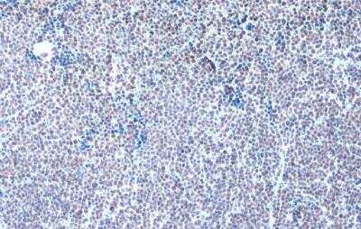
GABPA Antibody [NBP1-32105] - Mouse lymph node. GABPA stained by GABPA antibody [N2C2], Internal diluted at 1:500. Antigen Retrieval: Citrate buffer, pH 6.0, 15 min.

Western Blot: GABPA Antibody [NBP1-32105] -
Western Blot: GABPA Antibody [NBP1-32105] - Various whole cell extracts (30 ug) were separated by 10% SDS-PAGE, and the membrane was blotted with GABPA antibody [N2C2], Internal (NBP1-32105) diluted at 1:1000. The HRP-conjugated anti-rabbit IgG antibody was used to detect the primary antibody.Applications for GABPA Antibody - BSA Free
Application
Recommended Usage
Immunocytochemistry/ Immunofluorescence
1:100-1:1000
Immunoprecipitation
1:100-1:500
Knockdown Validated
Validated for Knockdown from a verified customer review.
Western Blot
1:500-1:3000
Application Notes
IHC-P-Assay dependent
Reviewed Applications
Read 1 review rated 5 using NBP1-32105 in the following applications:
Formulation, Preparation, and Storage
Purification
Antigen Affinity-purified
Formulation
PBS, 1% BSA, 20% Glycerol
Format
BSA Free
Preservative
0.025% Proclin 300
Concentration
Concentrations vary lot to lot. See vial label for concentration. If unlisted please contact technical services.
Shipping
The product is shipped with polar packs. Upon receipt, store it immediately at the temperature recommended below.
Stability & Storage
Aliquot and store at -20C or -80C. Avoid freeze-thaw cycles.
Background: GABPA
Alternate Names
E4TF1-60, E4TF1ATranscription factor E4TF1-60, GA binding protein transcription factor, alpha subunit 60kDa, GA-binding protein alpha chain, GA-binding protein transcription factor, alpha subunit (60kD), GABP subunit alpha, human nuclear respiratory factor-2 subunit alpha, 10, NFT2, NRF2, NRF2A, nuclear respiratory factor 2 alpha subunit, Nuclear respiratory factor 2 subunit alpha
Gene Symbol
GABPA
Additional GABPA Products
Product Documents for GABPA Antibody - BSA Free
Certificate of Analysis
To download a Certificate of Analysis, please enter a lot or batch number in the search box below.
Product Specific Notices for GABPA Antibody - BSA Free
This product is for research use only and is not approved for use in humans or in clinical diagnosis. Primary Antibodies are guaranteed for 1 year from date of receipt.
Customer Reviews for GABPA Antibody - BSA Free (1)
5 out of 5
1 Customer Rating
Have you used GABPA Antibody - BSA Free?
Submit a review and receive an Amazon gift card!
$25/€18/£15/$25CAN/¥2500 Yen for a review with an image
$10/€7/£6/$10CAN/¥1110 Yen for a review without an image
Submit a review
Customer Images
Showing
1
-
1 of
1 review
Showing All
Filter By:
-
Application: Western BlotSample Tested: primary neonatal ventricular cardiomyocytesSpecies: RatVerified Customer | Posted 08/29/2023Primary rat neonatal ventricular cardiomyocytes lysates with siRNA targeting Gabpa (siGabpa) and control (siCont).After 5 days of transfection with siRNA targeting rat Gabpa, the cells were lysed and subject to immunoblot analysis. Experimental conditions of immunoblot: Membrane: PDVF Blocking: 5% BSA/TBS-T for 0.5 hour First antibody (NBP1-32105) 1/5000 dilution (1 microL/5ml TBS-T) for overnight (16 hours) at 4 degrees. Second antibody HRP-anti-Rabbit IgG (Cell Signaling Technology 7074) 1/5000 dilution (1 microL/5ml TBS-T) for 1 hour at room temperature.

There are no reviews that match your criteria.
Protocols
Find general support by application which include: protocols, troubleshooting, illustrated assays, videos and webinars.
- Antigen Retrieval Protocol (PIER)
- Antigen Retrieval for Frozen Sections Protocol
- Appropriate Fixation of IHC/ICC Samples
- Cellular Response to Hypoxia Protocols
- Chromogenic IHC Staining of Formalin-Fixed Paraffin-Embedded (FFPE) Tissue Protocol
- Chromogenic Immunohistochemistry Staining of Frozen Tissue
- ClariTSA™ Fluorophore Kits
- Detection & Visualization of Antibody Binding
- Fluorescent IHC Staining of Frozen Tissue Protocol
- Graphic Protocol for Heat-induced Epitope Retrieval
- Graphic Protocol for the Preparation and Fluorescent IHC Staining of Frozen Tissue Sections
- Graphic Protocol for the Preparation and Fluorescent IHC Staining of Paraffin-embedded Tissue Sections
- Graphic Protocol for the Preparation of Gelatin-coated Slides for Histological Tissue Sections
- ICC Cell Smear Protocol for Suspension Cells
- ICC Immunocytochemistry Protocol Videos
- ICC for Adherent Cells
- IHC Sample Preparation (Frozen sections vs Paraffin)
- Immunocytochemistry (ICC) Protocol
- Immunocytochemistry Troubleshooting
- Immunofluorescence of Organoids Embedded in Cultrex Basement Membrane Extract
- Immunofluorescent IHC Staining of Formalin-Fixed Paraffin-Embedded (FFPE) Tissue Protocol
- Immunohistochemistry (IHC) and Immunocytochemistry (ICC) Protocols
- Immunohistochemistry Frozen Troubleshooting
- Immunohistochemistry Paraffin Troubleshooting
- Immunoprecipitation Protocol
- Preparing Samples for IHC/ICC Experiments
- Preventing Non-Specific Staining (Non-Specific Binding)
- Primary Antibody Selection & Optimization
- Protocol for Heat-Induced Epitope Retrieval (HIER)
- Protocol for Making a 4% Formaldehyde Solution in PBS
- Protocol for VisUCyte™ HRP Polymer Detection Reagent
- Protocol for the Fluorescent ICC Staining of Cell Smears - Graphic
- Protocol for the Fluorescent ICC Staining of Cultured Cells on Coverslips - Graphic
- Protocol for the Preparation & Fixation of Cells on Coverslips
- Protocol for the Preparation and Chromogenic IHC Staining of Frozen Tissue Sections
- Protocol for the Preparation and Chromogenic IHC Staining of Frozen Tissue Sections - Graphic
- Protocol for the Preparation and Chromogenic IHC Staining of Paraffin-embedded Tissue Sections
- Protocol for the Preparation and Chromogenic IHC Staining of Paraffin-embedded Tissue Sections - Graphic
- Protocol for the Preparation and Fluorescent ICC Staining of Cells on Coverslips
- Protocol for the Preparation and Fluorescent ICC Staining of Non-adherent Cells
- Protocol for the Preparation and Fluorescent ICC Staining of Stem Cells on Coverslips
- Protocol for the Preparation and Fluorescent IHC Staining of Frozen Tissue Sections
- Protocol for the Preparation and Fluorescent IHC Staining of Paraffin-embedded Tissue Sections
- Protocol for the Preparation of Gelatin-coated Slides for Histological Tissue Sections
- Protocol for the Preparation of a Cell Smear for Non-adherent Cell ICC - Graphic
- R&D Systems Quality Control Western Blot Protocol
- TUNEL and Active Caspase-3 Detection by IHC/ICC Protocol
- The Importance of IHC/ICC Controls
- Troubleshooting Guide: Immunohistochemistry
- Troubleshooting Guide: Western Blot Figures
- Western Blot Conditions
- Western Blot Protocol
- Western Blot Protocol for Cell Lysates
- Western Blot Troubleshooting
- Western Blot Troubleshooting Guide
- View all Protocols, Troubleshooting, Illustrated assays and Webinars
Loading...