gamma Tubulin Antibody (TU-30) - BSA Free
Novus Biologicals | Catalog # NB500-574
![Western Blot: gamma Tubulin Antibody (TU-30)BSA Free [NB500-574]](https://resources.rndsystems.com/images/products/gamma-Tubulin-Antibody-TU-30-Western-Blot-NB500-574-img0005.jpg "Western Blot: gamma Tubulin Antibody (TU-30)BSA Free [NB500-574]")
Key Product Details
Species Reactivity
Validated:
Human, Mouse, Rat, Porcine, Bovine, Chicken, Plant, Protozoa
Cited:
Human, Mouse
Applications
Validated:
Immunohistochemistry-Paraffin, Western Blot, Flow Cytometry, Flow (Intracellular), Immunocytochemistry/ Immunofluorescence
Cited:
Immunohistochemistry, Immunocytochemistry/ Immunofluorescence
Label
Unconjugated
Antibody Source
Monoclonal Mouse IgG1 Clone # TU-30
Format
BSA Free
Loading...
Product Specifications
Immunogen
C-terminal peptide of gamma-tubulin counjugated to KLH.
Epitope
The epitope was located in the amino acid sequence TRPDYI (aa439-444 in human), which is present on human gamma-tubulin 1 but not on human gamma-tubulin 2.
Reactivity Notes
Please note that this antibody is reactive to Mouse and derived from the same host, Mouse. Mouse-On-Mouse blocking reagent may be needed for IHC and ICC experiments to reduce high background signal. You can find these reagents under catalog numbers PK-2200-NB and MP-2400-NB. Please contact Technical Support if you have any questions.
Marker
Centrosome Marker
Specificity
The antibody TU-30 recognizes C-terminal peptide sequence EYHAATRPDYISWGTQ (aa 434-449) of gamma-tubulin, a 48 kDa structural constituent of cytoskeleton and microtubule organizing center (MTOC).
Clonality
Monoclonal
Host
Mouse
Isotype
IgG1
Theoretical MW
48 kDa.
Disclaimer note: The observed molecular weight of the protein may vary from the listed predicted molecular weight due to post translational modifications, post translation cleavages, relative charges, and other experimental factors.
Disclaimer note: The observed molecular weight of the protein may vary from the listed predicted molecular weight due to post translational modifications, post translation cleavages, relative charges, and other experimental factors.
Scientific Data Images for gamma Tubulin Antibody (TU-30) - BSA Free
Western Blot: gamma Tubulin Antibody (TU-30)BSA Free [NB500-574]
Western Blot: gamma Tubulin Antibody (TU-30) [NB500-574] - Western blotting analysis of differential reactivity of monoclonal antibodies to gamma-tubulin with human gamma-tubulin isotypes. (A) Immunoblots of total cell lysates from SH-SY5Y cells, expressing TagRFP-tagged human gamma-tubulin 1 (gamma-Tb1) or gamma-tubulin 2 (gamma-Tb2), probed with Abs to gamma-tubulin (TU-30, TU-32), TagRFP (RFP) and GAPDH. In control samples, only secondary anti-mouse Ab was applied. (B) Immunoblots of immobilized GST-tagged human C-terminal regions (a.a. 362-451) of gamma-Tb1 or gamma-Tb2 probed with Abs to gamma-tubulin (TU-30, TU-32) and GST. In control samples, only secondary anti-mouse Ab was applied.![Immunocytochemistry/ Immunofluorescence: gamma Tubulin Antibody (TU-30) - BSA Free [NB500-574]](https://resources.rndsystems.com/images/products/gamma-Tubulin-Antibody-TU-30-Immunofluorescence-NB500-574-img0004.jpg "Immunocytochemistry/ Immunofluorescence: gamma Tubulin Antibody (TU-30) - BSA Free [NB500-574]")
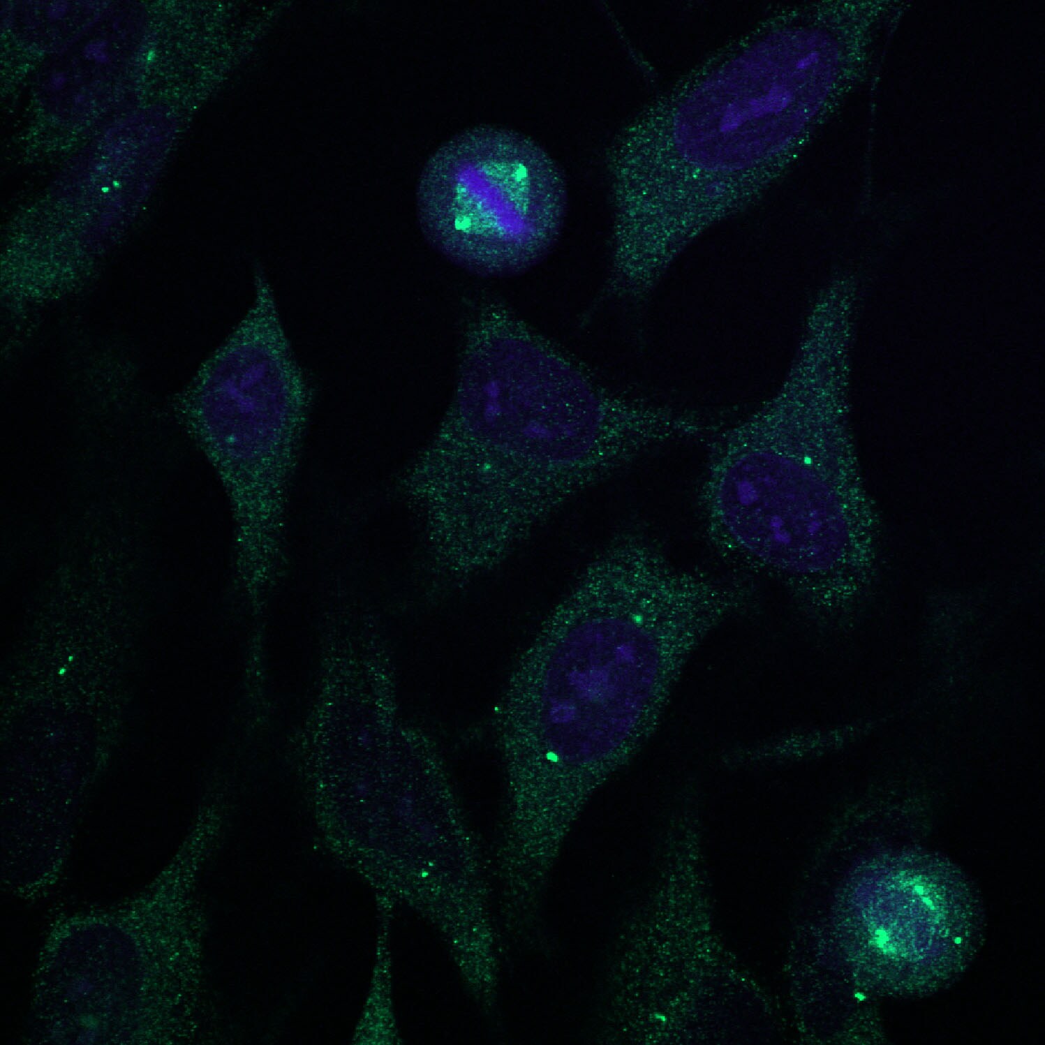
Immunocytochemistry/ Immunofluorescence: gamma Tubulin Antibody (TU-30) - BSA Free [NB500-574]
Immunocytochemistry/Immunofluorescence: gamma Tubulin Antibody (TU-30) [NB500-574] - Staining of HeLa cells using anti-gamma Tubulin antibody (green). Nuclei were stained with DAPI (blue). Image from verified customer review.![Immunohistochemistry-Paraffin: gamma Tubulin Antibody (TU-30) - BSA Free [NB500-574]](https://resources.rndsystems.com/images/products/gamma-Tubulin-Antibody-TU-30-BSA-Free-Immunohistochemistry-Paraffin-NB500-574-img0006.jpg "Immunohistochemistry-Paraffin: gamma Tubulin Antibody (TU-30) - BSA Free [NB500-574]")
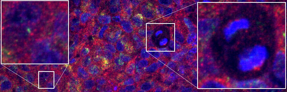
Immunohistochemistry-Paraffin: gamma Tubulin Antibody (TU-30) - BSA Free [NB500-574]
Immunohistochemistry-Paraffin: gamma Tubulin Antibody (TU-30) - BSA Free [NB500-574] - FFPE Glioblastoma PDX is stained with mouse-gamma tubulin antibody (NB500-574) (red) for centrosomes and rabbit Arl13B (green) for primary cilia. Gamma Tubulin antibody is diluted 1:25, incubated overnight at 4C. Image from verified customer review.![Immunocytochemistry/ Immunofluorescence: gamma Tubulin Antibody (TU-30) - BSA Free [NB500-574]](https://resources.rndsystems.com/images/products/gamma-Tubulin-Antibody-TU-30-Immunocytochemistry-Immunofluorescence-NB500-574-img0001.jpg "Immunocytochemistry/ Immunofluorescence: gamma Tubulin Antibody (TU-30) - BSA Free [NB500-574]")
Immunocytochemistry/ Immunofluorescence: gamma Tubulin Antibody (TU-30) - BSA Free [NB500-574]
Immunocytochemistry/Immunofluorescence: gamma Tubulin Antibody (TU-30) [NB500-574] - Staining of P19X1 mouse embryonal carcinoma cell line using anti-gamma-tubulin (TU-30) (detection by secondary antibody Goat anti-mouse Cy3). Nuclei were stained with DAPI (blue).![Immunocytochemistry/ Immunofluorescence: gamma Tubulin Antibody (TU-30) - BSA Free [NB500-574]](https://resources.rndsystems.com/images/products/gamma-Tubulin-Antibody-TU-30-Immunocytochemistry-Immunofluorescence-NB500-574-img0002.jpg "Immunocytochemistry/ Immunofluorescence: gamma Tubulin Antibody (TU-30) - BSA Free [NB500-574]")
Immunocytochemistry/ Immunofluorescence: gamma Tubulin Antibody (TU-30) - BSA Free [NB500-574]
Immunocytochemistry/Immunofluorescence: gamma Tubulin Antibody (TU-30) [NB500-574] - Staining of mouse fibroblasts using anti-gamma-tubulin (TU-30; direct conjugate with Dyomics 547, red). Nuclei were stained with DAPI (blue).![Immunocytochemistry/ Immunofluorescence: gamma Tubulin Antibody (TU-30) - BSA Free [NB500-574]](https://resources.rndsystems.com/images/products/gamma-Tubulin-Antibody-TU-30-Immunocytochemistry-Immunofluorescence-NB500-574-img0003.jpg "Immunocytochemistry/ Immunofluorescence: gamma Tubulin Antibody (TU-30) - BSA Free [NB500-574]")
Immunocytochemistry/ Immunofluorescence: gamma Tubulin Antibody (TU-30) - BSA Free [NB500-574]
Immunocytochemistry/Immunofluorescence: gamma Tubulin Antibody (TU-30) [NB500-574] - Staining of microtubular networks in 3T3 mouse fibroblasts. 3A - metaphase; 3B - anaphase; 3C - telophase Gamma-tubulin (red) stained with anti-gamma-tubulin (TU-30), alpha-tubulin (green) with polyclonal anti-alpha-tubulin antibody and nuclei with DAPI (blue).![gamma Tubulin Antibody (TU-30) - BSA Free Western Blot: gamma Tubulin Antibody (TU-30) - BSA Free [NB500-574]](https://resources.rndsystems.com/images/products/nb500-574_mouse-monoclonal-gamma-tubulin-antibody-tu-30-13120241629542.jpg "Western Blot: gamma Tubulin Antibody (TU-30) - BSA Free [NB500-574]")
Western Blot: gamma Tubulin Antibody (TU-30) - BSA Free [NB500-574]
Analysis of human gamma-tubulin using mouse monoclonal antibody TU-30 on lysates of various cell lines under reducing and non-reducing conditions. Nitrocellulose membrane was probed with 2 µg/ml of mouse anti-gamma-tubulin monoclonal antibody followed by IRDye800-conjugated anti-mouse secondary antibody. A specific band was detected for gamma-tubulin at approximately 46 kDa.Applications for gamma Tubulin Antibody (TU-30) - BSA Free
Application
Recommended Usage
Immunocytochemistry/ Immunofluorescence
1-2 ug/ml
Western Blot
1:1000
Application Notes
Immunocytochemistry - Staining technique: (a) Fix cells for 10 min in methanol at -20oC and for 6 min in acetone at -20oC; (b) Fix cells directly in methanol for 10 min at -20oC or in acetone for 10 min at -20oC. Incubation: 45 min in room temperature Application note: The antibody TU-30 stains only fixed cells. gamma Tubulin Antibody (TU-30) - BSA Free validated for Immunohistochemistry-Paraffin from a verified customer review.
Reviewed Applications
Read 2 reviews rated 4.5 using NB500-574 in the following applications:
Flow Cytometry Panel Builder
Bio-Techne Knows Flow Cytometry
Save time and reduce costly mistakes by quickly finding compatible reagents using the Panel Builder Tool.
Advanced Features
- Spectra Viewer - Custom analysis of spectra from multiple fluorochromes
- Spillover Popups - Visualize the spectra of individual fluorochromes
- Antigen Density Selector - Match fluorochrome brightness with antigen density

Formulation, Preparation, and Storage
Purification
Protein A purified
Formulation
Phosphate buffered saline (PBS), pH 7.4
Format
BSA Free
Preservative
15mM Sodium Azide
Concentration
1.0 mg/ml
Shipping
The product is shipped with polar packs. Upon receipt, store it immediately at the temperature recommended below.
Stability & Storage
Store at 4C. Do not freeze.
Background: gamma Tubulin
Long Name
Tubulin gamma-1 chain
Alternate Names
Gamma-1-tubulin, GCP-1, TUBG, TUBG1
Gene Symbol
TUBG1
Additional gamma Tubulin Products
Product Documents for gamma Tubulin Antibody (TU-30) - BSA Free
Certificate of Analysis
To download a Certificate of Analysis, please enter a lot or batch number in the search box below.
Product Specific Notices for gamma Tubulin Antibody (TU-30) - BSA Free
This product is for research use only and is not approved for use in humans or in clinical diagnosis. Primary Antibodies are guaranteed for 1 year from date of receipt.
Citations for gamma Tubulin Antibody (TU-30) - BSA Free
 Powered by Bioz
Customer Reviews for gamma Tubulin Antibody (TU-30) - BSA Free (2)
4.5 out of 5
2 Customer Ratings
Have you used gamma Tubulin Antibody (TU-30) - BSA Free?
Submit a review and receive an Amazon gift card!
$25/€18/£15/$25CAN/¥2500 Yen for a review with an image
$10/€7/£6/$10CAN/¥1110 Yen for a review without an image
Submit a review
Customer Images
Showing
1
-
2 of
2 reviews
Showing All
Filter By:
-
Application: Immunohistochemistry-ParaffinSample Tested: human glioblastomaSpecies: HumanVerified Customer | Posted 09/06/2022F-IHC. FFPE Glioblastoma PDX is stained with mouse-gamma tubulin antibody (NB500-574) (red) for centrosomes and rabbit Arl13B (green) for primary cilia. Gamma Tubulin antibody is diluted 1:25, incubated overnight at 4C.
 Bio-Techne ResponseThis review was submitted through the legacy Novus Innovators Program, reflecting a new species or application tested on a primary antibody.
 -
Application: ImmunofluorescenceSample Tested: HeLa cellsSpecies: HumanVerified Customer | Posted 08/16/2016Hela Cells with gamma Tubulin and DAPI
There are no reviews that match your criteria.
Protocols
Find general support by application which include: protocols, troubleshooting, illustrated assays, videos and webinars.
- 7-Amino Actinomycin D (7-AAD) Cell Viability Flow Cytometry Protocol
- Antigen Retrieval Protocol (PIER)
- Antigen Retrieval for Frozen Sections Protocol
- Appropriate Fixation of IHC/ICC Samples
- Cellular Response to Hypoxia Protocols
- Chromogenic IHC Staining of Formalin-Fixed Paraffin-Embedded (FFPE) Tissue Protocol
- Chromogenic Immunohistochemistry Staining of Frozen Tissue
- ClariTSA™ Fluorophore Kits
- Detection & Visualization of Antibody Binding
- Extracellular Membrane Flow Cytometry Protocol
- Flow Cytometry Protocol for Cell Surface Markers
- Flow Cytometry Protocol for Staining Membrane Associated Proteins
- Flow Cytometry Staining Protocols
- Flow Cytometry Troubleshooting Guide
- Fluorescent IHC Staining of Frozen Tissue Protocol
- Graphic Protocol for Heat-induced Epitope Retrieval
- Graphic Protocol for the Preparation and Fluorescent IHC Staining of Frozen Tissue Sections
- Graphic Protocol for the Preparation and Fluorescent IHC Staining of Paraffin-embedded Tissue Sections
- Graphic Protocol for the Preparation of Gelatin-coated Slides for Histological Tissue Sections
- ICC Cell Smear Protocol for Suspension Cells
- ICC Immunocytochemistry Protocol Videos
- ICC for Adherent Cells
- IHC Sample Preparation (Frozen sections vs Paraffin)
- Immunocytochemistry (ICC) Protocol
- Immunocytochemistry Troubleshooting
- Immunofluorescence of Organoids Embedded in Cultrex Basement Membrane Extract
- Immunofluorescent IHC Staining of Formalin-Fixed Paraffin-Embedded (FFPE) Tissue Protocol
- Immunohistochemistry (IHC) and Immunocytochemistry (ICC) Protocols
- Immunohistochemistry Frozen Troubleshooting
- Immunohistochemistry Paraffin Troubleshooting
- Intracellular Flow Cytometry Protocol Using Alcohol (Methanol)
- Intracellular Flow Cytometry Protocol Using Detergents
- Intracellular Nuclear Staining Flow Cytometry Protocol Using Detergents
- Intracellular Staining Flow Cytometry Protocol Using Alcohol Permeabilization
- Intracellular Staining Flow Cytometry Protocol Using Detergents to Permeabilize Cells
- Preparing Samples for IHC/ICC Experiments
- Preventing Non-Specific Staining (Non-Specific Binding)
- Primary Antibody Selection & Optimization
- Propidium Iodide Cell Viability Flow Cytometry Protocol
- Protocol for Heat-Induced Epitope Retrieval (HIER)
- Protocol for Liperfluo
- Protocol for Making a 4% Formaldehyde Solution in PBS
- Protocol for VisUCyte™ HRP Polymer Detection Reagent
- Protocol for the Characterization of Human Th22 Cells
- Protocol for the Characterization of Human Th9 Cells
- Protocol for the Fluorescent ICC Staining of Cell Smears - Graphic
- Protocol for the Fluorescent ICC Staining of Cultured Cells on Coverslips - Graphic
- Protocol for the Preparation & Fixation of Cells on Coverslips
- Protocol for the Preparation and Chromogenic IHC Staining of Frozen Tissue Sections
- Protocol for the Preparation and Chromogenic IHC Staining of Frozen Tissue Sections - Graphic
- Protocol for the Preparation and Chromogenic IHC Staining of Paraffin-embedded Tissue Sections
- Protocol for the Preparation and Chromogenic IHC Staining of Paraffin-embedded Tissue Sections - Graphic
- Protocol for the Preparation and Fluorescent ICC Staining of Cells on Coverslips
- Protocol for the Preparation and Fluorescent ICC Staining of Non-adherent Cells
- Protocol for the Preparation and Fluorescent ICC Staining of Stem Cells on Coverslips
- Protocol for the Preparation and Fluorescent IHC Staining of Frozen Tissue Sections
- Protocol for the Preparation and Fluorescent IHC Staining of Paraffin-embedded Tissue Sections
- Protocol for the Preparation of Gelatin-coated Slides for Histological Tissue Sections
- Protocol for the Preparation of a Cell Smear for Non-adherent Cell ICC - Graphic
- Protocol: Annexin V and PI Staining by Flow Cytometry
- Protocol: Annexin V and PI Staining for Apoptosis by Flow Cytometry
- R&D Systems Quality Control Western Blot Protocol
- TUNEL and Active Caspase-3 Detection by IHC/ICC Protocol
- The Importance of IHC/ICC Controls
- Troubleshooting Guide: Fluorokine Flow Cytometry Kits
- Troubleshooting Guide: Immunohistochemistry
- Troubleshooting Guide: Western Blot Figures
- Western Blot Conditions
- Western Blot Protocol
- Western Blot Protocol for Cell Lysates
- Western Blot Troubleshooting
- Western Blot Troubleshooting Guide
- View all Protocols, Troubleshooting, Illustrated assays and Webinars
Loading...