GFP Antibody (9F9.F9) - BSA Free
Novus Biologicals | Catalog # NB600-597
![Immunocytochemistry/ Immunofluorescence: GFP Antibody (9F9.F9) [NB600-597]](https://resources.rndsystems.com/images/products/GFP-Antibody-9F9-F9-Immunocytochemistry-Immunofluorescence-NB600-597-img0019.jpg "Immunocytochemistry/ Immunofluorescence: GFP Antibody (9F9.F9) [NB600-597]")
Key Product Details
Species Reactivity
Validated:
Non-species specific
Cited:
Human
Applications
Validated:
Immunohistochemistry, Immunohistochemistry-Paraffin, Western Blot, Immunoblotting, ELISA, Flow Cytometry, Immunocytochemistry/ Immunofluorescence, Immunoprecipitation, Dot Blot, In vitro assay, Knockdown Validated
Cited:
Immunohistochemistry-Paraffin, Immunohistochemistry-Frozen, Western Blot, Immunoblotting, ELISA, Immunocytochemistry/ Immunofluorescence, Immunoprecipitation, Cytometric Bead Assay Standard, In vitro assay, IF/IHC, Knockdown Validated
Label
Unconjugated
Antibody Source
Monoclonal Mouse IgG1 kappa Clone # 9F9.F9
Format
BSA Free
Loading...
Product Specifications
Immunogen
Recombinant Green Fluorescent Protein (GFP) fusion protein corresponding to the full length amino acid sequence (246 aa) derived from the jellyfish Aequorea victoria.
Reactivity Notes
Reactivity is observed against recombinant Green Fluorescent Protein from Aequorea victoria by both Western blot and ELISA. No reaction is seen against RFP.
Recognizes fusion proteins in Drosophila, reactivity reported in scientific literature (PMID: 26586808). Human reactivity reported in scientific literature (PMID: 28198010).
Recognizes fusion proteins in Drosophila, reactivity reported in scientific literature (PMID: 26586808). Human reactivity reported in scientific literature (PMID: 28198010).
Specificity
Reactivity is observed against recombinant Green Fluorescent Protein from Aequorea victoria by both Western blot and ELISA. No reaction is seen against RFP.
Clonality
Monoclonal
Host
Mouse
Isotype
IgG1 kappa
Description
GFP Monoclonal Antibody was prepared from tissue culture supernatant by Protein A affinity chromatography. Assay by Immunoelectrophoresis resulted in a single precipitin arc against anti-Mouse Serum
Store this antibody at -20C prior to opening. Aliquot contents and freeze at -20C or below for extended storage. Avoid cycles of freezing and thawing. Centrifuge product if not completely clear after standing at room temperature. This product is stable for several weeks at 4C as an undiluted liquid. Dilute only prior to immediate use.
Store this antibody at -20C prior to opening. Aliquot contents and freeze at -20C or below for extended storage. Avoid cycles of freezing and thawing. Centrifuge product if not completely clear after standing at room temperature. This product is stable for several weeks at 4C as an undiluted liquid. Dilute only prior to immediate use.
Scientific Data Images for GFP Antibody (9F9.F9) - BSA Free
Immunocytochemistry/ Immunofluorescence: GFP Antibody (9F9.F9) [NB600-597]
Immunocytochemistry/Immunofluorescence: GFP Antibody (9F9.F9) [NB600-597] - GFP-transducted 293T cells. Image from verified customer review.![Western Blot: GFP Antibody (9F9.F9) [NB600-597]](https://resources.rndsystems.com/images/products/GFP-Antibody-9F9-F9-Western-Blot-NB600-597-img0017.jpg "Western Blot: GFP Antibody (9F9.F9) [NB600-597]")
Western Blot: GFP Antibody (9F9.F9) [NB600-597]
Western Blot: GFP Antibody (9F9.F9) [NB600-597] - Lane 1: Molecular Weight Markers. Lane 2: GFP. Load: 10ng per lane. Primary antibody: Ms Anti-GFP antibody at 1:1000 for overnight at 4C. Secondary antibody: Mouse HRP secondary antibody at 1:40,000 for 30 min at RT. Block: 5% BLOTTO overnight at 4C. Predicted/Observed size: 27 kDa, 27 kDa for epitope tag GFP. Other band(s): None.![Western Blot: GFP Antibody (9F9.F9) [NB600-597]](https://resources.rndsystems.com/images/products/GFP-Antibody-9F9-F9-Western-Blot-NB600-597-img0015.jpg "Western Blot: GFP Antibody (9F9.F9) [NB600-597]")
Western Blot: GFP Antibody (9F9.F9) [NB600-597]
Western Blot: GFP Antibody (9F9.F9) [NB600-597] - All lanes have picogram amount recombinant GFP protein spiked into HeLa cell-derived lysates Lane 1: 64pg 2pg Lane 3: 16pg Lane 4: 8pg Lane 5: 4pg Lane 6: 2pg Lane 7: 1g Lane 8: 0pg. Primary antibody: anti-GFP monoclonal antibody at 1:400 for overnight at 4C. Secondary antibody: HRP-conjugated anti-Mouse IgG was performed at a dilution of 1:20,000 for 1h at 4C. Block: TTBS supplemented with 1% BSA for 1 h at 4C. Predicted/Observed size: 27 kDa for GFP. Other b&(s): none.![Immunohistochemistry: GFP Antibody (9F9.F9) [NB600-597]](https://resources.rndsystems.com/images/products/GFP-Antibody-9F9-F9-Immunohistochemistry-NB600-597-img0020.jpg "Immunohistochemistry: GFP Antibody (9F9.F9) [NB600-597]")
Immunohistochemistry: GFP Antibody (9F9.F9) [NB600-597]
Immunohistochemistry: GFP Antibody (9F9.F9) [NB600-597] - Used 1:5000 As referenced in: Lu S-M, Tremblay M-E`, King IL, Qi J, Reynolds HM, et al. (2011) HIV-1 Tat-Induced Microgliosis and Synaptic Damage via Interactions between Peripheral and Central Myeloid Cells. PLoS ONE 6(9): e23915. doi:10.1371/journal.pone.0023915 Image using the Biotin format of this antibody.![Western Blot: GFP Antibody (9F9.F9) [NB600-597]](https://resources.rndsystems.com/images/products/GFP-Antibody-9F9-F9-Western-Blot-NB600-597-img0007.jpg "Western Blot: GFP Antibody (9F9.F9) [NB600-597]")
Western Blot: GFP Antibody (9F9.F9) [NB600-597]
Western Blot: GFP Antibody (9F9.F9) [NB600-597] - Expression of GFP in CHO-K1 cells trasfected with GFP. Image from verified customer review.")
GFP Antibody (9F9.F9)
Immunoprecipitation/Western Blot using GFP Protein. Lane 1: Opal Prestained Molecular Weight Marker")
GFP Antibody (9F9.F9)
Western blot of GFP Antibody (9F9.F9). Lane 1: Opal Prestained Molecular Weight Marker")
GFP Antibody (9F9.F9)
Western blot of Mouse Anti-GFP Antibody. Lane 1: Thermo SuperSignal Molecular Weight Marker. Lane 2: GFP proteinApplications for GFP Antibody (9F9.F9) - BSA Free
Application
Recommended Usage
Dot Blot
1:100 - 1:2000
ELISA
1:10000-1:30000
Flow Cytometry
1:1:10 - 1:1000
Immunocytochemistry/ Immunofluorescence
1:10 - 1:500
Immunohistochemistry
1:1000-1:5000
Immunohistochemistry-Paraffin
1:10 - 1:500
Immunoprecipitation
1:10-1:500
Western Blot
1:3000-1:30000
Application Notes
This product is designed to detect enhanced GFP and GFP containing recombinant proteins. Tested in ELISA, IP, and WB and suitable in FACS, IHC, IF. This antibody can be used to detect GFP by ELISA (sandwich or capture) for the direct binding of antigen. Biotin conjugated monoclonal anti-GFP is well suited to titrate GFP in a sandwich ELISA in combination with polyclonal anti-GFP as the capture antibody. Only use the monoclonal form for the detection of enhanced or recombinant GFP. Polyclonal anti-GFP detects all variants of GFP tested to date. The biotin conjugated detection antibody is typically used with streptavidin conjugated HRP or other streptavidin conjugates. The use of polyclonal anti-GFP results in significant amplification of signal when fluorochrome conjugated polyclonal anti-GFP is used relative to the fluorescence of GFP alone. For immunoblotting use either alkaline phosphatase or peroxidase conjugated anti-GFP to detect GFP or GFP containing proteins on western blots. Optimal titers for applications should be determined by the researcher.
Use in In vitro assay reported in scientific literature (PMID 23125142).
Use in dot blot reported in scientific literature (PMID 24497578).
Use in immunoprecipitation reported in scientific literature (PMID 28198010).
Knockdown validation (Wolf et al).
Use in immunoblotting reported in scientific literature (PMID:31727786).
Reviewed Applications
Read 3 reviews rated 4.7 using NB600-597 in the following applications:
Flow Cytometry Panel Builder
Bio-Techne Knows Flow Cytometry
Save time and reduce costly mistakes by quickly finding compatible reagents using the Panel Builder Tool.
Advanced Features
- Spectra Viewer - Custom analysis of spectra from multiple fluorochromes
- Spillover Popups - Visualize the spectra of individual fluorochromes
- Antigen Density Selector - Match fluorochrome brightness with antigen density

Formulation, Preparation, and Storage
Purification
Protein A purified
Formulation
0.02 M Potassium Phosphate, 0.15 M Sodium Chloride, pH 7.2
Format
BSA Free
Preservative
0.01% Sodium Azide
Concentration
Please see the vial label for concentration. If unlisted please contact technical services.
Shipping
The product is shipped with polar packs. Upon receipt, store it immediately at the temperature recommended below.
Stability & Storage
Store at -20C. Avoid freeze-thaw cycles.
Background: GFP
References
1. Shi, C., Pan, F. C., Kim, J. N., Washington, M. K., Padmanabhan, C., Meyer, C. T.,... Means, A. L. (2019). Differential Cell Susceptibilities to Kras(G12D) in the Setting of Obstructive Chronic Pancreatitis. Cell Mol Gastroenterol Hepatol. doi:10.1016/j.jcmgh.2019.07.001
2. Zhao, S., Fortier, T. M., & Baehrecke, E. H. (2018). Autophagy Promotes Tumor-like Stem Cell Niche Occupancy. Curr Biol, 28(19), 3056-3064.e3053. doi:10.1016/j.cub.2018.07.075
3. Zusso, M., Lunardi, V., Franceschini, D., Pagetta, A., Lo, R., Stifani, S.,... Moro, S. (2019). Ciprofloxacin and levofloxacin attenuate microglia inflammatory response via TLR4/NF-kB pathway. J Neuroinflammation, 16(1), 148. doi:10.1186/s12974-019-1538-9
Long Name
Green Fluorescent Protein
Alternate Names
eGFP, GFPuv, clone 9F9.F9, GFP monoclonal
Additional GFP Products
Product Documents for GFP Antibody (9F9.F9) - BSA Free
Certificate of Analysis
To download a Certificate of Analysis, please enter a lot or batch number in the search box below.
Product Specific Notices for GFP Antibody (9F9.F9) - BSA Free
This product is for research use only and is not approved for use in humans or in clinical diagnosis. Primary Antibodies are guaranteed for 1 year from date of receipt.
Citations for GFP Antibody (9F9.F9) - BSA Free
 Powered by Bioz
Customer Reviews for GFP Antibody (9F9.F9) - BSA Free (3)
4.7 out of 5
3 Customer Ratings
Have you used GFP Antibody (9F9.F9) - BSA Free?
Submit a review and receive an Amazon gift card!
$25/€18/£15/$25CAN/¥2500 Yen for a review with an image
$10/€7/£6/$10CAN/¥1110 Yen for a review without an image
Submit a review
Customer Images
Showing
1
-
3 of
3 reviews
Showing All
Filter By:
-
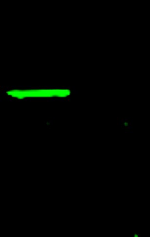
Application: Western BlotSample Tested: 293 cell extractSpecies: HumanVerified Customer | Posted 11/18/2019GFP fusion protein were overexpressed in 293T cells and can be detected by Nb600-597 antibody (Green). Specificity is good, but affinity is a little bit lower compared with Flag tag antibody.

-
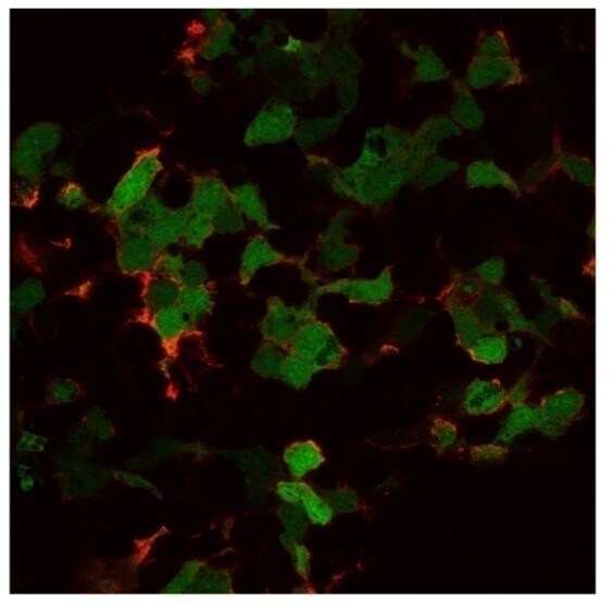
Application: ImmunocytochemistrySample Tested: HEK-293T cells transduced to have GFPSpecies: HumanVerified Customer | Posted 05/10/2018The 293T cells have been transduced to have GFP which was detected by an antibody after permeabilization. The green staining shows the anti-GFP antibody.
-
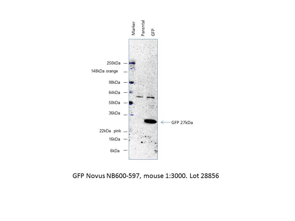
Application: Western BlotSample Tested: CHOK1 cells transfected with GFPSpecies: OtherVerified Customer | Posted 01/26/2015GFP NB600-597
There are no reviews that match your criteria.
Protocols
Find general support by application which include: protocols, troubleshooting, illustrated assays, videos and webinars.
- 7-Amino Actinomycin D (7-AAD) Cell Viability Flow Cytometry Protocol
- Antigen Retrieval Protocol (PIER)
- Antigen Retrieval for Frozen Sections Protocol
- Appropriate Fixation of IHC/ICC Samples
- Cellular Response to Hypoxia Protocols
- Chromogenic IHC Staining of Formalin-Fixed Paraffin-Embedded (FFPE) Tissue Protocol
- Chromogenic Immunohistochemistry Staining of Frozen Tissue
- ClariTSA™ Fluorophore Kits
- Detection & Visualization of Antibody Binding
- ELISA Sample Preparation & Collection Guide
- ELISA Troubleshooting Guide
- Extracellular Membrane Flow Cytometry Protocol
- Flow Cytometry Protocol for Cell Surface Markers
- Flow Cytometry Protocol for Staining Membrane Associated Proteins
- Flow Cytometry Staining Protocols
- Flow Cytometry Troubleshooting Guide
- Fluorescent IHC Staining of Frozen Tissue Protocol
- Graphic Protocol for Heat-induced Epitope Retrieval
- Graphic Protocol for the Preparation and Fluorescent IHC Staining of Frozen Tissue Sections
- Graphic Protocol for the Preparation and Fluorescent IHC Staining of Paraffin-embedded Tissue Sections
- Graphic Protocol for the Preparation of Gelatin-coated Slides for Histological Tissue Sections
- How to Run an R&D Systems DuoSet ELISA
- How to Run an R&D Systems Quantikine ELISA
- How to Run an R&D Systems Quantikine™ QuicKit™ ELISA
- ICC Cell Smear Protocol for Suspension Cells
- ICC Immunocytochemistry Protocol Videos
- ICC for Adherent Cells
- IHC Sample Preparation (Frozen sections vs Paraffin)
- Immunocytochemistry (ICC) Protocol
- Immunocytochemistry Troubleshooting
- Immunofluorescence of Organoids Embedded in Cultrex Basement Membrane Extract
- Immunofluorescent IHC Staining of Formalin-Fixed Paraffin-Embedded (FFPE) Tissue Protocol
- Immunohistochemistry (IHC) and Immunocytochemistry (ICC) Protocols
- Immunohistochemistry Frozen Troubleshooting
- Immunohistochemistry Paraffin Troubleshooting
- Immunoprecipitation Protocol
- Intracellular Flow Cytometry Protocol Using Alcohol (Methanol)
- Intracellular Flow Cytometry Protocol Using Detergents
- Intracellular Nuclear Staining Flow Cytometry Protocol Using Detergents
- Intracellular Staining Flow Cytometry Protocol Using Alcohol Permeabilization
- Intracellular Staining Flow Cytometry Protocol Using Detergents to Permeabilize Cells
- Preparing Samples for IHC/ICC Experiments
- Preventing Non-Specific Staining (Non-Specific Binding)
- Primary Antibody Selection & Optimization
- Propidium Iodide Cell Viability Flow Cytometry Protocol
- Protocol for Heat-Induced Epitope Retrieval (HIER)
- Protocol for Liperfluo
- Protocol for Making a 4% Formaldehyde Solution in PBS
- Protocol for VisUCyte™ HRP Polymer Detection Reagent
- Protocol for the Characterization of Human Th22 Cells
- Protocol for the Characterization of Human Th9 Cells
- Protocol for the Fluorescent ICC Staining of Cell Smears - Graphic
- Protocol for the Fluorescent ICC Staining of Cultured Cells on Coverslips - Graphic
- Protocol for the Preparation & Fixation of Cells on Coverslips
- Protocol for the Preparation and Chromogenic IHC Staining of Frozen Tissue Sections
- Protocol for the Preparation and Chromogenic IHC Staining of Frozen Tissue Sections - Graphic
- Protocol for the Preparation and Chromogenic IHC Staining of Paraffin-embedded Tissue Sections
- Protocol for the Preparation and Chromogenic IHC Staining of Paraffin-embedded Tissue Sections - Graphic
- Protocol for the Preparation and Fluorescent ICC Staining of Cells on Coverslips
- Protocol for the Preparation and Fluorescent ICC Staining of Non-adherent Cells
- Protocol for the Preparation and Fluorescent ICC Staining of Stem Cells on Coverslips
- Protocol for the Preparation and Fluorescent IHC Staining of Frozen Tissue Sections
- Protocol for the Preparation and Fluorescent IHC Staining of Paraffin-embedded Tissue Sections
- Protocol for the Preparation of Gelatin-coated Slides for Histological Tissue Sections
- Protocol for the Preparation of a Cell Smear for Non-adherent Cell ICC - Graphic
- Protocol: Annexin V and PI Staining by Flow Cytometry
- Protocol: Annexin V and PI Staining for Apoptosis by Flow Cytometry
- Quantikine HS ELISA Kit Assay Principle, Alkaline Phosphatase
- Quantikine HS ELISA Kit Principle, Streptavidin-HRP Polymer
- R&D Systems Quality Control Western Blot Protocol
- Sandwich ELISA (Colorimetric) – Biotin/Streptavidin Detection Protocol
- Sandwich ELISA (Colorimetric) – Direct Detection Protocol
- TUNEL and Active Caspase-3 Detection by IHC/ICC Protocol
- The Importance of IHC/ICC Controls
- Troubleshooting Guide: ELISA
- Troubleshooting Guide: Fluorokine Flow Cytometry Kits
- Troubleshooting Guide: Immunohistochemistry
- Troubleshooting Guide: Western Blot Figures
- Western Blot Conditions
- Western Blot Protocol
- Western Blot Protocol for Cell Lysates
- Western Blot Troubleshooting
- Western Blot Troubleshooting Guide
- View all Protocols, Troubleshooting, Illustrated assays and Webinars
FAQs for GFP Antibody (9F9.F9) - BSA Free
Showing
1
-
1 of
1 FAQ
Showing All
-
Q: We are looking for a rabbit anti-GFP antibody as a primary antibody for immunofluorescence and WB. I've seen that you carry several anti-GFP antibodies and I would like to ask which one you would recommend. The one with Cat.No NB600-308 has been used in a lot of publications and I reckon it is a solid antibody. Is this our best choice, or is there a better alternative?
A: NB600-308 is an excellent GFP antibody and I think it would work very well for you. It has been reviewed and published with which usually makes our customer feel much more confident, seeing as it worked in other people's hand.
Loading...