Glutamine Synthetase Antibody - BSA Free
Novus Biologicals | Catalog # NB110-41404
![Western Blot: Glutamine Synthetase Antibody [NB110-41404]](https://resources.rndsystems.com/images/products/Glutamine-Synthetase-Antibody-Western-Blot-NB110-41404-img0003.jpg "Western Blot: Glutamine Synthetase Antibody [NB110-41404]")
Key Product Details
Species Reactivity
Validated:
Cited:
Applications
Validated:
Cited:
Label
Antibody Source
Format
Product Specifications
Immunogen
Localization
Clonality
Host
Isotype
Scientific Data Images for Glutamine Synthetase Antibody - BSA Free
Western Blot: Glutamine Synthetase Antibody [NB110-41404]
Western Blot: Glutamine Synthetase Antibody [NB110-41404] - Analysis of glutamine synthase. 40ug of lysates from mouse (Lanes M), rat (Lane R), pig (Lane P), bovine (Lane B), or human (Lane Hu) retina were probed. A 42 kDa band was identified in lysates from retinas of all species.![Immunocytochemistry/ Immunofluorescence: Glutamine Synthetase Antibody [NB110-41404]](https://resources.rndsystems.com/images/products/Glutamine-Synthetase-Antibody-Immunocytochemistry-Immunofluorescence-NB110-41404-img0006.jpg "Immunocytochemistry/ Immunofluorescence: Glutamine Synthetase Antibody [NB110-41404]")
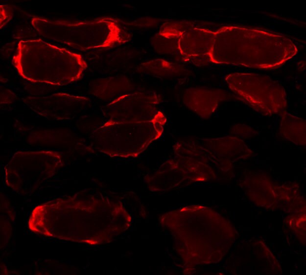
Immunocytochemistry/ Immunofluorescence: Glutamine Synthetase Antibody [NB110-41404]
Immunocytochemistry/Immunofluorescence: Glutamine Synthetase Antibody [NB110-41404] - Immunofluorescence using NB110-41404. Submitted via verified customer review.![Immunohistochemistry: Glutamine Synthetase Antibody [NB110-41404]](https://resources.rndsystems.com/images/products/Glutamine-Synthetase-Antibody-Immunohistochemistry-NB110-41404-img0002.jpg "Immunohistochemistry: Glutamine Synthetase Antibody [NB110-41404]")
Immunohistochemistry: Glutamine Synthetase Antibody [NB110-41404]
Immunohistochemistry: Glutamine Synthetase Antibody [NB110-41404] - Localization of glutamine synthase in the retina. Paraffin sections of mouse (A, B), rat (C, D, G-I), or human (E, F) retina fixed in 4% paraformaldehyde were reacted with anti-glutamine synthase (red fluorescence staining in B, D, G, I, and brown immuno-peroxidase reaction. Nuclei in IF experiments (A-D) were stained with DAPI (cyan), and with nuclear fast red in E and F. At low magnification, anti-glutamine synthase reacted with a single population of cells extending from the ganglion cell layer through the inner nuclear layer. No signal was detected in controls either pre-incubated with 100ug/ml of the immunizing peptide (A) or with pre-immune serum (C, E). This finding was confirmed by co-localization (indicated by yellow in I) of glutamine synthase (red in G) with antoher marker of glutamine synthase (green in H).Applications for Glutamine Synthetase Antibody - BSA Free
Immunocytochemistry/ Immunofluorescence
Immunohistochemistry
Western Blot
Reviewed Applications
Read 1 review rated 4 using NB110-41404 in the following applications:
Formulation, Preparation, and Storage
Purification
Formulation
Format
Preservative
Concentration
Shipping
Stability & Storage
Background: Glutamine Synthetase
Alternate Names
Gene Symbol
Additional Glutamine Synthetase Products
Product Documents for Glutamine Synthetase Antibody - BSA Free
Certificate of Analysis
To download a Certificate of Analysis, please enter a lot or batch number in the search box below.
Product Specific Notices for Glutamine Synthetase Antibody - BSA Free
This product is for research use only and is not approved for use in humans or in clinical diagnosis. Primary Antibodies are guaranteed for 1 year from date of receipt.
Citations for Glutamine Synthetase Antibody - BSA Free
 Powered by Bioz
Customer Reviews for Glutamine Synthetase Antibody - BSA Free (1)
Have you used Glutamine Synthetase Antibody - BSA Free?
Submit a review and receive an Amazon gift card!
$25/€18/£15/$25CAN/¥2500 Yen for a review with an image
$10/€7/£6/$10CAN/¥1110 Yen for a review without an image
Submit a review
Customer Images
-
Application: ImmunofluorescenceSample Tested: Mouse dorsal root gangliaSpecies: MouseVerified Customer | Posted 05/11/2016

There are no reviews that match your criteria.
Protocols
Find general support by application which include: protocols, troubleshooting, illustrated assays, videos and webinars.
- Antigen Retrieval Protocol (PIER)
- Antigen Retrieval for Frozen Sections Protocol
- Appropriate Fixation of IHC/ICC Samples
- Cellular Response to Hypoxia Protocols
- Chromogenic IHC Staining of Formalin-Fixed Paraffin-Embedded (FFPE) Tissue Protocol
- Chromogenic Immunohistochemistry Staining of Frozen Tissue
- ClariTSA™ Fluorophore Kits
- Detection & Visualization of Antibody Binding
- Fluorescent IHC Staining of Frozen Tissue Protocol
- Graphic Protocol for Heat-induced Epitope Retrieval
- Graphic Protocol for the Preparation and Fluorescent IHC Staining of Frozen Tissue Sections
- Graphic Protocol for the Preparation and Fluorescent IHC Staining of Paraffin-embedded Tissue Sections
- Graphic Protocol for the Preparation of Gelatin-coated Slides for Histological Tissue Sections
- ICC Cell Smear Protocol for Suspension Cells
- ICC Immunocytochemistry Protocol Videos
- ICC for Adherent Cells
- IHC Sample Preparation (Frozen sections vs Paraffin)
- Immunocytochemistry (ICC) Protocol
- Immunocytochemistry Troubleshooting
- Immunofluorescence of Organoids Embedded in Cultrex Basement Membrane Extract
- Immunofluorescent IHC Staining of Formalin-Fixed Paraffin-Embedded (FFPE) Tissue Protocol
- Immunohistochemistry (IHC) and Immunocytochemistry (ICC) Protocols
- Immunohistochemistry Frozen Troubleshooting
- Immunohistochemistry Paraffin Troubleshooting
- Preparing Samples for IHC/ICC Experiments
- Preventing Non-Specific Staining (Non-Specific Binding)
- Primary Antibody Selection & Optimization
- Protocol for Heat-Induced Epitope Retrieval (HIER)
- Protocol for Making a 4% Formaldehyde Solution in PBS
- Protocol for VisUCyte™ HRP Polymer Detection Reagent
- Protocol for the Fluorescent ICC Staining of Cell Smears - Graphic
- Protocol for the Fluorescent ICC Staining of Cultured Cells on Coverslips - Graphic
- Protocol for the Preparation & Fixation of Cells on Coverslips
- Protocol for the Preparation and Chromogenic IHC Staining of Frozen Tissue Sections
- Protocol for the Preparation and Chromogenic IHC Staining of Frozen Tissue Sections - Graphic
- Protocol for the Preparation and Chromogenic IHC Staining of Paraffin-embedded Tissue Sections
- Protocol for the Preparation and Chromogenic IHC Staining of Paraffin-embedded Tissue Sections - Graphic
- Protocol for the Preparation and Fluorescent ICC Staining of Cells on Coverslips
- Protocol for the Preparation and Fluorescent ICC Staining of Non-adherent Cells
- Protocol for the Preparation and Fluorescent ICC Staining of Stem Cells on Coverslips
- Protocol for the Preparation and Fluorescent IHC Staining of Frozen Tissue Sections
- Protocol for the Preparation and Fluorescent IHC Staining of Paraffin-embedded Tissue Sections
- Protocol for the Preparation of Gelatin-coated Slides for Histological Tissue Sections
- Protocol for the Preparation of a Cell Smear for Non-adherent Cell ICC - Graphic
- R&D Systems Quality Control Western Blot Protocol
- TUNEL and Active Caspase-3 Detection by IHC/ICC Protocol
- The Importance of IHC/ICC Controls
- Troubleshooting Guide: Immunohistochemistry
- Troubleshooting Guide: Western Blot Figures
- Western Blot Conditions
- Western Blot Protocol
- Western Blot Protocol for Cell Lysates
- Western Blot Troubleshooting
- Western Blot Troubleshooting Guide
- View all Protocols, Troubleshooting, Illustrated assays and Webinars
FAQs for Glutamine Synthetase Antibody - BSA Free
-
Q: I'm looking for a primary antibody specific to the cytosolic isoform of glutamine synthetase.
A:
I see no evidence that glutamine synthetase has multiple isoforms. Please see this link to UniProt (https://www.uniprot.org/uniprotkb/P15104/entry) for more details. NB110-41404 should work just fine for you as the only known isoform appears to be cytoplasmic.