GM130/GOLGA2 Antibody - BSA Free
Novus Biologicals | Catalog # NBP2-53420
![Immunocytochemistry/ Immunofluorescence: GM130/GOLGA2 Antibody - BSA Free [NBP2-53420]](https://resources.rndsystems.com/images/products/GM130-GOLGA2-Antibody-Immunocytochemistry-Immunofluorescence-NBP2-53420-img0007.jpg "Immunocytochemistry/ Immunofluorescence: GM130/GOLGA2 Antibody - BSA Free [NBP2-53420]")
Key Product Details
Species Reactivity
Validated:
Human, Mouse, Rat
Cited:
Human, Mouse, Primate - Macaca mulatta (Rhesus Macaque)
Applications
Validated:
Immunohistochemistry, Immunohistochemistry-Paraffin, Western Blot, Flow Cytometry, Immunocytochemistry/ Immunofluorescence
Cited:
Western Blot, Immunocytochemistry/ Immunofluorescence
Label
Unconjugated
Antibody Source
Polyclonal Rabbit IgG
Format
BSA Free
Loading...
Product Specifications
Immunogen
Partial recombinant human GM130/GOLGA2 protein (amino acids 528-606). [UniPro Q08379]
Clonality
Polyclonal
Host
Rabbit
Isotype
IgG
Scientific Data Images for GM130/GOLGA2 Antibody - BSA Free
Immunocytochemistry/ Immunofluorescence: GM130/GOLGA2 Antibody - BSA Free [NBP2-53420]
Immunocytochemistry/Immunofluorescence: GM130/GOLGA2 Antibody [NBP2-53420] - PC12 cells were fixed in 4% paraformaldehyde for 10 minutes and permeabilized in 0.05% Triton X-100 in PBS for 5 minutes. The cells were incubated with anti- NBP2-53420 at 1 ug/ml overnight at 4C and detected with an anti-rabbit Dylight 488 (Green) at a 1:1000 dilution for 60 minutes. Nuclei were counterstained with DAPI (Blue). Cells were imaged using a 100X objective and digitally deconvolved.![Immunocytochemistry/ Immunofluorescence: GM130/GOLGA2 Antibody - BSA Free [NBP2-53420]](https://resources.rndsystems.com/images/products/GM130-GOLGA2-Antibody-Immunocytochemistry-Immunofluorescence-NBP2-53420-img0008.jpg "Immunocytochemistry/ Immunofluorescence: GM130/GOLGA2 Antibody - BSA Free [NBP2-53420]")
Immunocytochemistry/ Immunofluorescence: GM130/GOLGA2 Antibody - BSA Free [NBP2-53420]
Immunocytochemistry/Immunofluorescence: GM130/GOLGA2 Antibody [NBP2-53420] - NIH3T3 cells were fixed in 4% paraformaldehyde for 10 minutes and permeabilized in 0.05% Triton X-100 in PBS for 5 minutes. The cells were incubated with anti-GM130/GOLGA2 Antibody NBP2-53420 at 1 ug/ml overnight at 4C and detected with an anti-rabbit Dylight 488 (Green) at a 1:1000 dilution for 60 minutes. Nuclei were counterstained with DAPI (Blue). Cells were imaged using a 100X objective and digitally deconvolved.![Immunocytochemistry/ Immunofluorescence: GM130/GOLGA2 Antibody - BSA Free [NBP2-53420]](https://resources.rndsystems.com/images/products/GM130-GOLGA2-Antibody-Immunocytochemistry-Immunofluorescence-NBP2-53420-img0006.jpg "Immunocytochemistry/ Immunofluorescence: GM130/GOLGA2 Antibody - BSA Free [NBP2-53420]")
Immunocytochemistry/ Immunofluorescence: GM130/GOLGA2 Antibody - BSA Free [NBP2-53420]
Immunocytochemistry/Immunofluorescence: GM130/GOLGA2 Antibody [NBP2-53420] - HeLa cells were fixed in 4% paraformaldehyde for 10 minutes and permeabilized in 0.05% Triton X-100 in PBS for 5 minutes. The cells were incubated with anti-GM130/GOLGA2 Antibody NBP2-53420 at 1 ug/ml overnight at 4C and detected with an anti-rabbit Dylight 488 (Green) at a 1:1000 dilution for 60 minutes. Nuclei were counterstained with DAPI (Blue). Cells were imaged using a 100X objective and digitally deconvolved.![Immunocytochemistry/ Immunofluorescence: GM130/GOLGA2 Antibody - BSA Free [NBP2-53420]](https://resources.rndsystems.com/images/products/GM130-GOLGA2-Antibody-Immunocytochemistry-Immunofluorescence-NBP2-53420-img0001.jpg "Immunocytochemistry/ Immunofluorescence: GM130/GOLGA2 Antibody - BSA Free [NBP2-53420]")
Immunocytochemistry/ Immunofluorescence: GM130/GOLGA2 Antibody - BSA Free [NBP2-53420]
Immunocytochemistry/Immunofluorescence: GM130/GOLGA2 Antibody [NBP2-53420] - HeLa cells were fixed for 10 min using 10% formalin and then permeabilized for 5 min using 1X TBS + 0.5% Triton X-100. The cells were incubated with anti-GOLGA2 at 1:200 overnight at 4C and detected with an anti-rabbit DyLight 488 (Green) at 1:500. Alpha tubulin (DM1A) NB100-690 was used as a co-stain at 1:1000 and detected with an anti-mouse DyLight 550 (Red) at 1:500. Nuclei were counterstained with DAPI (Blue). Cells were imaged using a 40X objective.![Western Blot: GM130/GOLGA2 AntibodyBSA Free [NBP2-53420]](https://resources.rndsystems.com/images/products/GM130-GOLGA2-Antibody-Western-Blot-NBP2-53420-img0002.jpg "Western Blot: GM130/GOLGA2 AntibodyBSA Free [NBP2-53420]")
Western Blot: GM130/GOLGA2 AntibodyBSA Free [NBP2-53420]
Western Blot: GM130/GOLGA2 Antibody [NBP2-53420] - Total protein from human HeLa and mouse Neuro2A cells was separated on a 7.5% gel by SDS-PAGE, transferred to PVDF membrane and blocked in 5% non-fat milk in TBST. The membrane was probed with 0.5 ug/mL anti-GOLGA2 in 1% non-fat milk in TBST and detected with an anti-rabbit HRP secondary antibody using chemiluminescence.![Immunohistochemistry-Paraffin: GM130/GOLGA2 Antibody - BSA Free [NBP2-53420]](https://resources.rndsystems.com/images/products/GM130-GOLGA2-Antibody-Immunohistochemistry-Paraffin-NBP2-53420-img0003.jpg "Immunohistochemistry-Paraffin: GM130/GOLGA2 Antibody - BSA Free [NBP2-53420]")
Immunohistochemistry-Paraffin: GM130/GOLGA2 Antibody - BSA Free [NBP2-53420]
Immunohistochemistry-Paraffin: GM130/GOLGA2 Antibody [NBP2-53420] - Analysis of a FFPE tissue section of mouse intesting using GM130/GOLGA2 antibody at 1:1500. The antibody generated staining in the the Golgi apparatus in the perinuclear region.![Immunocytochemistry/ Immunofluorescence: GM130/GOLGA2 Antibody - BSA Free [NBP2-53420]](https://resources.rndsystems.com/images/products/GM130-GOLGA2-Antibody-Immunocytochemistry-Immunofluorescence-NBP2-53420-img0013.jpg "Immunocytochemistry/ Immunofluorescence: GM130/GOLGA2 Antibody - BSA Free [NBP2-53420]")
Immunocytochemistry/ Immunofluorescence: GM130/GOLGA2 Antibody - BSA Free [NBP2-53420]
Immunocytochemistry/Immunofluorescence: GM130/GOLGA2 Antibody [NBP2-53420] - HeLa cells were fixed in 4% paraformaldehyde for 10 minutes and permeabilized in 0.05% Triton X-100 in PBS for 5 minutes. The cells were incubated with GM130/GOLGA2 Antibody conjugated to Biotin (NBP2-53420B) at 5 ug/ml for 1 hour at room temperature and detected with Streptavidin conjugated to DyLight 488. Nuclei were counterstained with DAPI (Blue). Cells were imaged using a 100X objective and digitally deconvolved.![Immunocytochemistry/ Immunofluorescence: GM130/GOLGA2 Antibody - BSA Free [NBP2-53420]](https://resources.rndsystems.com/images/products/GM130-GOLGA2-Antibody-Immunocytochemistry-Immunofluorescence-NBP2-53420-img0015.jpg "Immunocytochemistry/ Immunofluorescence: GM130/GOLGA2 Antibody - BSA Free [NBP2-53420]")
Immunocytochemistry/ Immunofluorescence: GM130/GOLGA2 Antibody - BSA Free [NBP2-53420]
Immunocytochemistry/Immunofluorescence: GM130/GOLGA2 Antibody [NBP2-53420] - A431 cells were fixed in 4% paraformaldehyde for 10 minutes and permeabilized in 0.05% Triton X-100 in PBS for 5 minutes. The cells were incubated with GM130/GOLGA2 Antibody conjugated to FITC (NBP2-53420F) at 2 ug/ml for 1 hour at room temperature. Nuclei were counterstained with DAPI (Blue). Cells were imaged using a 100X objective and digitally deconvolved.![Flow Cytometry: GM130/GOLGA2 Antibody - BSA Free [NBP2-53420]](https://resources.rndsystems.com/images/products/GM130-GOLGA2-Antibody-Flow-Cytometry-NBP2-53420-img0012.jpg "Flow Cytometry: GM130/GOLGA2 Antibody - BSA Free [NBP2-53420]")
Flow Cytometry: GM130/GOLGA2 Antibody - BSA Free [NBP2-53420]
Flow Cytometry: GM130/GOLGA2 Antibody [NBP2-53420] - An intracellular stain was performed on NIH3T3 cells with NBP2-53420 (blue) and a matched isotype control NBP2-24891 (orange). Cells were fixed with 4% PFA and then permeabilized with 0.1% saponin. Cells were incubated in an antibody dilution of 2.5 ug/mL for 30 minutes at room temperature, followed by Rabbit IgG (H+L) Cross-Adsorbed Secondary Antibody, Dylight 550 (SA5-10033, Thermo Fisher).![Immunocytochemistry/ Immunofluorescence: GM130/GOLGA2 Antibody - BSA Free [NBP2-53420]](https://resources.rndsystems.com/images/products/GM130-GOLGA2-Antibody-Immunocytochemistry-Immunofluorescence-NBP2-53420-img0014.jpg "Immunocytochemistry/ Immunofluorescence: GM130/GOLGA2 Antibody - BSA Free [NBP2-53420]")
Immunocytochemistry/ Immunofluorescence: GM130/GOLGA2 Antibody - BSA Free [NBP2-53420]
Immunocytochemistry/Immunofluorescence: GM130/GOLGA2 Antibody [NBP2-53420] - HeLa cells were fixed in 4% paraformaldehyde for 10 minutes and permeabilized in 0.05% Triton X-100 in PBS for 5 minutes. The cells were incubated with GM130/GOLGA2 Antibody conjugated to FITC (NBP2-53420F) at 5 ug/ml for 1 hour at room temperature. Nuclei were counterstained with DAPI (Blue). Cells were imaged using a 100X objective and digitally deconvolved.![Flow Cytometry: GM130/GOLGA2 Antibody - BSA Free [NBP2-53420]](https://resources.rndsystems.com/images/products/GM130-GOLGA2-Antibody-Flow-Cytometry-NBP2-53420-img0004.jpg "Flow Cytometry: GM130/GOLGA2 Antibody - BSA Free [NBP2-53420]")
Flow Cytometry: GM130/GOLGA2 Antibody - BSA Free [NBP2-53420]
Flow Cytometry: GM130/GOLGA2 Antibody [NBP2-53420] - An intracellular stain was performed on HeLa cells with NBP2-53420AF647 (blue) and a matched isotype control (orange). Cells were fixed with 4% PFA and then permeabilized with 0.1% saponin. Cells were incubated in an antibody dilution of 2.5 ug/mL for 30 min at room temperature. Both antibodies were conjugated to Alexa Fluor 647.![Flow Cytometry: GM130/GOLGA2 Antibody - BSA Free [NBP2-53420]](https://resources.rndsystems.com/images/products/GM130-GOLGA2-Antibody-Flow-Cytometry-NBP2-53420-img0005.jpg "Flow Cytometry: GM130/GOLGA2 Antibody - BSA Free [NBP2-53420]")
Flow Cytometry: GM130/GOLGA2 Antibody - BSA Free [NBP2-53420]
Flow Cytometry: GM130/GOLGA2 Antibody [NBP2-53420] - An intracellular stain was performed on MCF7 cells with GM130/GOLGA2 Antibody NBP2-53420AF647 (blue) and a matched isotype control (orange). Cells were fixed with 4% PFA and then permeabilized with 0.1% saponin. Cells were incubated in an antibody dilution of 2.5 ug/mL for 30 minutes at room temperature. Both antibodies were conjugated to Alexa Fluor 647.![Flow Cytometry: GM130/GOLGA2 Antibody - BSA Free [NBP2-53420]](https://resources.rndsystems.com/images/products/GM130-GOLGA2-Antibody-Flow-Cytometry-NBP2-53420-img0011.jpg "Flow Cytometry: GM130/GOLGA2 Antibody - BSA Free [NBP2-53420]")
Flow Cytometry: GM130/GOLGA2 Antibody - BSA Free [NBP2-53420]
Flow Cytometry: GM130/GOLGA2 Antibody [NBP2-53420] - An intracellular stain was performed on HeLa cells with NBP2-53420 (blue) and a matched isotype control NBP2-24891 (orange). Cells were fixed with 4% PFA and then permeabilized with 0.1% saponin. Cells were incubated in an antibody dilution of 2.5 ug/mL for 30 minutes at room temperature, followed by Rabbit IgG (H+L) Cross-Adsorbed Secondary Antibody, Dylight 550 (SA5-10033, Thermo Fisher).
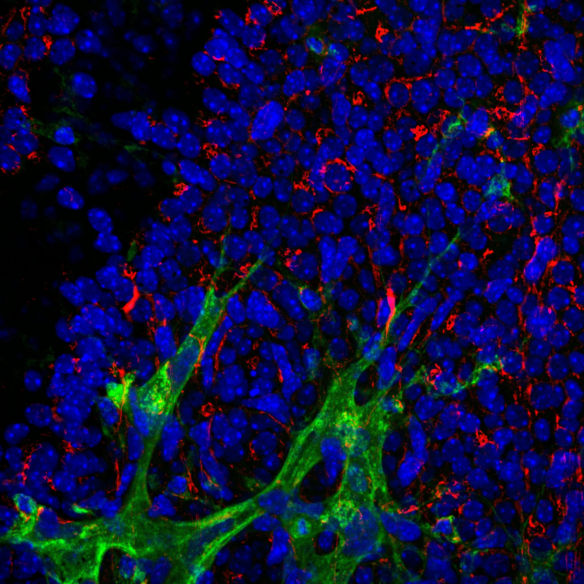
Immunohistochemistry: Rabbit Polyclonal GM130/GOLGA2 Antibody [NBP2-53420] -
Immunohistochemistry: Rabbit Polyclonal GM130/GOLGA2 Antibody [NBP2-53420] - Staining of mouse C57BL/6 retina using GM130/GOLGA2 Antibody. GOLGA (red), BS-1 lectin (green), DAPI (blue). Primary antibody dilution: 1:200. Image from a verified customer review.
Western Blot: GM130/GOLGA2 Antibody - BSA Free [NBP2-53420] -
Extracellular vesicle secretion induced by Sorafenib and their miRNA content. (A) Particle size and concentration analysis of Large, Small, and Very Small EVs obtained at 6 and 24 h. (B) Expression of EV markers and cellular contaminants in Large, Small, and Very Small EVs and cell lysates obtained at 24 h after Sorafenib treatment. Total lane protein content was used as the loading control. (C) Representative images of cryo-EM of Small and Very Small EVs obtained at 6 and 24 h from the control and Sorafenib-treated cells. (D) Assessment of EV size (nm) in the cryo-EM images. (E) Number of EVs quantified in the cryo-EM images. (F) miRNA expression in the three fractions of EVs at 6 h. (G) miRNA expression in the three fractions of EVs at 24 h. Fold-change values were calculated between Sorafenib and the control treated samples. Results are expressed as the mean +/- SEM of six independent experiments. Ns, non-significant; * p ≤0.05, ** p ≤ 0.01, *** p ≤ 0.001, and **** p ≤ 0.0001 between the miRNA expression in the control and Sorafenib derived EVs. Multiple comparison test statistics are expressed with lower case a–i. Image collected and cropped by CiteAb from the following open publication (https://pubmed.ncbi.nlm.nih.gov/36078082), licensed under a CC-BY license. Not internally tested by Novus Biologicals.Applications for GM130/GOLGA2 Antibody - BSA Free
Application
Recommended Usage
Immunocytochemistry/ Immunofluorescence
1-5 ug/mL
Immunohistochemistry
1:1000 - 1:1500
Immunohistochemistry-Paraffin
1:1000 - 1:1500
Western Blot
0.5 ug/mL
Reviewed Applications
Read 1 review rated 5 using NBP2-53420 in the following applications:
Flow Cytometry Panel Builder
Bio-Techne Knows Flow Cytometry
Save time and reduce costly mistakes by quickly finding compatible reagents using the Panel Builder Tool.
Advanced Features
- Spectra Viewer - Custom analysis of spectra from multiple fluorochromes
- Spillover Popups - Visualize the spectra of individual fluorochromes
- Antigen Density Selector - Match fluorochrome brightness with antigen density

Formulation, Preparation, and Storage
Purification
Immunogen affinity purified
Formulation
PBS
Format
BSA Free
Preservative
0.02% Sodium Azide
Concentration
1.0 mg/ml
Shipping
The product is shipped with polar packs. Upon receipt, store it immediately at the temperature recommended below.
Stability & Storage
Store at 4C short term. Aliquot and store at -20C long term. Avoid freeze-thaw cycles.
Background: GM130/GOLGA2
Long Name
Golgin A2
Alternate Names
GOLGA2, Golgin-95, SY11 Protein
Gene Symbol
GOLGA2
Additional GM130/GOLGA2 Products
Product Documents for GM130/GOLGA2 Antibody - BSA Free
Certificate of Analysis
To download a Certificate of Analysis, please enter a lot or batch number in the search box below.
Product Specific Notices for GM130/GOLGA2 Antibody - BSA Free
This product is for research use only and is not approved for use in humans or in clinical diagnosis. Primary Antibodies are guaranteed for 1 year from date of receipt.
Related Research Areas
Citations for GM130/GOLGA2 Antibody - BSA Free
 Powered by Bioz
Customer Reviews for GM130/GOLGA2 Antibody - BSA Free (1)
5 out of 5
1 Customer Rating
Have you used GM130/GOLGA2 Antibody - BSA Free?
Submit a review and receive an Amazon gift card!
$25/€18/£15/$25CAN/¥2500 Yen for a review with an image
$10/€7/£6/$10CAN/¥1110 Yen for a review without an image
Submit a review
Customer Images
Showing
1
-
1 of
1 review
Showing All
Filter By:
-
Application: IF- fixed tissueSample Tested: C57BL/6 mouse retinaSpecies: MouseVerified Customer | Posted 05/09/2024GOLGA (red), BS-1 lectin (green), DAPI (blue)1:200

There are no reviews that match your criteria.
Protocols
Find general support by application which include: protocols, troubleshooting, illustrated assays, videos and webinars.
- 7-Amino Actinomycin D (7-AAD) Cell Viability Flow Cytometry Protocol
- Antigen Retrieval Protocol (PIER)
- Antigen Retrieval for Frozen Sections Protocol
- Appropriate Fixation of IHC/ICC Samples
- Cellular Response to Hypoxia Protocols
- Chromogenic IHC Staining of Formalin-Fixed Paraffin-Embedded (FFPE) Tissue Protocol
- Chromogenic Immunohistochemistry Staining of Frozen Tissue
- ClariTSA™ Fluorophore Kits
- Detection & Visualization of Antibody Binding
- Extracellular Membrane Flow Cytometry Protocol
- Flow Cytometry Protocol for Cell Surface Markers
- Flow Cytometry Protocol for Staining Membrane Associated Proteins
- Flow Cytometry Staining Protocols
- Flow Cytometry Troubleshooting Guide
- Fluorescent IHC Staining of Frozen Tissue Protocol
- Graphic Protocol for Heat-induced Epitope Retrieval
- Graphic Protocol for the Preparation and Fluorescent IHC Staining of Frozen Tissue Sections
- Graphic Protocol for the Preparation and Fluorescent IHC Staining of Paraffin-embedded Tissue Sections
- Graphic Protocol for the Preparation of Gelatin-coated Slides for Histological Tissue Sections
- ICC Cell Smear Protocol for Suspension Cells
- ICC Immunocytochemistry Protocol Videos
- ICC for Adherent Cells
- IHC Sample Preparation (Frozen sections vs Paraffin)
- Immunocytochemistry (ICC) Protocol
- Immunocytochemistry Troubleshooting
- Immunofluorescence of Organoids Embedded in Cultrex Basement Membrane Extract
- Immunofluorescent IHC Staining of Formalin-Fixed Paraffin-Embedded (FFPE) Tissue Protocol
- Immunohistochemistry (IHC) and Immunocytochemistry (ICC) Protocols
- Immunohistochemistry Frozen Troubleshooting
- Immunohistochemistry Paraffin Troubleshooting
- Intracellular Flow Cytometry Protocol Using Alcohol (Methanol)
- Intracellular Flow Cytometry Protocol Using Detergents
- Intracellular Nuclear Staining Flow Cytometry Protocol Using Detergents
- Intracellular Staining Flow Cytometry Protocol Using Alcohol Permeabilization
- Intracellular Staining Flow Cytometry Protocol Using Detergents to Permeabilize Cells
- Preparing Samples for IHC/ICC Experiments
- Preventing Non-Specific Staining (Non-Specific Binding)
- Primary Antibody Selection & Optimization
- Propidium Iodide Cell Viability Flow Cytometry Protocol
- Protocol for Heat-Induced Epitope Retrieval (HIER)
- Protocol for Liperfluo
- Protocol for Making a 4% Formaldehyde Solution in PBS
- Protocol for VisUCyte™ HRP Polymer Detection Reagent
- Protocol for the Characterization of Human Th22 Cells
- Protocol for the Characterization of Human Th9 Cells
- Protocol for the Fluorescent ICC Staining of Cell Smears - Graphic
- Protocol for the Fluorescent ICC Staining of Cultured Cells on Coverslips - Graphic
- Protocol for the Preparation & Fixation of Cells on Coverslips
- Protocol for the Preparation and Chromogenic IHC Staining of Frozen Tissue Sections
- Protocol for the Preparation and Chromogenic IHC Staining of Frozen Tissue Sections - Graphic
- Protocol for the Preparation and Chromogenic IHC Staining of Paraffin-embedded Tissue Sections
- Protocol for the Preparation and Chromogenic IHC Staining of Paraffin-embedded Tissue Sections - Graphic
- Protocol for the Preparation and Fluorescent ICC Staining of Cells on Coverslips
- Protocol for the Preparation and Fluorescent ICC Staining of Non-adherent Cells
- Protocol for the Preparation and Fluorescent ICC Staining of Stem Cells on Coverslips
- Protocol for the Preparation and Fluorescent IHC Staining of Frozen Tissue Sections
- Protocol for the Preparation and Fluorescent IHC Staining of Paraffin-embedded Tissue Sections
- Protocol for the Preparation of Gelatin-coated Slides for Histological Tissue Sections
- Protocol for the Preparation of a Cell Smear for Non-adherent Cell ICC - Graphic
- Protocol: Annexin V and PI Staining by Flow Cytometry
- Protocol: Annexin V and PI Staining for Apoptosis by Flow Cytometry
- R&D Systems Quality Control Western Blot Protocol
- TUNEL and Active Caspase-3 Detection by IHC/ICC Protocol
- The Importance of IHC/ICC Controls
- Troubleshooting Guide: Fluorokine Flow Cytometry Kits
- Troubleshooting Guide: Immunohistochemistry
- Troubleshooting Guide: Western Blot Figures
- Western Blot Conditions
- Western Blot Protocol
- Western Blot Protocol for Cell Lysates
- Western Blot Troubleshooting
- Western Blot Troubleshooting Guide
- View all Protocols, Troubleshooting, Illustrated assays and Webinars
Loading...