GOLGB1/Giantin Antibody - BSA Free
Novus Biologicals | Catalog # NBP2-22321
![Western Blot: GOLGB1/Giantin Antibody [NBP2-22321]](https://resources.rndsystems.com/images/products/GOLGB1-Giantin-Antibody-Western-Blot-NBP2-22321-img0010.jpg "Western Blot: GOLGB1/Giantin Antibody [NBP2-22321]")
Key Product Details
Validated by
Independent Antibodies, Biological Validation
Species Reactivity
Validated:
Human
Cited:
Human
Applications
Validated:
Western Blot, Immunocytochemistry/ Immunofluorescence, Immunoprecipitation
Cited:
Western Blot, Immunocytochemistry/ Immunofluorescence, Immunoprecipitation
Label
Unconjugated
Antibody Source
Polyclonal Rabbit IgG
Format
BSA Free
Loading...
Product Specifications
Immunogen
The immunogen this antibody was made to, maps to a region between residue 425 to 475 of human Golgin B1 using the numbering given in entry NP_004478.3 (GeneID 2804).
Marker
Golgi Apparatus Marker
Clonality
Polyclonal
Host
Rabbit
Isotype
IgG
Scientific Data Images for GOLGB1/Giantin Antibody - BSA Free
![Immunocytochemistry/ Immunofluorescence: GOLGB1/Giantin Antibody [NBP2-22321]](https://resources.rndsystems.com/images/products/GOLGB1-Giantin-Antibody-Immunocytochemistry-Immunofluorescence-NBP2-22321-img0011.jpg "Immunocytochemistry/ Immunofluorescence: GOLGB1/Giantin Antibody [NBP2-22321]")
Immunocytochemistry/ Immunofluorescence: GOLGB1/Giantin Antibody [NBP2-22321]
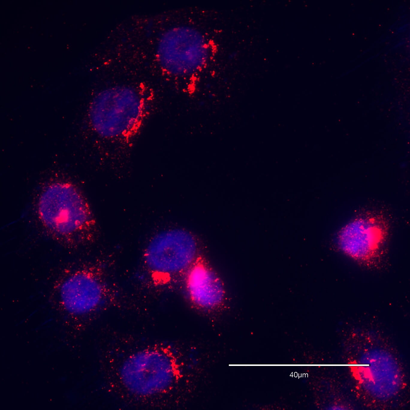
Immunocytochemistry/Immunofluorescence: GOLGB1/Giantin Antibody [NBP2-22321] - PC-3 human prostate cancer cell line. Image was captured using EVOS M5000 microscope. Red - giantin; blue - nucleus, DAPI; bar 40 micron. ICC/IF image submitted by a verified customer review.![Western Blot: GOLGB1/Giantin Antibody [NBP2-22321]](https://resources.rndsystems.com/images/products/GOLGB1-Giantin-Antibody-Western-Blot-NBP2-22321-img0004.jpg "Western Blot: GOLGB1/Giantin Antibody [NBP2-22321]")
Western Blot: GOLGB1/Giantin Antibody [NBP2-22321]
Western Blot: GOLGB1/Giantin Antibody [NBP2-22321] - Whole cell lysate (50 ug) from 293T, HeLa, and Jurkat cells. NBP2-22321 used for WB at 0.1 ug/mL. Detection: Chemiluminescence with an exposure time of 10 seconds.![Immunocytochemistry/ Immunofluorescence: GOLGB1/Giantin Antibody [NBP2-22321]](https://resources.rndsystems.com/images/products/GOLGB1-Giantin-Antibody-Immunofluorescence-NBP2-22321-img0005.jpg "Immunocytochemistry/ Immunofluorescence: GOLGB1/Giantin Antibody [NBP2-22321]")
![Immunocytochemistry/ Immunofluorescence: GOLGB1/Giantin Antibody [NBP2-22321]](https://resources.rndsystems.com/images/products/GOLGB1-Giantin-Antibody-Immunofluorescence-NBP2-22321-img0009.jpg "Immunocytochemistry/ Immunofluorescence: GOLGB1/Giantin Antibody [NBP2-22321]")
Immunocytochemistry/ Immunofluorescence: GOLGB1/Giantin Antibody [NBP2-22321]
GOLGB1-Giantin-Antibody-Immunofluorescence-NBP2-22321-img0009.jpg![Immunocytochemistry/ Immunofluorescence: GOLGB1/Giantin Antibody [NBP2-22321]](https://resources.rndsystems.com/images/products/GOLGB1-Giantin-Antibody-Immunofluorescence-NBP2-22321-img0008.jpg "Immunocytochemistry/ Immunofluorescence: GOLGB1/Giantin Antibody [NBP2-22321]")
Immunocytochemistry/ Immunofluorescence: GOLGB1/Giantin Antibody [NBP2-22321]
GOLGB1-Giantin-Antibody-Immunofluorescence-NBP2-22321-img0008.jpg![Immunoprecipitation: GOLGB1/Giantin Antibody [NBP2-22321]](https://resources.rndsystems.com/images/products/GOLGB1-Giantin-Antibody-Immunoprecipitation-NBP2-22321-img0003.jpg "Immunoprecipitation: GOLGB1/Giantin Antibody [NBP2-22321]")
Immunoprecipitation: GOLGB1/Giantin Antibody [NBP2-22321]
Immunoprecipitation: GOLGB1/Giantin Antibody [NBP2-22321] - Whole cell lysate (1 mg for IP; 20% of IP loaded) from 293T cells. Antibodies: NBP2-22321 used for IP at 6 ug/mg lysate. GOLGB1 was also immunoprecipitated by rabbit anti-GOLGB1 antibodies NBP2-22322 and NBP2-22323. For blotting immunoprecipitated GOLGB1, NBP2-22321 was used at 1 ug/ml. Detection: Chemiluminescence with an exposure time of 10 seconds.
Immunocytochemistry/ Immunofluorescence: GOLGB1/Giantin Antibody - BSA Free [NBP2-22321] -
Effect of Golgi disrupting treatments on the Golgi apparatus of MDA-MB-231 cells.Cells were left untreated (A; Control), or transfected to transiently express the HA-epitope-tagged ARF1 constitutively-activated mutant for 16 h (B; HA-ARF1-Q71L), or treated for 60 min either with 5 μg/ml Brefeldin A (C; BFA) or 10 μM Golgicide A (D; GCA). Cells were fixed, permeabilized, and immunolabeled with mouse monoclonal antibody to GM130, rabbit polyclonal antibody to Giantin, and sheep antibody to TGN46. Secondary antibodies were Alexa-594-conjugated donkey anti-mouse IgG (red channel), Alexa-488-conjugated donkey anti-rabbit IgG (green channel), and Alexa-647-conjugated donkey anti-sheep IgG (blue channel). Nuclei were stained with DAPI (gray channel). Stained cells were examined by fluorescence microscopy. Merging red, green, blue, and grey channels generated the fourth image on each row; yellow indicates overlapping localization of the red and green channels, cyan indicates overlapping localization of the green and blue channels, magenta indicates overlapping localization of the red and blue channels, and white indicates overlapping localization of all three channels. Bar, 10 μm. Image collected and cropped by CiteAb from the following open publication (https://pubmed.ncbi.nlm.nih.gov/29614107), licensed under a CC-BY license. Not internally tested by Novus Biologicals.
Immunocytochemistry/ Immunofluorescence: GOLGB1/Giantin Antibody - BSA Free [NBP2-22321] -
Effect of Actinomycin D and Vinblastine on the Golgi apparatus of MDA-MB-231 cells.Cells were left untreated (A; Control), or treated for 60 min either with 10 ng/ml Actinomycin D (B; ActD) or 25 nM Vinblastine (C; VLB). Cells were fixed, permeabilized, and immunolabeled with mouse monoclonal antibody to GM130, rabbit polyclonal antibody to Giantin, and sheep antibody to TGN46. Secondary antibodies were Alexa-594-conjugated donkey anti-mouse IgG (red channel), Alexa-488-conjugated donkey anti-rabbit IgG (green channel), and Alexa-647-conjugated donkey anti-sheep IgG (blue channel). Nuclei were stained with DAPI (gray channel). Stained cells were examined by fluorescence microscopy. Merging red, green, blue, and grey channels generated the fourth image on each row; yellow indicates overlapping localization of the red and green channels, cyan indicates overlapping localization of the green and blue channels, magenta indicates overlapping localization of the red and blue channels, and white indicates overlapping localization of all three channels. Bar, 10 μm. Image collected and cropped by CiteAb from the following open publication (https://pubmed.ncbi.nlm.nih.gov/29614107), licensed under a CC-BY license. Not internally tested by Novus Biologicals.Applications for GOLGB1/Giantin Antibody - BSA Free
Application
Recommended Usage
Immunocytochemistry/ Immunofluorescence
Validated from a verified customer review.
Immunoprecipitation
2 - 10 ug/mg
Western Blot
1:2000 - 1:10000
Reviewed Applications
Read 2 reviews rated 5 using NBP2-22321 in the following applications:
Formulation, Preparation, and Storage
Purification
Immunogen affinity purified
Formulation
Tris-Citrate/Phosphate (pH 7.0 - 8.0)
Format
BSA Free
Preservative
0.09% Sodium Azide
Concentration
1.0 mg/ml
Shipping
The product is shipped with polar packs. Upon receipt, store it immediately at the temperature recommended below.
Stability & Storage
Store at 4C. Do not freeze.
Background: GOLGB1/Giantin
Long Name
Golgin B1
Alternate Names
GCP372, Giantin, GOLIM1, Macrogolgin
Gene Symbol
GOLGB1
UniProt
Additional GOLGB1/Giantin Products
Product Documents for GOLGB1/Giantin Antibody - BSA Free
Certificate of Analysis
To download a Certificate of Analysis, please enter a lot or batch number in the search box below.
Product Specific Notices for GOLGB1/Giantin Antibody - BSA Free
This product is for research use only and is not approved for use in humans or in clinical diagnosis. Primary Antibodies are guaranteed for 1 year from date of receipt.
Related Research Areas
Citations for GOLGB1/Giantin Antibody - BSA Free
 Powered by Bioz
Customer Reviews for GOLGB1/Giantin Antibody - BSA Free (2)
5 out of 5
2 Customer Ratings
Have you used GOLGB1/Giantin Antibody - BSA Free?
Submit a review and receive an Amazon gift card!
$25/€18/£15/$25CAN/¥2500 Yen for a review with an image
$10/€7/£6/$10CAN/¥1110 Yen for a review without an image
Submit a review
Customer Images
Showing
1
-
2 of
2 reviews
Showing All
Filter By:
-
Application: ImmunofluorescenceSample Tested: PC-3 cells and PC-3 human prostate cancer cell lineSpecies: Human and PC-3 cellsVerified Customer | Posted 08/19/2020Image was captured using EVOS M5000 microscope. Red - giantin; blue - nucleus, DAPI; bar 40 mcm.

-
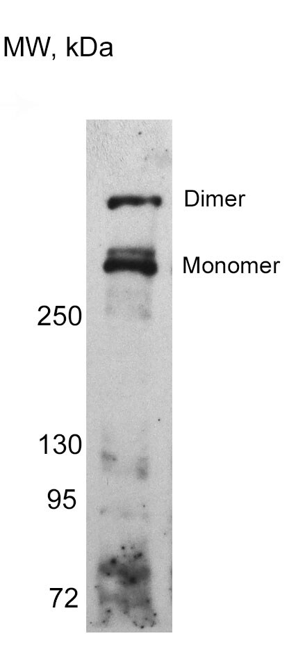
Application: Western BlotSample Tested: HeLa Cell LysateSpecies: HumanVerified Customer | Posted 11/20/2018HeLa cell lysate sample treated with DMSO was run on 8% SDS-PAGE. Sample was prepared in Laemmli buffer containing 10% beta-mercaptoethanol and heated for 10 min at 100 C. Both giantin monomer and dimer are indicated.
There are no reviews that match your criteria.
Protocols
Find general support by application which include: protocols, troubleshooting, illustrated assays, videos and webinars.
- Appropriate Fixation of IHC/ICC Samples
- Cellular Response to Hypoxia Protocols
- ClariTSA™ Fluorophore Kits
- Detection & Visualization of Antibody Binding
- ICC Cell Smear Protocol for Suspension Cells
- ICC Immunocytochemistry Protocol Videos
- ICC for Adherent Cells
- Immunocytochemistry (ICC) Protocol
- Immunocytochemistry Troubleshooting
- Immunofluorescence of Organoids Embedded in Cultrex Basement Membrane Extract
- Immunohistochemistry (IHC) and Immunocytochemistry (ICC) Protocols
- Immunoprecipitation Protocol
- Preparing Samples for IHC/ICC Experiments
- Preventing Non-Specific Staining (Non-Specific Binding)
- Primary Antibody Selection & Optimization
- Protocol for VisUCyte™ HRP Polymer Detection Reagent
- Protocol for the Fluorescent ICC Staining of Cell Smears - Graphic
- Protocol for the Fluorescent ICC Staining of Cultured Cells on Coverslips - Graphic
- Protocol for the Preparation and Fluorescent ICC Staining of Cells on Coverslips
- Protocol for the Preparation and Fluorescent ICC Staining of Non-adherent Cells
- Protocol for the Preparation and Fluorescent ICC Staining of Stem Cells on Coverslips
- Protocol for the Preparation of a Cell Smear for Non-adherent Cell ICC - Graphic
- R&D Systems Quality Control Western Blot Protocol
- TUNEL and Active Caspase-3 Detection by IHC/ICC Protocol
- The Importance of IHC/ICC Controls
- Troubleshooting Guide: Western Blot Figures
- Western Blot Conditions
- Western Blot Protocol
- Western Blot Protocol for Cell Lysates
- Western Blot Troubleshooting
- Western Blot Troubleshooting Guide
- View all Protocols, Troubleshooting, Illustrated assays and Webinars
Loading...