![Knockout Validated: GOLM1 Antibody [NBP1-50627]](https://resources.rndsystems.com/images/products/GOLM1-Antibody-Knockout-Validated-NBP1-50627-img0009.jpg "Western Blot: GOLM1 Antibody [NBP1-50627]")
Loading...
Key Product Details
Validated by
Knockout/Knockdown
Species Reactivity
Validated:
Human, Mouse
Cited:
Human
Applications
Validated:
Knockout Validated, Western Blot, Immunocytochemistry/ Immunofluorescence
Cited:
Western Blot, Immunocytochemistry/ Immunofluorescence, Knockdown Validated
Label
Unconjugated
Antibody Source
Polyclonal Rabbit IgG
Loading...
Product Specifications
Immunogen
A genomic peptide made to an internal region of the human GOLM1 protein (within residues 250-400). [Swiss-Prot Q8NBJ4]
Localization
Golgi apparatus - cis-Golgi network membrane
Marker
Golgi Apparatus Marker
Clonality
Polyclonal
Host
Rabbit
Isotype
IgG
Scientific Data Images for GOLM1 Antibody
Western Blot: GOLM1 Antibody [NBP1-50627]
Western Blot: GOLM1 Antibody [NBP1-50627] - Western blot shows lysates of HEK293 human embryonic kidney parental cell line and GOLM1 knockout (KO) HEK293 human embryonic kidney cell line. PVDF membrane was probed with 1:5,000 of Rabbit Anti-Human GOLM1 polyclonal Antibody (Catalog # NBP1-50627) followed by HRP-conjugated Anti-Rabbit IgG Secondary Antibody (Catalog #HAF008). Specific band was detected for GOLM1 at approximately 75 kDa (as indicated) in the parental HEK293 celll line, but is not detectable in the knockout HEK293 cell line. This experiment was conducted under reducing conditions.![Western Blot: GOLM1 Antibody [NBP1-50627]](https://resources.rndsystems.com/images/products/GOLM1-Antibody-Western-Blot-NBP1-50627-img0008.jpg "Western Blot: GOLM1 Antibody [NBP1-50627]")
Western Blot: GOLM1 Antibody [NBP1-50627]
Western Blot: GOLM1 Antibody [NBP1-50627] - Western Blot Image of GOLM1. Whole cell protein from HeLa (lane 1), 3T3 (lane 2), MEF (lane 3), Raw264.7 (lane 4), L929 (lane 5), Neuro2A (lane 6) and PC12 (lane 7) were separated on a 12% gel by SDS-PAGE, transferred to PVDF membrane and blocked in 5% non-fat milk in TBST. The membrane was probed with 0.5 mg/ml anti-GOLM1 in 1% milk and detected with an anti-rabbit HRP secondary antibody using chemiluminescence.![Immunocytochemistry/ Immunofluorescence: GOLM1 Antibody [NBP1-50627]](https://resources.rndsystems.com/images/products/GOLM1-Antibody-Immunocytochemistry-Immunofluorescence-NBP1-50627-img0007.jpg "Immunocytochemistry/ Immunofluorescence: GOLM1 Antibody [NBP1-50627]")
Immunocytochemistry/ Immunofluorescence: GOLM1 Antibody [NBP1-50627]
Immunocytochemistry/Immunofluorescence: GOLM1 Antibody [NBP1-50627] - Confocal immunofluorescent analysis of HeLa cells using GOLM1 antibody (NBP1-50627, 1:25). An Alexa Fluor 488-conjugated Goat to rabbit IgG was used as secondary antibody (green). Actin filaments were labeled with Alexa Fluor 568 phalloidin (red). DAPI was used to stain the cell nuclei (blue).![Immunocytochemistry/ Immunofluorescence: GOLM1 Antibody [NBP1-50627]](https://resources.rndsystems.com/images/products/GOLM1-Antibody-Immunocytochemistry-Immunofluorescence-NBP1-50627-img0004.jpg "Immunocytochemistry/ Immunofluorescence: GOLM1 Antibody [NBP1-50627]")
Immunocytochemistry/ Immunofluorescence: GOLM1 Antibody [NBP1-50627]
Immunocytochemistry/Immunofluorescence: GOLM1 Antibody [NBP1-50627] - ICC staining of GOLM1 in HEK293T cells with FITC (green). Nuclei (blue) were counterstained with Dapi.![Immunocytochemistry/ Immunofluorescence: GOLM1 Antibody [NBP1-50627]](https://resources.rndsystems.com/images/products/GOLM1-Antibody-Immunocytochemistry-Immunofluorescence-NBP1-50627-img0003.jpg "Immunocytochemistry/ Immunofluorescence: GOLM1 Antibody [NBP1-50627]")
Immunocytochemistry/ Immunofluorescence: GOLM1 Antibody [NBP1-50627]
Immunocytochemistry/Immunofluorescence: GOLM1 Antibody [NBP1-50627] - GOLM1 antibody was tested in HEK-293 cells with Dylight 488 (green). Nuclei and alpha-tubulin were counterstained with DAPI (blue) and Dylight 550 (red).Applications for GOLM1 Antibody
Application
Recommended Usage
Immunocytochemistry/ Immunofluorescence
1:500
Western Blot
1:5000
Reviewed Applications
Read 3 reviews rated 4.7 using NBP1-50627 in the following applications:
Formulation, Preparation, and Storage
Purification
Immunogen affinity purified
Formulation
PBS, 0.1% BSA, 50% Glycerol
Preservative
0.05% Sodium Azide
Concentration
0.61 mg/ml
Shipping
The product is shipped with polar packs. Upon receipt, store it immediately at the temperature recommended below.
Stability & Storage
Store at 4C short term. Aliquot and store at -20C long term. Avoid freeze-thaw cycles.
Background: GOLM1
Alternate Names
bA379P1.3, FLJ23608, golgi membrane protein 1, Golgi membrane protein GP73, Golgi phosphoprotein 2C9orf155, golgi protein, 73-kD, GOLPH2 FLJ22634, GP73, GP73 chromosome 9 open reading frame 155, PSEC0257
Entrez Gene IDs
51280 (Human)
Gene Symbol
GOLM1
UniProt
Additional GOLM1 Products
Product Documents for GOLM1 Antibody
Certificate of Analysis
To download a Certificate of Analysis, please enter a lot or batch number in the search box below.
Product Specific Notices for GOLM1 Antibody
Manufactured by Genomic Antibody Technology™. GAT FAQs
This product is for research use only and is not approved for use in humans or in clinical diagnosis. Primary Antibodies are guaranteed for 1 year from date of receipt.
Citations for GOLM1 Antibody
 Powered by Bioz
Customer Reviews for GOLM1 Antibody (3)
4.7 out of 5
3 Customer Ratings
Have you used GOLM1 Antibody?
Submit a review and receive an Amazon gift card!
$25/€18/£15/$25CAN/¥2500 Yen for a review with an image
$10/€7/£6/$10CAN/¥1110 Yen for a review without an image
Submit a review
Customer Images
-(01-ml)_NBP1-50627_8246.bmp)
-(01-ml)_NBP1-50627_8241.jpg)
Showing
1
-
3 of
3 reviews
Showing All
Filter By:
-
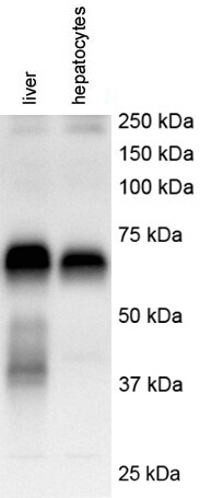
Application: Western BlotSample Tested: human liver and primary hepatocytesSpecies: HumanVerified Customer | Posted 12/15/2017human liver and primary hepatocyteshuman liver and primary hepatocytes

-
Application: ImmunocytochemistrySample Tested:Species: HumanVerified Customer | Posted 06/12/2014Confocal immunofluorescent analysis of HeLa cells using GOLM1 antibody (NBP1-50627, 1:25).
-
Application: Western BlotSample Tested:Species: HumanVerified Customer | Posted 06/12/2014Western blot analysis of extracts from LNCaP cells using GOLM1 antibody (NBP1-50627, 1:5000).
There are no reviews that match your criteria.
Protocols
View specific protocols for GOLM1 Antibody (NBP1-50627):
GOLM1 Antibody:
Immunocytochemistry Protocol
Culture cells to appropriate density in 35 mm culture dishes or 6-well plates.
1. Remove culture medium and add 10% formalin to the dish. Fix at room temperature for 30 minutes.
2. Remove the formalin and add ice cold methanol. Incubate for 5-10 minutes.
3. Remove methanol and add washing solution (i.e. PBS). Be sure to not let the specimen dry out. Wash three times for 10 minutes.
4. To block nonspecific antibody binding incubate in 10% normal goat serum from 1 hour to overnight at room temperature.
5. Add primary antibody at appropriate dilution and incubate at room temperature from 2 hours to overnight at room temperature.
6. Remove primary antibody and replace with washing solution. Wash three times for 10 minutes.
7. Add secondary antibody at appropriate dilution. Incubate for 1 hour at room temperature.
8. Remove antibody and replace with wash solution, then wash for 10 minutes. Add Hoechst 33258 to wash solution at 1:25,0000 and incubate for 10 minutes. Wash a third time for 10 minutes.
9. Cells can be viewed directly after washing. The plates can also be stored in PBS containing Azide covered in Parafilm (TM). Cells can also be cover-slipped using Fluoromount, with appropriate sealing.
*The above information is only intended as a guide. The researcher should determine what protocol best meets their needs. Please follow safe laboratory procedures.
GOLM1 Antibody:
Western Blot Protocol
1. Perform SDS-PAGE on samples to be analyzed, loading 40 ug of total protein per lane.
2. Transfer proteins to membrane according to the instructions provided by the manufacturer of the membrane and transfer apparatus.
3. Stain according to standard Ponceau S procedure (or similar product) to assess transfer success, and mark molecular weight standards where appropriate.
4. Rinse the blot.
5. Block the membrane using standard blocking buffer for at least 1 hour.
6. Wash the membrane in wash buffer three times for 10 minutes each.
7. Dilute the primary antibody in blocking buffer and incubate 1 hour at room temperature.
8. Wash the membrane in wash buffer three times for 10 minutes each.
9. Apply diluted rabbit-IgG HRP-conjugated secondary antibody in blocking buffer (as per manufacturers instructions) and incubate 1 hour at room temperature.
10. Wash the blot in wash buffer three times for 10 minutes each (this step can be repeated as required to reduce background).
11. Apply the detection reagent of choice in accordance with the manufacturers instructions.
*Note: Tween-20 can be added to the blocking or antibody dilution buffer at a final concentration of 0.05-0.2%.
Find general support by application which include: protocols, troubleshooting, illustrated assays, videos and webinars.
- Appropriate Fixation of IHC/ICC Samples
- Cellular Response to Hypoxia Protocols
- ClariTSA™ Fluorophore Kits
- Detection & Visualization of Antibody Binding
- ICC Cell Smear Protocol for Suspension Cells
- ICC Immunocytochemistry Protocol Videos
- ICC for Adherent Cells
- Immunocytochemistry (ICC) Protocol
- Immunocytochemistry Troubleshooting
- Immunofluorescence of Organoids Embedded in Cultrex Basement Membrane Extract
- Immunohistochemistry (IHC) and Immunocytochemistry (ICC) Protocols
- Preparing Samples for IHC/ICC Experiments
- Preventing Non-Specific Staining (Non-Specific Binding)
- Primary Antibody Selection & Optimization
- Protocol for VisUCyte™ HRP Polymer Detection Reagent
- Protocol for the Fluorescent ICC Staining of Cell Smears - Graphic
- Protocol for the Fluorescent ICC Staining of Cultured Cells on Coverslips - Graphic
- Protocol for the Preparation and Fluorescent ICC Staining of Cells on Coverslips
- Protocol for the Preparation and Fluorescent ICC Staining of Non-adherent Cells
- Protocol for the Preparation and Fluorescent ICC Staining of Stem Cells on Coverslips
- Protocol for the Preparation of a Cell Smear for Non-adherent Cell ICC - Graphic
- R&D Systems Quality Control Western Blot Protocol
- TUNEL and Active Caspase-3 Detection by IHC/ICC Protocol
- The Importance of IHC/ICC Controls
- Troubleshooting Guide: Western Blot Figures
- Western Blot Conditions
- Western Blot Protocol
- Western Blot Protocol for Cell Lysates
- Western Blot Troubleshooting
- Western Blot Troubleshooting Guide
- View all Protocols, Troubleshooting, Illustrated assays and Webinars
Loading...