GPR18 Antibody - BSA Free
Novus Biologicals | Catalog # NBP2-24918
![Western Blot: GPR18 AntibodyBSA Free [NBP2-24918]](https://resources.rndsystems.com/images/products/GPR18-Antibody-Western-Blot-NBP2-24918-img0001.jpg "Western Blot: GPR18 AntibodyBSA Free [NBP2-24918]")
Key Product Details
Species Reactivity
Validated:
Cited:
Applications
Validated:
Cited:
Label
Antibody Source
Format
Product Specifications
Immunogen
Clonality
Host
Isotype
Scientific Data Images for GPR18 Antibody - BSA Free
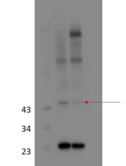
Western Blot: GPR18 AntibodyBSA Free [NBP2-24918]
Western Blot: GPR18 Antibody [NBP2-24918] - Analysis of human spleen lysate (4 ug/mL) in the 1) absence and 2) presence of immunizing peptide 3) mouse spleen (6 ug/mL) and 4) rat spleen lysate (3 ug/mL) using GPR18 antibody.![Immunocytochemistry/ Immunofluorescence: GPR18 Antibody - BSA Free [NBP2-24918]](https://resources.rndsystems.com/images/products/GPR18-Antibody-Immunocytochemistry-Immunofluorescence-NBP2-24918-img0006.jpg "Immunocytochemistry/ Immunofluorescence: GPR18 Antibody - BSA Free [NBP2-24918]")
Immunocytochemistry/ Immunofluorescence: GPR18 Antibody - BSA Free [NBP2-24918]
Immunocytochemistry/Immunofluorescence: GPR18 Antibody [NBP2-24918] - Ntera2 cells were fixed in 4% paraformaldehyde for 10 minutes and permeabilized in 0.05% Triton X-100 in PBS for 5 minutes. The cells were incubated with anti-GPR18 Antibody NBP2-24918 at 2 ug/ml overnight at 4C and detected with an anti-rabbit Dylight 488 (Green) at a 1:1000 dilution for 60 minutes. Nuclei were counterstained with DAPI (Blue). Cells were imaged using a 100X objective and digitally deconvolved.![Immunohistochemistry-Paraffin: GPR18 Antibody - BSA Free [NBP2-24918]](https://resources.rndsystems.com/images/products/GPR18-Antibody-Immunohistochemistry-Paraffin-NBP2-24918-img0003.jpg "Immunohistochemistry-Paraffin: GPR18 Antibody - BSA Free [NBP2-24918]")
Immunohistochemistry-Paraffin: GPR18 Antibody - BSA Free [NBP2-24918]
Immunohistochemistry-Paraffin: GPR18 Antibody [NBP2-24918] - Analysis of a FFPE tissue section of human tonsil using 1:200 dilution of GPR18 antibody. The staining was developed using HRP labeled anti-rabbit secondary antibody and DAB reagent, and nuclei of cells were counter-stained with hematoxylin.![Flow Cytometry: GPR18 Antibody - BSA Free [NBP2-24918]](https://resources.rndsystems.com/images/products/GPR18-Antibody-Flow-Cytometry-NBP2-24918-img0007.jpg "Flow Cytometry: GPR18 Antibody - BSA Free [NBP2-24918]")
Flow Cytometry: GPR18 Antibody - BSA Free [NBP2-24918]
Flow Cytometry: GPR18 Antibody [NBP2-24918] - An intracellular stain was performed on Daudi cells with GPR18 Antibody NBP2-24918 (blue) and a matched isotype control NBP2-24891 (orange). Cells were fixed with 4% PFA and then permeabilized with 0.1% saponin. Cells were incubated in an antibody dilution of 1.0 ug/mL for 30 minutes at room temperature, followed by Rabbit IgG (H+L) Cross-Adsorbed Secondary Antibody, Dylight 550 (SA5-10033, Thermo Fisher).![Immunohistochemistry-Paraffin: GPR18 Antibody - BSA Free [NBP2-24918]](https://resources.rndsystems.com/images/products/GPR18-Antibody-Immunohistochemistry-Paraffin-NBP2-24918-img0002.jpg "Immunohistochemistry-Paraffin: GPR18 Antibody - BSA Free [NBP2-24918]")
Immunohistochemistry-Paraffin: GPR18 Antibody - BSA Free [NBP2-24918]
Immunohistochemistry-Paraffin: GPR18 Antibody [NBP2-24918] - Analysis of human breast tumor tissue using an isotype control (top) and GPR18 antibody (bottom) at 5 ug/mL.![Flow Cytometry: GPR18 Antibody - BSA Free [NBP2-24918]](https://resources.rndsystems.com/images/products/GPR18-Antibody-Flow-Cytometry-NBP2-24918-img0004.jpg "Flow Cytometry: GPR18 Antibody - BSA Free [NBP2-24918]")
Flow Cytometry: GPR18 Antibody - BSA Free [NBP2-24918]
Flow Cytometry: GPR18 Antibody [NBP2-24918] - An intracellular stain was performed on Ntera2 cells with GPR18 Antibody NBP2-24918 (blue) and a matched isotype control (orange). Cells were fixed with 4% PFA and then permeabilized with 0.1% saponin. Cells were incubated in an antibody dilution of 1.0 ug/mL for 30 minutes at room temperature, followed by Rabbit IgG (H+L) Cross-Adsorbed Secondary Antibody, Dylight 550 (SA5-10033, Thermo Fisher).![Flow Cytometry: GPR18 Antibody - BSA Free [NBP2-24918]](https://resources.rndsystems.com/images/products/GPR18-Antibody-Flow-Cytometry-NBP2-24918-img0005.jpg "Flow Cytometry: GPR18 Antibody - BSA Free [NBP2-24918]")
Flow Cytometry: GPR18 Antibody - BSA Free [NBP2-24918]
Flow Cytometry: GPR18 Antibody [NBP2-24918] - An intracellular stain was performed on THP-1 cells with GPR18 Antibody NBP2-24918 (blue) and a matched isotype control (orange). Cells were fixed with 4% PFA and then permeabilized with 0.1% saponin. Cells were incubated in an antibody dilution of 1.0 ug/mL for 30 minutes at room temperature, followed by Rabbit IgG (H+L) Cross-Adsorbed Secondary Antibody, Dylight 550 (SA5-10033, Thermo Fisher).Applications for GPR18 Antibody - BSA Free
Flow Cytometry
Immunocytochemistry/ Immunofluorescence
Immunohistochemistry
Immunohistochemistry-Paraffin
Western Blot
Reviewed Applications
Read 1 review rated 3 using NBP2-24918 in the following applications:
Flow Cytometry Panel Builder
Bio-Techne Knows Flow Cytometry
Save time and reduce costly mistakes by quickly finding compatible reagents using the Panel Builder Tool.
Advanced Features
- Spectra Viewer - Custom analysis of spectra from multiple fluorochromes
- Spillover Popups - Visualize the spectra of individual fluorochromes
- Antigen Density Selector - Match fluorochrome brightness with antigen density

Formulation, Preparation, and Storage
Purification
Formulation
Format
Preservative
Concentration
Shipping
Stability & Storage
Background: GPR18
Long Name
Alternate Names
Gene Symbol
Additional GPR18 Products
Product Documents for GPR18 Antibody - BSA Free
Certificate of Analysis
To download a Certificate of Analysis, please enter a lot or batch number in the search box below.
Product Specific Notices for GPR18 Antibody - BSA Free
This product is for research use only and is not approved for use in humans or in clinical diagnosis. Primary Antibodies are guaranteed for 1 year from date of receipt.
Related Research Areas
Citations for GPR18 Antibody - BSA Free
 Powered by Bioz
Customer Reviews for GPR18 Antibody - BSA Free (1)
Have you used GPR18 Antibody - BSA Free?
Submit a review and receive an Amazon gift card!
$25/€18/£15/$25CAN/¥2500 Yen for a review with an image
$10/€7/£6/$10CAN/¥1110 Yen for a review without an image
Submit a review
Customer Images
-
Application: Western BlotSample Tested: Jurkat and HT-29 human colon adenocarcinoma cell lineSpecies: HumanVerified Customer | Posted 01/23/2019Ladder - HT29 - Jurkat

There are no reviews that match your criteria.
Protocols
View specific protocols for GPR18 Antibody - BSA Free (NBP2-24918):
Culture cells to appropriate density in 35 mm culture dishes or 6-well plates.
1. Remove culture medium and wash the cells briefly in PBS. Add 10% formalin to the dish and fix at room temperature for 10 minutes.
2. Remove the formalin and wash the cells in PBS.
3. Permeablize the cells with 0.1% Triton X100 or other suitable detergent for 10 min.
4. Remove the permeablization buffer and wash three times for 10 minutes each in PBS. Be sure to not let the specimen dry out.
5. To block nonspecific antibody binding, incubate in 10% normal goat serum from 1 hour to overnight at room temperature.
6. Add primary antibody at appropriate dilution and incubate overnight at 4C.
7. Remove primary antibody and replace with PBS. Wash three times for 10 minutes each.
8. Add secondary antibody at appropriate dilution. Incubate for 1 hour at room temperature.
9. Remove secondary antibody and replace with PBS. Wash three times for 10 minutes each.
10. Counter stain DNA with DAPi if required.
Antigen Unmasking:
Bring slides to a boil in 10 mM sodium citrate buffer (pH 6.0) then maintain at a sub-boiling temperature for 10 minutes. Cool slides on bench-top for 30 minutes (keep slides in the sodium citrate buffer all the time).
Staining:
1. Wash sections in deionized water three times for 5 minutes each.
2. Wash sections in PBS for 5 minutes.
3. Block each section with 100-400 ul blocking solution (1% BSA in PBS) for 1 hour at room temperature.
4. Remove blocking solution and add 100-400 ul diluted primary antibody. Incubate overnight at 4 C.
5. Remove antibody solution and wash sections in wash buffer three times for 5 minutes each.
6. Add 100-400 ul HRP polymer conjugated secondary antibody. Incubate 30 minutes at room temperature.
7. Wash sections three times in wash buffer for 5 minutes each.
8. Add 100-400 ul DAB substrate to each section and monitor staining closely.
9. As soon as the sections develop, immerse slides in deionized water.
10. Counterstain sections in hematoxylin.
11. Wash sections in deionized water two times for 5 minutes each.
12. Dehydrate sections.
13. Mount coverslips.
1. Perform SDS-PAGE on samples to be analyzed, loading 10-25 ug of total protein per lane.
2. Transfer proteins to PVDF membrane according to the instructions provided by the manufacturer of the membrane and transfer apparatus.
3. Stain the membrane with Ponceau S (or similar product) to assess transfer success, and mark molecular weight standards where appropriate.
4. Rinse the blot TBS -0.05% Tween 20 (TBST).
5. Block the membrane in 5% Non-fat milk in TBST (blocking buffer) for at least 1 hour.
6. Wash the membrane in TBST three times for 10 minutes each.
7. Dilute primary antibody in blocking buffer and incubate overnight at 4C with gentle rocking.
8. Wash the membrane in TBST three times for 10 minutes each.
9. Incubate the membrane in diluted HRP conjugated secondary antibody in blocking buffer (as per manufacturer's instructions) for 1 hour at room temperature.
10. Wash the blot in TBST three times for 10 minutes each (this step can be repeated as required to reduce background).
11. Apply the detection reagent of choice in accordance with the manufacturers instructions.
Find general support by application which include: protocols, troubleshooting, illustrated assays, videos and webinars.
- 7-Amino Actinomycin D (7-AAD) Cell Viability Flow Cytometry Protocol
- Antigen Retrieval Protocol (PIER)
- Antigen Retrieval for Frozen Sections Protocol
- Appropriate Fixation of IHC/ICC Samples
- Cellular Response to Hypoxia Protocols
- Chromogenic IHC Staining of Formalin-Fixed Paraffin-Embedded (FFPE) Tissue Protocol
- Chromogenic Immunohistochemistry Staining of Frozen Tissue
- ClariTSA™ Fluorophore Kits
- Detection & Visualization of Antibody Binding
- Extracellular Membrane Flow Cytometry Protocol
- Flow Cytometry Protocol for Cell Surface Markers
- Flow Cytometry Protocol for Staining Membrane Associated Proteins
- Flow Cytometry Staining Protocols
- Flow Cytometry Troubleshooting Guide
- Fluorescent IHC Staining of Frozen Tissue Protocol
- Graphic Protocol for Heat-induced Epitope Retrieval
- Graphic Protocol for the Preparation and Fluorescent IHC Staining of Frozen Tissue Sections
- Graphic Protocol for the Preparation and Fluorescent IHC Staining of Paraffin-embedded Tissue Sections
- Graphic Protocol for the Preparation of Gelatin-coated Slides for Histological Tissue Sections
- ICC Cell Smear Protocol for Suspension Cells
- ICC Immunocytochemistry Protocol Videos
- ICC for Adherent Cells
- IHC Sample Preparation (Frozen sections vs Paraffin)
- Immunocytochemistry (ICC) Protocol
- Immunocytochemistry Troubleshooting
- Immunofluorescence of Organoids Embedded in Cultrex Basement Membrane Extract
- Immunofluorescent IHC Staining of Formalin-Fixed Paraffin-Embedded (FFPE) Tissue Protocol
- Immunohistochemistry (IHC) and Immunocytochemistry (ICC) Protocols
- Immunohistochemistry Frozen Troubleshooting
- Immunohistochemistry Paraffin Troubleshooting
- Intracellular Flow Cytometry Protocol Using Alcohol (Methanol)
- Intracellular Flow Cytometry Protocol Using Detergents
- Intracellular Nuclear Staining Flow Cytometry Protocol Using Detergents
- Intracellular Staining Flow Cytometry Protocol Using Alcohol Permeabilization
- Intracellular Staining Flow Cytometry Protocol Using Detergents to Permeabilize Cells
- Preparing Samples for IHC/ICC Experiments
- Preventing Non-Specific Staining (Non-Specific Binding)
- Primary Antibody Selection & Optimization
- Propidium Iodide Cell Viability Flow Cytometry Protocol
- Protocol for Heat-Induced Epitope Retrieval (HIER)
- Protocol for Liperfluo
- Protocol for Making a 4% Formaldehyde Solution in PBS
- Protocol for VisUCyte™ HRP Polymer Detection Reagent
- Protocol for the Characterization of Human Th22 Cells
- Protocol for the Characterization of Human Th9 Cells
- Protocol for the Fluorescent ICC Staining of Cell Smears - Graphic
- Protocol for the Fluorescent ICC Staining of Cultured Cells on Coverslips - Graphic
- Protocol for the Preparation & Fixation of Cells on Coverslips
- Protocol for the Preparation and Chromogenic IHC Staining of Frozen Tissue Sections
- Protocol for the Preparation and Chromogenic IHC Staining of Frozen Tissue Sections - Graphic
- Protocol for the Preparation and Chromogenic IHC Staining of Paraffin-embedded Tissue Sections
- Protocol for the Preparation and Chromogenic IHC Staining of Paraffin-embedded Tissue Sections - Graphic
- Protocol for the Preparation and Fluorescent ICC Staining of Cells on Coverslips
- Protocol for the Preparation and Fluorescent ICC Staining of Non-adherent Cells
- Protocol for the Preparation and Fluorescent ICC Staining of Stem Cells on Coverslips
- Protocol for the Preparation and Fluorescent IHC Staining of Frozen Tissue Sections
- Protocol for the Preparation and Fluorescent IHC Staining of Paraffin-embedded Tissue Sections
- Protocol for the Preparation of Gelatin-coated Slides for Histological Tissue Sections
- Protocol for the Preparation of a Cell Smear for Non-adherent Cell ICC - Graphic
- Protocol: Annexin V and PI Staining by Flow Cytometry
- Protocol: Annexin V and PI Staining for Apoptosis by Flow Cytometry
- R&D Systems Quality Control Western Blot Protocol
- TUNEL and Active Caspase-3 Detection by IHC/ICC Protocol
- The Importance of IHC/ICC Controls
- Troubleshooting Guide: Fluorokine Flow Cytometry Kits
- Troubleshooting Guide: Immunohistochemistry
- Troubleshooting Guide: Western Blot Figures
- Western Blot Conditions
- Western Blot Protocol
- Western Blot Protocol for Cell Lysates
- Western Blot Troubleshooting
- Western Blot Troubleshooting Guide
- View all Protocols, Troubleshooting, Illustrated assays and Webinars
FAQs for GPR18 Antibody - BSA Free
-
Q: Has the antibody been tested in Flow Cytometry?
A: Our GPR18 antibody with catalogue number NBP2-24918 has been tested for Western blotting and for IHC with paraffin-embedded tissue samples, but it has not yet been tested in any other applications. For your future reference, if we have tested one of our antibodies in a particular application and found it not to work, we would indicate this by writing (negative) after that application. In this instance you might wish to participate in our Innovator's Reward programme. This was created to allow researchers the opportunity to try our primary antibodies in an untested species or application, without the financial risk of failure. To participate you simply submit an online review detailing your positive or negative results. In return, you receive a discount voucher for 100% of the purchase price of the reviewed product. Further details can be found at the following link: Innovators Reward.