GPR55 Antibody - BSA Free
Novus Biologicals | Catalog # NB110-55498
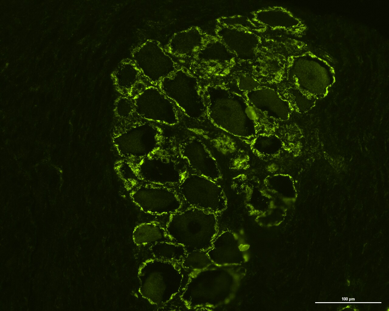
![Immunohistochemistry: GPR55 Antibody - BSA Free [NB110-55498]](https://resources.rndsystems.com/images/products/GPR55-Antibody-Immunohistochemistry-NB110-55498-img0005.jpg "Immunohistochemistry: GPR55 Antibody - BSA Free [NB110-55498]")
Key Product Details
Species Reactivity
Validated:
Cited:
Predicted:
Applications
Validated:
Cited:
Label
Antibody Source
Format
Product Specifications
Immunogen
Epitope
Reactivity Notes
Specificity
Clonality
Host
Isotype
Description
Scientific Data Images for GPR55 Antibody - BSA Free
Immunohistochemistry: GPR55 Antibody - BSA Free [NB110-55498]
GPR55-Antibody-Immunohistochemistry-NB110-55498-img0005.jpg![Western Blot: GPR55 AntibodyBSA Free [NB110-55498]](https://resources.rndsystems.com/images/products/GPR55-Antibody-Western-Blot-NB110-55498-img0004.jpg "Western Blot: GPR55 AntibodyBSA Free [NB110-55498]")
Western Blot: GPR55 AntibodyBSA Free [NB110-55498]
GPR55-Antibody-Western-Blot-NB110-55498-img0004.jpg![Immunohistochemistry-Paraffin: GPR55 Antibody - BSA Free [NB110-55498]](https://resources.rndsystems.com/images/products/GPR55-Antibody-Immunohistochemistry-Paraffin-NB110-55498-img0002.jpg "Immunohistochemistry-Paraffin: GPR55 Antibody - BSA Free [NB110-55498]")
Immunohistochemistry-Paraffin: GPR55 Antibody - BSA Free [NB110-55498]
Immunohistochemistry-Paraffin: GPR55 Antibody [NB110-55498] - Analysis of anti-GPR55 antibody with human pancreas, islet of langerhans.![Immunohistochemistry-Frozen: GPR55 Antibody - BSA Free [NB110-55498]](https://resources.rndsystems.com/images/products/GPR55-Antibody-Immunohistochemistry-Frozen-NB110-55498-img0003.jpg "Immunohistochemistry-Frozen: GPR55 Antibody - BSA Free [NB110-55498]")
Immunohistochemistry-Frozen: GPR55 Antibody - BSA Free [NB110-55498]
Immunohistochemistry-Frozen: GPR55 Antibody [NB110-55498] - Cryosection of canine cervical dorsal root ganglion in which satellite glial cells and some sensory neurons expressed GPR55 immunoreactivity. Image submitted by a verified customer review.
Immunocytochemistry/ Immunofluorescence: GPR55 Antibody - BSA Free [NB110-55498] -
Immunocytochemistry/ Immunofluorescence: GPR55 Antibody - BSA Free [NB110-55498] - Photomicrographs of cryosections of canine cervical (C8) dorsal root ganglion showing GPR55 (a–f) & PPARalpha (g–i) immunolabeling. (a–c) Arrows indicate the Neurotrace-labeled nuclei of satellite glial cells (a) which showed bright GPR55 immunolabelling (b). White stars indicate unlabeled sensory neurons; open stars indicate empty spaces in which sensory neurons were no more evident. (d–f) White arrows indicate satellite glial cells which co-expressed bright GPR55- (d) & glial fibrillary acidic protein (GFAP) immunoreactivity; open arrows indicate SGCs which were GPR55 immunoreactive & GFAP negative (e). Stars indicate sensory neurons of different dimension, which expressed faint –to-moderate GPR55 immunoreactivity. (g–i) White arrows indicate the Neurotrace labeled nuclei of SGCs which showed PPARalpha immunoreactivity (h). Open arrow indicate autofluorescent pigment. Bar: a–i = 50 μm. Image collected & cropped by CiteAb from the following publication (https://pubmed.ncbi.nlm.nih.gov/31608295), licensed under a CC-BY license. Not internally tested by Novus Biologicals.
Immunocytochemistry/ Immunofluorescence: GPR55 Antibody - BSA Free [NB110-55498] -
Immunocytochemistry/ Immunofluorescence: GPR55 Antibody - BSA Free [NB110-55498] - Photomicrographs of cryosections of canine cervical (C8) dorsal root ganglion showing GPR55 (a–f) & PPARalpha (g–i) immunolabeling. (a–c) Arrows indicate the Neurotrace-labeled nuclei of satellite glial cells (a) which showed bright GPR55 immunolabelling (b). White stars indicate unlabeled sensory neurons; open stars indicate empty spaces in which sensory neurons were no more evident. (d–f) White arrows indicate satellite glial cells which co-expressed bright GPR55- (d) & glial fibrillary acidic protein (GFAP) immunoreactivity; open arrows indicate SGCs which were GPR55 immunoreactive & GFAP negative (e). Stars indicate sensory neurons of different dimension, which expressed faint –to-moderate GPR55 immunoreactivity. (g–i) White arrows indicate the Neurotrace labeled nuclei of SGCs which showed PPARalpha immunoreactivity (h). Open arrow indicate autofluorescent pigment. Bar: a–i = 50 μm. Image collected & cropped by CiteAb from the following publication (https://pubmed.ncbi.nlm.nih.gov/31608295), licensed under a CC-BY license. Not internally tested by Novus Biologicals.Applications for GPR55 Antibody - BSA Free
Immunohistochemistry-Paraffin
Reviewed Applications
Read 1 review rated 5 using NB110-55498 in the following applications:
Formulation, Preparation, and Storage
Purification
Formulation
Format
Preservative
Concentration
Shipping
Stability & Storage
Background: GPR55
Long Name
Alternate Names
Entrez Gene IDs
Gene Symbol
UniProt
Additional GPR55 Products
Product Documents for GPR55 Antibody - BSA Free
Certificate of Analysis
To download a Certificate of Analysis, please enter a lot or batch number in the search box below.
Product Specific Notices for GPR55 Antibody - BSA Free
This product is for research use only and is not approved for use in humans or in clinical diagnosis. Primary Antibodies are guaranteed for 1 year from date of receipt.
Related Research Areas
Citations for GPR55 Antibody - BSA Free
 Powered by Bioz
Customer Reviews for GPR55 Antibody - BSA Free (1)
Have you used GPR55 Antibody - BSA Free?
Submit a review and receive an Amazon gift card!
$25/€18/£15/$25CAN/¥2500 Yen for a review with an image
$10/€7/£6/$10CAN/¥1110 Yen for a review without an image
Submit a review
Customer Images
-
Application: Immunohistochemistry-FrozenSample Tested: Canine sensory neurons and Canine peripheral sensory neuronsSpecies: CanineVerified Customer | Posted 07/08/2019Cryosection of canine cervical dorsal root ganglion in which satellite glial cells and some sensory neurons expressed GPR55 immunoreactivity.

There are no reviews that match your criteria.
Protocols
Find general support by application which include: protocols, troubleshooting, illustrated assays, videos and webinars.
- Antigen Retrieval Protocol (PIER)
- Antigen Retrieval for Frozen Sections Protocol
- Appropriate Fixation of IHC/ICC Samples
- Cellular Response to Hypoxia Protocols
- Chromogenic IHC Staining of Formalin-Fixed Paraffin-Embedded (FFPE) Tissue Protocol
- Chromogenic Immunohistochemistry Staining of Frozen Tissue
- ClariTSA™ Fluorophore Kits
- Detection & Visualization of Antibody Binding
- Fluorescent IHC Staining of Frozen Tissue Protocol
- Graphic Protocol for Heat-induced Epitope Retrieval
- Graphic Protocol for the Preparation and Fluorescent IHC Staining of Frozen Tissue Sections
- Graphic Protocol for the Preparation and Fluorescent IHC Staining of Paraffin-embedded Tissue Sections
- Graphic Protocol for the Preparation of Gelatin-coated Slides for Histological Tissue Sections
- IHC Sample Preparation (Frozen sections vs Paraffin)
- Immunofluorescent IHC Staining of Formalin-Fixed Paraffin-Embedded (FFPE) Tissue Protocol
- Immunohistochemistry (IHC) and Immunocytochemistry (ICC) Protocols
- Immunohistochemistry Frozen Troubleshooting
- Immunohistochemistry Paraffin Troubleshooting
- Preparing Samples for IHC/ICC Experiments
- Preventing Non-Specific Staining (Non-Specific Binding)
- Primary Antibody Selection & Optimization
- Protocol for Heat-Induced Epitope Retrieval (HIER)
- Protocol for Making a 4% Formaldehyde Solution in PBS
- Protocol for VisUCyte™ HRP Polymer Detection Reagent
- Protocol for the Preparation & Fixation of Cells on Coverslips
- Protocol for the Preparation and Chromogenic IHC Staining of Frozen Tissue Sections
- Protocol for the Preparation and Chromogenic IHC Staining of Frozen Tissue Sections - Graphic
- Protocol for the Preparation and Chromogenic IHC Staining of Paraffin-embedded Tissue Sections
- Protocol for the Preparation and Chromogenic IHC Staining of Paraffin-embedded Tissue Sections - Graphic
- Protocol for the Preparation and Fluorescent IHC Staining of Frozen Tissue Sections
- Protocol for the Preparation and Fluorescent IHC Staining of Paraffin-embedded Tissue Sections
- Protocol for the Preparation of Gelatin-coated Slides for Histological Tissue Sections
- R&D Systems Quality Control Western Blot Protocol
- TUNEL and Active Caspase-3 Detection by IHC/ICC Protocol
- The Importance of IHC/ICC Controls
- Troubleshooting Guide: Immunohistochemistry
- Troubleshooting Guide: Western Blot Figures
- Western Blot Conditions
- Western Blot Protocol
- Western Blot Protocol for Cell Lysates
- Western Blot Troubleshooting
- Western Blot Troubleshooting Guide
- View all Protocols, Troubleshooting, Illustrated assays and Webinars
FAQs for GPR55 Antibody - BSA Free
-
Q: Do you have any data about the use of the following antibody in Immunofluorescence and Confocal Microscopy:
A:
We have only validated product NB110-55498 for use in IHC Paraffin. Please see our images on the datasheet. The recommended dilution for this antibody in IHC-P is 5 ug/ml.
-
Q: I'm interested in one of your products: GPR55 Antibody (NB110-55498). I'd like to know if it has ever been used in non permeabilised cells, indicating that it can efficiently recognize an extracellular epitope.
A: We have done validation of this product in IHC-P of pancreas as well as spleen tissues after heat-induced antigen retrieval and yes, permeabilization was executed before blocking/primary antibody incubation step in the protocol. Have you seen our another GPR55 antibody (NLS6817)? This antibody has been generated employing an immunogen which was a synthetic peptide made to the 3rd extracellular domain of GPR55 protein. Because this antibody should technically be detecting extra-cellular domain of the target, you may try skipping permeabilization step for this antibody, however, you will still need to perform antigen retrieval.
-
Q: Do you have any data about the use of the following antibody in Immunofluorescence and Confocal Microscopy:
A:
We have only validated product NB110-55498 for use in IHC Paraffin. Please see our images on the datasheet. The recommended dilution for this antibody in IHC-P is 5 ug/ml.
-
Q: I'm interested in one of your products: GPR55 Antibody (NB110-55498). I'd like to know if it has ever been used in non permeabilised cells, indicating that it can efficiently recognize an extracellular epitope.
A: We have done validation of this product in IHC-P of pancreas as well as spleen tissues after heat-induced antigen retrieval and yes, permeabilization was executed before blocking/primary antibody incubation step in the protocol. Have you seen our another GPR55 antibody (NLS6817)? This antibody has been generated employing an immunogen which was a synthetic peptide made to the 3rd extracellular domain of GPR55 protein. Because this antibody should technically be detecting extra-cellular domain of the target, you may try skipping permeabilization step for this antibody, however, you will still need to perform antigen retrieval.