GRP78/HSPA5 Antibody - BSA Free
Novus Biologicals | Catalog # NBP1-06274
![Simple Western: GRP78/HSPA5 AntibodyBSA Free [NBP1-06274]](https://resources.rndsystems.com/images/products/GRP78-HSPA5-Antibody-Simple-Western-NBP1-06274-img0006.jpg "Simple Western: GRP78/HSPA5 AntibodyBSA Free [NBP1-06274]")
Key Product Details
Validated by
Species Reactivity
Validated:
Cited:
Predicted:
Applications
Validated:
Cited:
Label
Antibody Source
Format
Product Specifications
Immunogen
Reactivity Notes
Localization
Marker
Clonality
Host
Isotype
Theoretical MW
Disclaimer note: The observed molecular weight of the protein may vary from the listed predicted molecular weight due to post translational modifications, post translation cleavages, relative charges, and other experimental factors.
Scientific Data Images for GRP78/HSPA5 Antibody - BSA Free
Simple Western: GRP78/HSPA5 AntibodyBSA Free [NBP1-06274]
Simple Western: GRP78/HSPA5 Antibody [NBP1-06274] - Image shows a specific band for GRP78 in 0.1 mg/mL of HeLa lysate. This experiment was performed under reducing conditions using the 12-230 kDa separation system.![Western Blot: GRP78/HSPA5 AntibodyBSA Free [NBP1-06274]](https://resources.rndsystems.com/images/products/GRP78-HSPA5-Antibody-Western-Blot-NBP1-06274-img0016.jpg "Western Blot: GRP78/HSPA5 AntibodyBSA Free [NBP1-06274]")
![Western Blot: GRP78/HSPA5 AntibodyBSA Free [NBP1-06274]](https://resources.rndsystems.com/images/products/GRP78-HSPA5-Antibody-Western-Blot-NBP1-06274-img0017.jpg "Western Blot: GRP78/HSPA5 AntibodyBSA Free [NBP1-06274]")
Western Blot: GRP78/HSPA5 AntibodyBSA Free [NBP1-06274]
Western Blot: GRP78/HSPA5 Antibody [NBP1-06274] - Analysis of MAM-associated protein levels in control and patients' fibroblasts.(A) Equal amounts from one control and the five patients were loaded (50 ug of total cell lysates) and subjected to Western Blot with anti-Grp75, anti-sigma-1R and anti-Mfn2 antibodies. We used anti-GAPDH antibody to ensure equal amounts of protein loaded in each lane. This result is representative of three independent experiments. PLoS One. 2016 Mar 9;11(3):e0150357. doi: 10.1371/journal.pone.0150357![Immunocytochemistry/ Immunofluorescence: GRP78/HSPA5 Antibody - BSA Free [NBP1-06274]](https://resources.rndsystems.com/images/products/GRP78-HSPA5-Antibody-Immunocytochemistry-Immunofluorescence-NBP1-06274-img0013.jpg "Immunocytochemistry/ Immunofluorescence: GRP78/HSPA5 Antibody - BSA Free [NBP1-06274]")
Immunocytochemistry/ Immunofluorescence: GRP78/HSPA5 Antibody - BSA Free [NBP1-06274]
Immunocytochemistry/Immunofluorescence: GRP78/HSPA5 Antibody [NBP1-06274] - NIH-3T3 cells were fixed and permeabilized for 10 minutes using -20C MeOH. The cells were incubated with anti-GRP78/HSPA5 at 2 ug/mL overnight at 4C and detected with an anti-rabbit Dylight 488 (Green) at a 1:500 dilution. Nuclei were counterstained with DAPI (Blue). Cells were imaged using a 40X objective.![Western Blot: GRP78/HSPA5 AntibodyBSA Free [NBP1-06274]](https://resources.rndsystems.com/images/products/GRP78-HSPA5-Antibody-Western-Blot-NBP1-06274-img0010.jpg "Western Blot: GRP78/HSPA5 AntibodyBSA Free [NBP1-06274]")
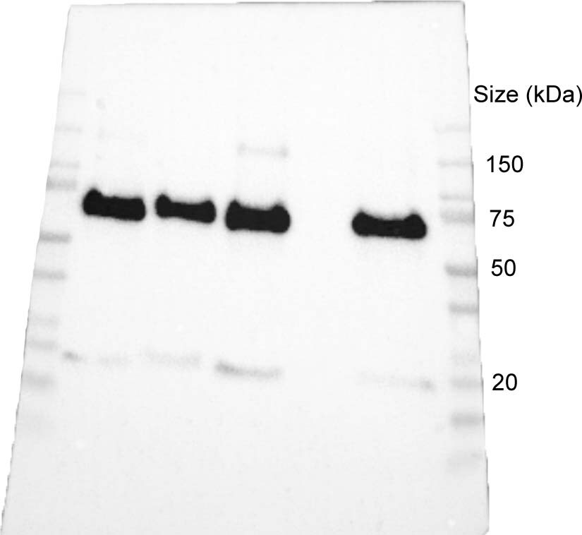
Western Blot: GRP78/HSPA5 AntibodyBSA Free [NBP1-06274]
Western Blot: GRP78/HSPA5 Antibody [NBP1-06274] - Total protein from human HeLa and A431 cells, mouse 3T3 cells and rat PC12 cells was separated on a 7.5% gel by SDS-PAGE, transferred to PVDF membrane and blocked in 5% non-fat milk in TBST. The membrane was probed with 1.0 ug/mL anti-GPR78 in blocking buffer and detected with an anti-rabbit HRP secondary antibody using chemiluminescence.![Immunocytochemistry/ Immunofluorescence: GRP78/HSPA5 Antibody - BSA Free [NBP1-06274]](https://resources.rndsystems.com/images/products/GRP78-HSPA5-Antibody-Immunocytochemistry-Immunofluorescence-NBP1-06274-img0015.jpg "Immunocytochemistry/ Immunofluorescence: GRP78/HSPA5 Antibody - BSA Free [NBP1-06274]")
Immunocytochemistry/ Immunofluorescence: GRP78/HSPA5 Antibody - BSA Free [NBP1-06274]
Immunocytochemistry/Immunofluorescence: GRP78/HSPA5 Antibody [NBP1-06274] - HeLa cells were fixed for 10 minutes using 10% formalin and then permeabilized for 5 minutes using 1X PBS + 0.05% Triton-X100. The cells were incubated with anti-GRP78/HSPA5 at 5 ug/ml overnight at 4C and detected with an anti-rabbit Dylight 488 (Green) at a 1:500 dilution. Alpha tubulin (DM1A) NB100-690 was used as a co-stain at a 1:1000 dilution and detected with an anti-mouse Dylight 550 (Red) at a 1:500 dilution. Nuclei were counterstained with DAPI (Blue). Cells were imaged using a 40X objective.![Flow Cytometry: GRP78/HSPA5 Antibody - BSA Free [NBP1-06274]](https://resources.rndsystems.com/images/products/GRP78-HSPA5-Antibody-Flow-Cytometry-NBP1-06274-img0014.jpg "Flow Cytometry: GRP78/HSPA5 Antibody - BSA Free [NBP1-06274]")
Flow Cytometry: GRP78/HSPA5 Antibody - BSA Free [NBP1-06274]
Flow Cytometry: GRP78/HSPA5 Antibody [NBP1-06274] - An intracellular stain was performed on NIH3T3 cells with GRP78/HSPA5 Antibody NBP1-06274AF488 (blue) and a matched isotype control (orange). Cells were fixed with 4% PFA and then permeabilized with 0.1% saponin. Cells were incubated in an antibody dilution of 5 ug/mL for 30 minutes at room temperature. Both antibodies were conjugated to Alexa Fluor 488.![Western Blot: GRP78/HSPA5 AntibodyBSA Free [NBP1-06274]](https://resources.rndsystems.com/images/products/GRP78-HSPA5-Antibody-Western-Blot-NBP1-06274-img0004.jpg "Western Blot: GRP78/HSPA5 AntibodyBSA Free [NBP1-06274]")
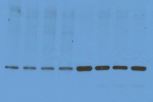
Western Blot: GRP78/HSPA5 AntibodyBSA Free [NBP1-06274]
Western Blot: GRP78/HSPA5 Antibody [NBP1-06274] - Detection of Bip/Grp78 on HeLa whole cell extracts using NBP1-06274.![Flow Cytometry: GRP78/HSPA5 Antibody - BSA Free [NBP1-06274]](https://resources.rndsystems.com/images/products/GRP78-HSPA5-Antibody-Flow-Cytometry-NBP1-06274-img0007.jpg "Flow Cytometry: GRP78/HSPA5 Antibody - BSA Free [NBP1-06274]")
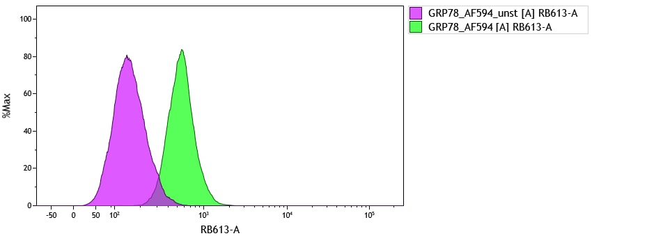
Flow Cytometry: GRP78/HSPA5 Antibody - BSA Free [NBP1-06274]
Flow Cytometry: GRP78/HSPA5 Antibody [NBP1-06274] - Analysis using Alexa Fluor (R) 488 conjugate of NBP1-06274. An intracellular stain was performed on HeLa cells with GPR78/HSPA5 antibody NBP1-06274 (blue) and a matched isotype control NBP2-24893 (orange). Cells were fixed with 4% PFA and then permeablized with 0.1% saponin. 1 ug of antibody was added to 100 uL of staining buffer and cells were incubated for 30 minutes at room temperature. Both antibodies were conjugated to Alexa Fluor 488.![Flow Cytometry: GRP78/HSPA5 Antibody - BSA Free [NBP1-06274]](https://resources.rndsystems.com/images/products/GRP78-HSPA5-Antibody-Flow-Cytometry-NBP1-06274-img0008.jpg "Flow Cytometry: GRP78/HSPA5 Antibody - BSA Free [NBP1-06274]")
Flow Cytometry: GRP78/HSPA5 Antibody - BSA Free [NBP1-06274]
Flow Cytometry: GRP78/HSPA5 Antibody [NBP1-06274] - An intracellular stain was performed on Jurkat cells with Alexa Fluor 647 conjugate of GPR78/HSPA5 antibody NBP1-06274AF647 (blue) and a matched isotype control NBP2-24893AF647 (orange). Cells were fixed with 4% PFA and then permeablized with 0.1% saponin. Cells were incubated in an antibody dilution of 2 ug/mL for 30 minutes at room temperature. Both antibodies were conjugated to Alexa Fluor 647.![Flow Cytometry: GRP78/HSPA5 Antibody - BSA Free [NBP1-06274]](https://resources.rndsystems.com/images/products/GRP78-HSPA5-Antibody-Flow-Cytometry-NBP1-06274-img0012.jpg "Flow Cytometry: GRP78/HSPA5 Antibody - BSA Free [NBP1-06274]")
Flow Cytometry: GRP78/HSPA5 Antibody - BSA Free [NBP1-06274]
Flow Cytometry: GRP78/HSPA5 Antibody [NBP1-06274] - An intracellular stain was performed on HeLa with NBP1-06274 and a matched isotype control. Cells were fixed with 4% PFA and then permeablized with 0.1% saponin. Cells were incubated in an antibody dilution of 1 ug/mL for 30 minutes at room temperature, followed by Rabbit IgG (H+L) Cross-Adsorbed Secondary Antibody, Dylight 550.
Western Blot: GRP78/HSPA5 Antibody - BSA Free [NBP1-06274] -
Western Blot: GRP78/HSPA5 Antibody - BSA Free [NBP1-06274] - Hotair promotes the expression of GRP78 & PD-L1, mediates the proliferation, & helps to regulate the T cell-mediated immune responses in Tu212 & Hep-2 cells. Western blot analysis of GRP78 & PD-L1 after mi-NC or has-miR-30a-5p & vehicle or Hotair transfection in Hep-2 cells (a). The relative expression level of GRP78 & PD-L1 in Hep-2 cells (b). (c–e) The cell viability, apoptosis, & colony formation after mi-NC or has-miR-30a-5p & vehicle or Hotair transfection in Hep-2 cells. (f–h) T cell killing assay of target Tu212 & Hep-2 cells cocultured with PBMC, & ELISA detects the concentration of cytokines (g) IFN-gamma & (h) IL-2. The targeted Tu212 & Hep-2 cells after 24 h transfected with mi-NC or has-miR-30a-5p & vehicle or Hotair were cocultured with PBMC, & the cytotoxicity was measured by the LDH release assay. Data are mean ± SD of at least three independent experiments; ∗p < 0.05; ∗∗p < 0.01; ∗∗∗p < 0.001. Image collected & cropped by CiteAb from the following publication (https://pubmed.ncbi.nlm.nih.gov/35419461), licensed under a CC-BY license. Not internally tested by Novus Biologicals.
Western Blot: GRP78/HSPA5 Antibody - BSA Free [NBP1-06274] -
Western Blot: GRP78/HSPA5 Antibody - BSA Free [NBP1-06274] - Analysis of protein levels involved in ER stress & Ca2+ homeostasis & processing of mRNA XBP1 in control & patients-derived fibroblasts.(A) Equal amounts from controls & patients were loaded (50 μg of total cell lysates) & subjected to Western Blot with anti-Herp, anti-Grp78 & anti-IP3R1 antibodies. We used anti-Hsp60 antibody to ensure equal amounts of protein loaded in each lane. This result is representative of three independent experiments. Protein quantification was performed by laser densitometry. The ratios between proteins/Hsp60 for each cell line were calculated to determine the expression fold-change relative to control. (B) Data represent mean ± standard deviation of three independent experiments. (C) Equal amounts from controls were loaded (50 μg of total cell lysates) & subjected to Western Blot with anti-Herp & anti-Grp78 antibodies. We used anti-GAPDH antibody to ensure equal amounts of protein loaded in each lane. This result is representative of two independent experiments. (D) Equal amounts from control & patients were loaded (50 μg of total cell lysates) & subjected to Western Blot with anti-phospho-PERK antibody. We used anti-GAPDH antibody to ensure equal amounts of protein loaded in each lane. This result is representative of two independent experiments. (E) RT-PCR analysis of the processing of mRNA XBP1 transcription factor. Tm: tunicamycin; u: XBP1 unspliced form; s: XBP1 spliced form. Image collected & cropped by CiteAb from the following publication (https://dx.plos.org/10.1371/journal.pone.0150357), licensed under a CC-BY license. Not internally tested by Novus Biologicals.
Western Blot: GRP78/HSPA5 Antibody - BSA Free [NBP1-06274] -
Western Blot: GRP78/HSPA5 Antibody - BSA Free [NBP1-06274] - UPR Components Activated in A beta -Expressing Flies Are Induced Even Further by Glut1 Overexpression(A) Western blot of eIF2 phosphorylation levels in heads of A beta - & A beta Glut1-expressing flies (+RU) & in controls (-RU), showing no significant difference. Bottom: plotted as means ± SEM (n = 3). Top: a representative gel from the same samples.(B) Grp78 mRNA levels in heads of 18-day-old flies expressing A beta or A beta Glut1 in neurons (+RU) & uninduced controls (-RU), measured by qPCR (relative to eIF1A), plotted as means ± SEM. Genotypes: UAS A beta ; elavGS, UAS A beta /UAS Glut1; elavGS.(C) Quantification of GFP fluorescence in fly brains expressing an Xbp1GFP splicing reporter, plotted as means ± SEM (n = 6–13). Genotypes: elavGS/UAS-Xbp1GFP, UAS A beta ; elavGS/UAS-Xbp1GFP, UAS A beta /UAS Glut1; elavGS/UAS-Xbp1GFP.(D) Spliced Xbp1 mRNA levels in heads of 18-day-old flies expressing A beta or A beta Glut1 in neurons (+RU) & uninduced controls (-RU), measured by qPCR (relative to eIF1A), plotted as means ± SEM(E) Western blot of Grp78 in 14-day-old flies of the same genotypes, plotted below as means ± SEM (n = 6–16). The image is a representative gel of the same samples. Genotypes: UAS A beta ; elavGS, UAS A beta /UAS Glut1; elavGS.∗p ≤ 0.05; ∗∗p ≤ 0.01, by ANOVA. See also Figure S2. Image collected & cropped by CiteAb from the following publication (https://pubmed.ncbi.nlm.nih.gov/27524482), licensed under a CC-BY license. Not internally tested by Novus Biologicals.
Immunocytochemistry/ Immunofluorescence: GRP78/HSPA5 Antibody - BSA Free [NBP1-06274] -
Immunocytochemistry/ Immunofluorescence: GRP78/HSPA5 Antibody - BSA Free [NBP1-06274] - C. elegans granulins impair organismal fitness & resistance to ER stress.(A) Wild-type (N2) & pgrn-1(-) animals with & without granulin expression were subjected to ER stress with tunicamycin (5 μg / ml). The fraction developing to L4 stage was quantified (n = 50, 3 biological replicates). (B) Wild-type (N2) & pgrn-1(-) animals with & without C. elegans progranulin over-expression (OE) were subjected to ER stress with tunicamycin (5 μg / ml). The fraction developing to L4 stage was quantified (n = 50, 3 biological replicates). (C) Total worm lysates from synchronized day 1 adult granulin-expressing animals were immunoblotted with an anti-HSP-4/BiP antibody (3 biological replicates). Anti-actin was used as a loading control. (D) Wild-type & pgrn-1(-) animals with & without granulin expression were staged as embryos. Animals were scored for development to L4 stage (n = 50, 12 biological replicates). (E) Measurement of body length at day 1 adulthood (n = 12). (F) Measurement of short-term associative learning (three biological replicates). The glutamate receptor mutant nmr-1(ak4) was used as a positive control. Throughout, error bars show mean ± SEM, one or two-way ANOVA with post-hoc Tukey multiple comparisons test. Comparisons are to wild-type unless otherwise indicated (*P<0.05, ***P<0.001, ****P<0.0001, ns = not significant, wt = wild-type). Image collected & cropped by CiteAb from the following publication (https://pubmed.ncbi.nlm.nih.gov/31398187), licensed under a CC-BY license. Not internally tested by Novus Biologicals.
Flow Cytometry: Rabbit Polyclonal GRP78/HSPA5 Antibody [NBP1-06274]
Flow cytometry of GRP78/HSPA5 expression on breast cell cancer line. Image from a verified customer review.
Immunohistochemistry: GRP78/HSPA5 Antibody - BSA Free [NBP1-06274] -
Beta cell TFs, ER stress, and autophagy markers in aging beta cells.Immunohistochemistry and confocal microscopy of human pancreas formalin-fixed paraffin-embedded samples from adults (22 to 35 years old) and old adults (61 to 79 years old). Human islets were stained with (A) insulin, PDX1, NKX6-1, and/or NKX2-2; (B) insulin and HSPA5 or XBP1; and (C) insulin, LC3A/B, and LAMP1. (D) Quantification of LC3-LAMP1 colocalization, area of beta cell cytosol covered by LAMP1+ granules, and LAMP1+ granule size. Each dot represents data from a single region of ~140 μm2 of human islets. Here, 1 region per islet, 10 regions per donor were quantified. In (A) and (B), the islet region is demarcated by the dotted yellow line. Scale bars, 50 μm (A and B), 20 μm (C), and 5 μm (inset of C). CI, confidence interval. Image collected and cropped by CiteAb from the following open publication (https://pubmed.ncbi.nlm.nih.gov/36197983), licensed under a CC-BY license. Not internally tested by Novus Biologicals.Applications for GRP78/HSPA5 Antibody - BSA Free
Flow Cytometry
Immunocytochemistry/ Immunofluorescence
Immunohistochemistry
Immunohistochemistry Free-Floating
Immunohistochemistry-Paraffin
Simple Western
Western Blot
In Simple Western only 10 - 15 uL of the recommended dilution is used per data point.
See Simple Western Antibody Database for Simple Western validation: Tested in HeLa lysate 0.1 mg/mL, separated by Size, antibody dilution of 1:25, apparent MW was 77 kDa. Separated by Size-Wes, Sally Sue/Peggy Sue.
The observed molecular weight of the protein may vary from the listed predicted molecular weight due to post translational modifications, post translation cleavages, relative charges, and other experimental factors.
Reviewed Applications
Read 3 reviews rated 4.3 using NBP1-06274 in the following applications:
Flow Cytometry Panel Builder
Bio-Techne Knows Flow Cytometry
Save time and reduce costly mistakes by quickly finding compatible reagents using the Panel Builder Tool.
Advanced Features
- Spectra Viewer - Custom analysis of spectra from multiple fluorochromes
- Spillover Popups - Visualize the spectra of individual fluorochromes
- Antigen Density Selector - Match fluorochrome brightness with antigen density

Formulation, Preparation, and Storage
Purification
Formulation
Format
Preservative
Concentration
Shipping
Stability & Storage
Background: GRP78/HSPA5
Long Name
Alternate Names
Gene Symbol
Additional GRP78/HSPA5 Products
Product Documents for GRP78/HSPA5 Antibody - BSA Free
Certificate of Analysis
To download a Certificate of Analysis, please enter a lot or batch number in the search box below.
Product Specific Notices for GRP78/HSPA5 Antibody - BSA Free
This product is for research use only and is not approved for use in humans or in clinical diagnosis. Primary Antibodies are guaranteed for 1 year from date of receipt.
Related Research Areas
Citations for GRP78/HSPA5 Antibody - BSA Free
 Powered by Bioz
Customer Reviews for GRP78/HSPA5 Antibody - BSA Free (3)
Have you used GRP78/HSPA5 Antibody - BSA Free?
Submit a review and receive an Amazon gift card!
$25/€18/£15/$25CAN/¥2500 Yen for a review with an image
$10/€7/£6/$10CAN/¥1110 Yen for a review without an image
Submit a review
Customer Images
-
Application: Flow CytometrySample Tested: Human breast cancer cellsSpecies: HumanVerified Customer | Posted 01/16/2026Flow cytometry of GRP78 expression on breast cell cancer line

-
Application: Western BlotSample Tested: Mouse dorsal root gangliaSpecies: MouseVerified Customer | Posted 03/23/2019Primary antibody BiP incubated at 1:1000 at 4 degrees overnight. Secondary antibody goat anti-rabbit HRP incubated at 1:10,000 for 1 hour at room temperature. Visualized using ECL.
-
Application: Western BlotSample Tested: Whole embryos lysateSpecies: OtherVerified Customer | Posted 07/27/2016Grp78 WB using zebrafish embryos
There are no reviews that match your criteria.
Protocols
View specific protocols for GRP78/HSPA5 Antibody - BSA Free (NBP1-06274):
Sample Preparation.
1. Grow cells to 60-85% confluency. Flow cytometry requires between 2 x 10⁵ and 1 x 10⁶ cells for optimal performance.
2. If cells are adherent, harvest gently by washing once with staining buffer and then scraping. Avoid using trypsin as this can disrupt certain epitopes of interest. If enzymatic harvest is required, use Accutase, Collagenase, or TrypLE Express for a less damaging option.
3. Reserve 100 uL for counting, then transfer cell volume into a 50 mL conical tube and centrifuge for 8 minutes at 400 RCF.
a. Count cells using a hemocytometer and a 1:1 trypan blue exclusion stain to determine cell viability before starting the flow protocol. If cells appear blue, do not proceed.
4. Re-suspend cells to a concentration of 1 x 10⁶ cells/mL in staining buffer (NBP2-26247).
5. Aliquot out 100 uL samples in accordance with your experimental samples.
Tip: When cell surface and intracellular staining are required in the same sample, it is advisable that the cell surface staining be performed first since the fixation and permeabilization steps might reduce the availability of surface antigens.
Intracellular Staining.
Tip: When performing intracellular staining, it is important to use appropriate fixation and permeabilization reagents based upon the target and its subcellular location. Generally, our Intracellular Flow Assay Kit (NBP2-29450) is a good place to start as it contains an optimized combination of reagents for intracellular staining as well as an inhibitor of intracellular protein transport (necessary if staining secreted proteins). Certain targets may require more gentle or transient permeabilization protocols such as the commonly employed methanol or saponin-based methods.
Protocol for Cytoplasmic Targets:
1. Fix the cells by adding 100 uL fixation solution (such as 4% PFA) to each sample for 10-15 minutes.
2. Permeabilize cells by adding 100 uL of a permeabilization buffer to every 1 x 10⁶ cells present in the sample. Mix well and incubate at room temperature for 15 minutes.
a. For cytoplasmic targets, use a gentle permeabilization solution such as 1X PBS + 0.5% Saponin or 1X PBS + 0.5% Tween-20.
b. To maintain the permeabilized state throughout your experiment, use staining buffer + 0.1% of the permeabilization reagent (i.e. 0.1% Tween-20 or 0.1% Saponin).
3. Following the 15 minute incubation, add 2 mL of the staining buffer + 0.1% permeabilizer to each sample.
4. Centrifuge for 1 minute at 400 RCF.
5. Discard supernatant and re-suspend in 100 uL of staining buffer + 0.1% permeabilizer.
6. Add appropriate amount of each antibody (eg. 1 test or 1 ug per sample, as experimentally determined).
7. Mix well and incubate at room temperature for 30 minutes- 1 hour. Gently mix samples every 10-15 minutes.
8. Following the primary/conjugate incubation, add 1-2 mL/sample of staining buffer +0.1% permeabilizer and centrifuge for 1 minute at 400 RCF.
9. Wash twice by re-suspending cells in staining buffer (2 mL for tubes or 200 uL for wells) and centrifuging at 400 RCF for 5 minutes. Discard supernatant.
10. Add appropriate amount of secondary antibody (as experimentally determined) to each sample.
11. Incubate at room temperature in dark for 20 minutes.
12. Add 1-2 mL of staining buffer and centrifuge at 400 RCF for 1 minute and discard supernatant.
13. Wash twice by re-suspending cells in staining buffer (2 mL for tubes or 200 uL for wells) and centrifuging at 400 RCF for 5 minutes. Discard supernatant.
14. Resuspend in an appropriate volume of staining buffer (usually 500 uL per sample) and proceed with analysis on your flow cytometer.
Culture cells to appropriate density in 35 mm culture dishes or 6-well plates.
1. Remove culture medium and wash the cells briefly in PBS. Add 10% formalin to the dish and fix at room temperature for 10 minutes.
2. Remove the formalin and wash the cells in PBS.
3. Permeablize the cells with 0.1% Triton X100 or other suitable detergent for 10 min.
4. Remove the permeablization buffer and wash three times for 10 minutes each in PBS. Be sure to not let the specimen dry out.
5. To block nonspecific antibody binding, incubate in 10% normal goat serum from 1 hour to overnight at room temperature.
6. Add primary antibody at appropriate dilution and incubate overnight at 4C.
7. Remove primary antibody and replace with PBS. Wash three times for 10 minutes each.
8. Add secondary antibody at appropriate dilution. Incubate for 1 hour at room temperature.
9. Remove secondary antibody and replace with PBS. Wash three times for 10 minutes each.
10. Counter stain DNA with DAPi if required.
Antigen Unmasking:
Bring slides to a boil in 10 mM sodium citrate buffer (pH 6.0) then maintain at a sub-boiling temperature for 10 minutes. Cool slides on bench-top for 30 minutes (keep slides in the sodium citrate buffer all the time).
Staining:
1. Wash sections in deionized water three times for 5 minutes each.
2. Wash sections in PBS for 5 minutes.
3. Block each section with 100-400 ul blocking solution (1% BSA in PBS) for 1 hour at room temperature.
4. Remove blocking solution and add 100-400 ul diluted primary antibody. Incubate overnight at 4 C.
5. Remove antibody solution and wash sections in wash buffer three times for 5 minutes each.
6. Add 100-400 ul HRP polymer conjugated secondary antibody. Incubate 30 minutes at room temperature.
7. Wash sections three times in wash buffer for 5 minutes each.
8. Add 100-400 ul DAB substrate to each section and monitor staining closely.
9. As soon as the sections develop, immerse slides in deionized water.
10. Counterstain sections in hematoxylin.
11. Wash sections in deionized water two times for 5 minutes each.
12. Dehydrate sections.
13. Mount coverslips.
1. Perform SDS-PAGE on samples to be analyzed, loading 10-25 ug of total protein per lane.
2. Transfer proteins to PVDF membrane according to the instructions provided by the manufacturer of the membrane and transfer apparatus.
3. Stain the membrane with Ponceau S (or similar product) to assess transfer success, and mark molecular weight standards where appropriate.
4. Rinse the blot TBS -0.05% Tween 20 (TBST).
5. Block the membrane in 5% Non-fat milk in TBST (blocking buffer) for at least 1 hour.
6. Wash the membrane in TBST three times for 10 minutes each.
7. Dilute primary antibody in blocking buffer and incubate overnight at 4C with gentle rocking.
8. Wash the membrane in TBST three times for 10 minutes each.
9. Incubate the membrane in diluted HRP conjugated secondary antibody in blocking buffer (as per manufacturer's instructions) for 1 hour at room temperature.
10. Wash the blot in TBST three times for 10 minutes each (this step can be repeated as required to reduce background).
11. Apply the detection reagent of choice in accordance with the manufacturers instructions.
Find general support by application which include: protocols, troubleshooting, illustrated assays, videos and webinars.
- 7-Amino Actinomycin D (7-AAD) Cell Viability Flow Cytometry Protocol
- Antigen Retrieval Protocol (PIER)
- Antigen Retrieval for Frozen Sections Protocol
- Appropriate Fixation of IHC/ICC Samples
- Cellular Response to Hypoxia Protocols
- Chromogenic IHC Staining of Formalin-Fixed Paraffin-Embedded (FFPE) Tissue Protocol
- Chromogenic Immunohistochemistry Staining of Frozen Tissue
- ClariTSA™ Fluorophore Kits
- Detection & Visualization of Antibody Binding
- Extracellular Membrane Flow Cytometry Protocol
- Flow Cytometry Protocol for Cell Surface Markers
- Flow Cytometry Protocol for Staining Membrane Associated Proteins
- Flow Cytometry Staining Protocols
- Flow Cytometry Troubleshooting Guide
- Fluorescent IHC Staining of Frozen Tissue Protocol
- Graphic Protocol for Heat-induced Epitope Retrieval
- Graphic Protocol for the Preparation and Fluorescent IHC Staining of Frozen Tissue Sections
- Graphic Protocol for the Preparation and Fluorescent IHC Staining of Paraffin-embedded Tissue Sections
- Graphic Protocol for the Preparation of Gelatin-coated Slides for Histological Tissue Sections
- ICC Cell Smear Protocol for Suspension Cells
- ICC Immunocytochemistry Protocol Videos
- ICC for Adherent Cells
- IHC Sample Preparation (Frozen sections vs Paraffin)
- Immunocytochemistry (ICC) Protocol
- Immunocytochemistry Troubleshooting
- Immunofluorescence of Organoids Embedded in Cultrex Basement Membrane Extract
- Immunofluorescent IHC Staining of Formalin-Fixed Paraffin-Embedded (FFPE) Tissue Protocol
- Immunohistochemistry (IHC) and Immunocytochemistry (ICC) Protocols
- Immunohistochemistry Frozen Troubleshooting
- Immunohistochemistry Paraffin Troubleshooting
- Intracellular Flow Cytometry Protocol Using Alcohol (Methanol)
- Intracellular Flow Cytometry Protocol Using Detergents
- Intracellular Nuclear Staining Flow Cytometry Protocol Using Detergents
- Intracellular Staining Flow Cytometry Protocol Using Alcohol Permeabilization
- Intracellular Staining Flow Cytometry Protocol Using Detergents to Permeabilize Cells
- Preparing Samples for IHC/ICC Experiments
- Preventing Non-Specific Staining (Non-Specific Binding)
- Primary Antibody Selection & Optimization
- Propidium Iodide Cell Viability Flow Cytometry Protocol
- Protocol for Heat-Induced Epitope Retrieval (HIER)
- Protocol for Liperfluo
- Protocol for Making a 4% Formaldehyde Solution in PBS
- Protocol for VisUCyte™ HRP Polymer Detection Reagent
- Protocol for the Characterization of Human Th22 Cells
- Protocol for the Characterization of Human Th9 Cells
- Protocol for the Fluorescent ICC Staining of Cell Smears - Graphic
- Protocol for the Fluorescent ICC Staining of Cultured Cells on Coverslips - Graphic
- Protocol for the Preparation & Fixation of Cells on Coverslips
- Protocol for the Preparation and Chromogenic IHC Staining of Frozen Tissue Sections
- Protocol for the Preparation and Chromogenic IHC Staining of Frozen Tissue Sections - Graphic
- Protocol for the Preparation and Chromogenic IHC Staining of Paraffin-embedded Tissue Sections
- Protocol for the Preparation and Chromogenic IHC Staining of Paraffin-embedded Tissue Sections - Graphic
- Protocol for the Preparation and Fluorescent ICC Staining of Cells on Coverslips
- Protocol for the Preparation and Fluorescent ICC Staining of Non-adherent Cells
- Protocol for the Preparation and Fluorescent ICC Staining of Stem Cells on Coverslips
- Protocol for the Preparation and Fluorescent IHC Staining of Frozen Tissue Sections
- Protocol for the Preparation and Fluorescent IHC Staining of Paraffin-embedded Tissue Sections
- Protocol for the Preparation of Gelatin-coated Slides for Histological Tissue Sections
- Protocol for the Preparation of a Cell Smear for Non-adherent Cell ICC - Graphic
- Protocol: Annexin V and PI Staining by Flow Cytometry
- Protocol: Annexin V and PI Staining for Apoptosis by Flow Cytometry
- R&D Systems Quality Control Western Blot Protocol
- TUNEL and Active Caspase-3 Detection by IHC/ICC Protocol
- The Importance of IHC/ICC Controls
- Troubleshooting Guide: Fluorokine Flow Cytometry Kits
- Troubleshooting Guide: Immunohistochemistry
- Troubleshooting Guide: Western Blot Figures
- Western Blot Conditions
- Western Blot Protocol
- Western Blot Protocol for Cell Lysates
- Western Blot Troubleshooting
- Western Blot Troubleshooting Guide
- View all Protocols, Troubleshooting, Illustrated assays and Webinars
FAQs for GRP78/HSPA5 Antibody - BSA Free
-
Q: I have received the Novus Cat # for a human GRP78-specific rabbit Ab from a lab that had used it and achieved some nice results with it. They suggest we use the same Ab to reproduce their data. However, I have not been able to find a product with that Cat # in your online search engine. Perhaps the product has been discontinued, or was given a different Cat #? Could you please look into this? The Ab they used was: GRP78 Cat # NBP1-49339, Lot # A113911. In case the product is no longer available, could you please recommend something that should be equivalent or very similar to it?
A: NBP1-49339 has been discontinued but NBP1-06274 uses a similar antigen that falls between amino acids 250 and 300 and works in Western blot and ICC. ICC and flow use essentially the same techniques, so it should work just fine if you were using it in flow or Western blot before.