![Western Blot: HA Tag Antibody [NBP2-21581]](https://resources.rndsystems.com/images/products/HA-Tag-Antibody-Western-Blot-NBP2-21581-img0017.jpg "Western Blot: HA Tag Antibody [NBP2-21581]")
Key Product Details
Species Reactivity
Epitope Tag
Applications
Western Blot, ELISA, Flow Cytometry, Immunocytochemistry/ Immunofluorescence, Immunoprecipitation, Chromatin Immunoprecipitation (ChIP)
Label
Unconjugated
Antibody Source
Polyclonal Rabbit IgG
Loading...
Product Specifications
Immunogen
The immunogen used to generate this antibody corresponds to HA Tag
Clonality
Polyclonal
Host
Rabbit
Isotype
IgG
Scientific Data Images for HA Tag Antibody
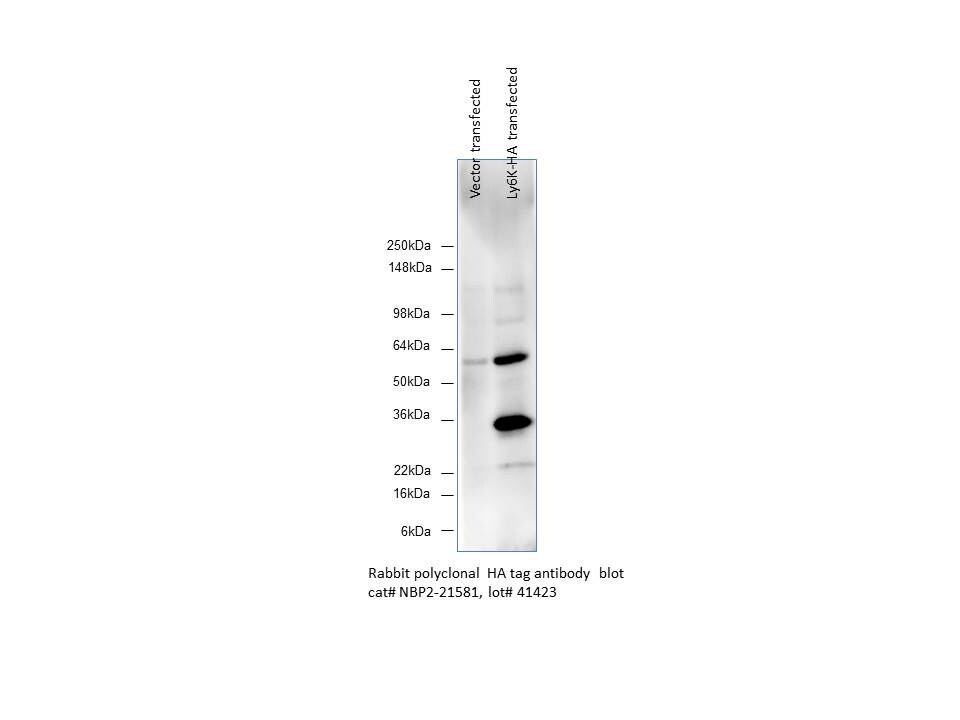
Western Blot: HA Tag Antibody [NBP2-21581]
Western Blot: HA Tag Antibody [NBP2-21581] - Analysis in human HEK293T cells overexpressed with Ly6K-HA. Western blot image submitted by a verified customer review.![Flow Cytometry: HA Tag Antibody [NBP2-21581]](https://resources.rndsystems.com/images/products/HA-Tag-Antibody-Flow-Cytometry-NBP2-21581-img0013.jpg "Flow Cytometry: HA Tag Antibody [NBP2-21581]")
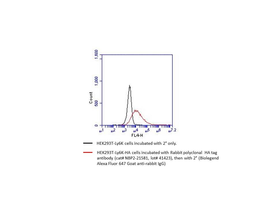
Flow Cytometry: HA Tag Antibody [NBP2-21581]
Flow Cytometry: HA Tag Antibody [NBP2-21581] - HEK293T cells overexpressed with Ly6K-HA stained with HA Epitope Tag Antibody (red) or secondary only (black). Flow cytometry image submitted by a verified customer review.
Western Blot: HA Tag Antibody [NBP2-21581] -
Non-transfected (-) and transfected (+) 293T whole cell extracts (30 ug) were separated by 12% SDS-PAGE, and the membrane was blotted with HA tag antibody (NBP2-21581) diluted at 1:3000. The HRP-conjugated anti-rabbit IgG antibody was used to detect the primary antibody.
ELISA: HA Tag Antibody [NBP2-21581] -
An ELISA plate is coated with 50 uL of a HA-Tag conjugated BSA at concentration of 5 ug/mL. The peptide conjugated carrier protein is coated and detected with anti-HA tag antibody (NBP2-21581) at concentration ranged from 3.1 to 200 ng/mL.
ELISA: HA Tag Antibody [NBP2-21581] -
Indirect ELISA analysis performed by coating plate with recombinant HA-tag tagged protein (1000-15.63 ng/mL). Coated protein was probed with HA tag antibody (HRP) (NBP2-21581-01) (1 ug/mL).
Western Blot: HA Tag Antibody [NBP2-21581] -
Non-transfected (-) and transfected (+) 293T whole cell extracts (30 ug) were separated by 12% SDS-PAGE, and the membrane was blotted with HA tag antibody (NBP2-21581-01) diluted at 1:3000. The HRP-conjugated anti-rabbit IgG antibody was used to detect the primary antibody.Applications for HA Tag Antibody
Application
Recommended Usage
Chromatin Immunoprecipitation (ChIP)
Assay Dependent
ELISA
1:1000 - 1:10000
Flow Cytometry
Validated by a verified customer review
Immunocytochemistry/ Immunofluorescence
1:100 - 1:1000
Immunoprecipitation
1:100 - 1:500
Western Blot
1:1000 - 1:10000
Reviewed Applications
Read 2 reviews rated 5 using NBP2-21581 in the following applications:
Flow Cytometry Panel Builder
Bio-Techne Knows Flow Cytometry
Save time and reduce costly mistakes by quickly finding compatible reagents using the Panel Builder Tool.
Advanced Features
- Spectra Viewer - Custom analysis of spectra from multiple fluorochromes
- Spillover Popups - Visualize the spectra of individual fluorochromes
- Antigen Density Selector - Match fluorochrome brightness with antigen density

Formulation, Preparation, and Storage
Purification
Antigen Affinity-purified
Formulation
PBS, 20% Glycerol
Preservative
0.025% Proclin 300
Concentration
Concentrations vary lot to lot. See vial label for concentration. If unlisted please contact technical services.
Shipping
The product is shipped with polar packs. Upon receipt, store it immediately at the temperature recommended below.
Stability & Storage
Aliquot and store at -20C or -80C. Avoid freeze-thaw cycles.
Background: HA Tag
References
1. Wilks, S., Graaf, M. D., Smith, D. J., & Burke, D. F. (2012). A review of influenza haemagglutinin receptor binding as it relates to pandemic properties. Vaccine, 30(29), 4369-4376. doi:10.1016/j.vaccine.2012.02.076
2. Wu, N. C., & Wilson, I. A. (2019). Influenza hemagglutinin structures and antibody recognition. Cold Spring Harbor Perspectives in Medicine, 10(8). doi:10.1101/cshperspect.a038778
3. Zhao, X., Li, G., & Liang, S. (2013). Several affinity tags commonly used in chromatographic purification. Journal of Analytical Methods in Chemistry, 2013, 1-8. doi:10.1155/2013/581093
4. Kimple, M. E., Brill, A. L., & Pasker, R. L. (2013). Overview of affinity tags for protein purification. Current Protocols in Protein Science, 73(1). doi:10.1002/0471140864.ps0909s73
5. Schembri, L., Dalibart, R., Tomasello, F., Legembre, P., Ichas, F., & Giorgi, F. D. (2007). The HA tag is cleaved and loses immunoreactivity during apoptosis. Nature Methods, 4(2), 107-108. doi:10.1038/nmeth0207-107
Alternate Names
HA Epitope Tag
Additional HA Tag Products
Product Documents for HA Tag Antibody
Certificate of Analysis
To download a Certificate of Analysis, please enter a lot or batch number in the search box below.
Product Specific Notices for HA Tag Antibody
This product is for research use only and is not approved for use in humans or in clinical diagnosis. Primary Antibodies are guaranteed for 1 year from date of receipt.
Related Research Areas
Customer Reviews for HA Tag Antibody (2)
5 out of 5
2 Customer Ratings
Have you used HA Tag Antibody?
Submit a review and receive an Amazon gift card!
$25/€18/£15/$25CAN/¥2500 Yen for a review with an image
$10/€7/£6/$10CAN/¥1110 Yen for a review without an image
Submit a review
Customer Images
Showing
1
-
2 of
2 reviews
Showing All
Filter By:
-
Application: Flow CytometrySample Tested: human HEK293T cells overexpressed with Ly6K-HASpecies: HumanVerified Customer | Posted 07/06/2016HA Epitope Tag Antibody (NBP2-21581) for flow cytometry

-
Application: Western BlotSample Tested: human HEK293T cells overexoressed with Ly6K-HASpecies: HumanVerified Customer | Posted 06/17/2016HA Epitope Tag Antibody (NBP2-21581)
There are no reviews that match your criteria.
Protocols
Find general support by application which include: protocols, troubleshooting, illustrated assays, videos and webinars.
- 7-Amino Actinomycin D (7-AAD) Cell Viability Flow Cytometry Protocol
- Appropriate Fixation of IHC/ICC Samples
- Cellular Response to Hypoxia Protocols
- ChIP Protocol Video
- Chromatin Immunoprecipitation (ChIP) Protocol
- Chromatin Immunoprecipitation Protocol
- ClariTSA™ Fluorophore Kits
- Detection & Visualization of Antibody Binding
- ELISA Sample Preparation & Collection Guide
- ELISA Troubleshooting Guide
- Extracellular Membrane Flow Cytometry Protocol
- Flow Cytometry Protocol for Cell Surface Markers
- Flow Cytometry Protocol for Staining Membrane Associated Proteins
- Flow Cytometry Staining Protocols
- Flow Cytometry Troubleshooting Guide
- How to Run an R&D Systems DuoSet ELISA
- How to Run an R&D Systems Quantikine ELISA
- How to Run an R&D Systems Quantikine™ QuicKit™ ELISA
- ICC Cell Smear Protocol for Suspension Cells
- ICC Immunocytochemistry Protocol Videos
- ICC for Adherent Cells
- Immunocytochemistry (ICC) Protocol
- Immunocytochemistry Troubleshooting
- Immunofluorescence of Organoids Embedded in Cultrex Basement Membrane Extract
- Immunohistochemistry (IHC) and Immunocytochemistry (ICC) Protocols
- Immunoprecipitation Protocol
- Intracellular Flow Cytometry Protocol Using Alcohol (Methanol)
- Intracellular Flow Cytometry Protocol Using Detergents
- Intracellular Nuclear Staining Flow Cytometry Protocol Using Detergents
- Intracellular Staining Flow Cytometry Protocol Using Alcohol Permeabilization
- Intracellular Staining Flow Cytometry Protocol Using Detergents to Permeabilize Cells
- Preparing Samples for IHC/ICC Experiments
- Preventing Non-Specific Staining (Non-Specific Binding)
- Primary Antibody Selection & Optimization
- Propidium Iodide Cell Viability Flow Cytometry Protocol
- Protocol for Liperfluo
- Protocol for VisUCyte™ HRP Polymer Detection Reagent
- Protocol for the Characterization of Human Th22 Cells
- Protocol for the Characterization of Human Th9 Cells
- Protocol for the Fluorescent ICC Staining of Cell Smears - Graphic
- Protocol for the Fluorescent ICC Staining of Cultured Cells on Coverslips - Graphic
- Protocol for the Preparation and Fluorescent ICC Staining of Cells on Coverslips
- Protocol for the Preparation and Fluorescent ICC Staining of Non-adherent Cells
- Protocol for the Preparation and Fluorescent ICC Staining of Stem Cells on Coverslips
- Protocol for the Preparation of a Cell Smear for Non-adherent Cell ICC - Graphic
- Protocol: Annexin V and PI Staining by Flow Cytometry
- Protocol: Annexin V and PI Staining for Apoptosis by Flow Cytometry
- Quantikine HS ELISA Kit Assay Principle, Alkaline Phosphatase
- Quantikine HS ELISA Kit Principle, Streptavidin-HRP Polymer
- R&D Systems Quality Control Western Blot Protocol
- Sandwich ELISA (Colorimetric) – Biotin/Streptavidin Detection Protocol
- Sandwich ELISA (Colorimetric) – Direct Detection Protocol
- TUNEL and Active Caspase-3 Detection by IHC/ICC Protocol
- The Importance of IHC/ICC Controls
- Troubleshooting Guide: ELISA
- Troubleshooting Guide: Fluorokine Flow Cytometry Kits
- Troubleshooting Guide: Western Blot Figures
- Western Blot Conditions
- Western Blot Protocol
- Western Blot Protocol for Cell Lysates
- Western Blot Troubleshooting
- Western Blot Troubleshooting Guide
- View all Protocols, Troubleshooting, Illustrated assays and Webinars
FAQs for HA Tag Antibody
Showing
1
-
1 of
1 FAQ
Showing All
-
Q: We are looking for a NON-RABBIT polyclonal anti-HA (hemagglutinin - YPYDVPDYA ) antibody for immunohistochemistry that works in 4% paraformaldehyde-fixed tissue. We have a transgenic mouse expressing an HA-tagged protein and want to perform double-labeling with another antibody made in rabbit. We have had no luck using mouse or rat monoclonal anti-HA antibodies. The non-rabbit polyclonal antibody would preferably be made in goat, although if you have other species let us know. It could be unconjugated or conjugated (eg fluorochrome or HRP). However, we are only interested in a product with demonstrated effectiveness for IHC in a published paper. Do you have any such products?
A:
We do have other host species for this target and some that have been mentioned in publications. Unfortunately for our goat antibodies against this tag none of them have been validated in IHC-P, and for two we have shown they do not work. Please see this link to our goat anti-HA Epitope Tag antibodies. Since they work in ICC/IF I would assume as long as the tag is expressed in your tissues that you should still pick it up. In this case we have an Innovators Reward Program you can take advantage of for testing a new application to save some money. Here are the chicken anti-HA Epitope Tag antibodies. None of them have been tested in IHC-P as before, or that would be indicated with a positive result and the application on the datasheet or a negative results with the (-) beside the application. Another option for you as well would be going with a Rabbit polyclonal that we have available that has been well publicized and reviewed by customers (cat# NB600-363). I know you plan on double staining, but one way around this would be to use your other antibody and incubate with the secondary for detection first. After doing that you could have this one directly conjugated to your preferred detection probe and then pick up both of their signals since you would already have bound secondary to your first rabbit primary, there would not be any non-specific binding.
Loading...