HDAC1 Antibody - BSA Free
Novus Biologicals | Catalog # NB100-56340
![Knockout Validated: HDAC1 Antibody [NB100-56340]](https://resources.rndsystems.com/images/products/HDAC1-Antibody-Knockout-Validated-NB100-56340-img0007.jpg "Western Blot: HDAC1 Antibody [NB100-56340]")
Loading...
Key Product Details
Validated by
Knockout/Knockdown
Species Reactivity
Validated:
Human, Mouse, Rat
Cited:
Human, Mouse, Rat
Applications
Validated:
Knockout Validated, Immunohistochemistry, Immunohistochemistry-Paraffin, Immunohistochemistry-Frozen, Western Blot, Simple Western
Cited:
Immunohistochemistry-Paraffin, Immunohistochemistry-Frozen, Western Blot, Immunocytochemistry/ Immunofluorescence, Simple Western, IF/IHC, Knockdown Validated
Label
Unconjugated
Antibody Source
Polyclonal Rabbit IgG
Format
BSA Free
Loading...
Product Specifications
Immunogen
This antibody was generated by immunizing rabbits with a mixture of synthetic peptides corresponding to amino acids 1-5, 433-448 and 467-482 of human HDAC1 (Genbank Accession no. Q13547).
Reactivity Notes
Mouse reactivity reported in scientific literature (PMID: 25648995). Rat reactivity reported in scientific literature (PMID: 26551542)
Clonality
Polyclonal
Host
Rabbit
Isotype
IgG
Scientific Data Images for HDAC1 Antibody - BSA Free
Western Blot: HDAC1 Antibody [NB100-56340]
Western Blot: HDAC1 Antibody [NB100-56340] - Western blot shows lysates of HeLa human cervical epithelial carcinoma parental cell line and HDAC1 knockout (KO) HeLa cell line. PVDF membrane was probed with 2.0 ug/ml of Rabbit Anti-Human HDAC1 Polyclonal Antibody (Catalog # NB100-56340) followed by HRP-conjugated Anti-Rabbit IgG Secondary Antibody (Catalog #HAF008). Specific band was detected for HDAC1 at approximately 65 kDa (as indicated) in the parental HeLa cell line, but is not detectable in the knockout HeLa cell line. This experiment was conducted under reducing conditions.![Western Blot: HDAC1 Antibody [NB100-56340]](https://resources.rndsystems.com/images/products/HDAC1-Antibody-Western-Blot-NB100-56340-img0002.jpg "Western Blot: HDAC1 Antibody [NB100-56340]")
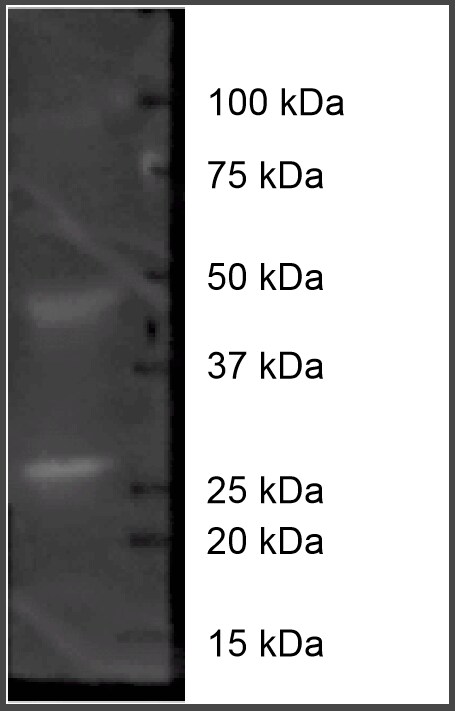
Western Blot: HDAC1 Antibody [NB100-56340]
Western Blot: HDAC1 Antibody [NB100-56340] - Analysis of HDAC1 in HEK293 cell lysate with this antibody.![Immunohistochemistry-Paraffin: HDAC1 Antibody [NB100-56340]](https://resources.rndsystems.com/images/products/HDAC1-Antibody-Immunohistochemistry-Paraffin-NB100-56340-img0005.jpg "Immunohistochemistry-Paraffin: HDAC1 Antibody [NB100-56340]")
Immunohistochemistry-Paraffin: HDAC1 Antibody [NB100-56340]
Immunohistochemistry-Paraffin: HDAC1 Antibody [NB100-56340] - Human testis using HDAC1 antibody at 1:250 on a Bond Rx autostainer (Leica Biosystems). The assay involved 20 minutes of heat induced antigen retrieval (HIER) using 10mM sodium citrate buffer (pH 6.0) and endogenous peroxidase quenching with peroxide block. The sections were incubated with primary antibody for 30 minutes and Bond Polymer Refine Detection (Leica Biosystems) with DAB was used for signal development followed by counterstaining with hematoxylin. Whole slide scanning and capturing of representative images was performed using Aperio AT2 (Leica Biosystems). Staining was performed by Histowiz.![Simple Western: HDAC1 Antibody [NB100-56340]](https://resources.rndsystems.com/images/products/HDAC1-Antibody-Simple-Western-NB100-56340-img0003.jpg "Simple Western: HDAC1 Antibody [NB100-56340]")
Simple Western: HDAC1 Antibody [NB100-56340]
Simple Western: HDAC1 Antibody [NB100-56340] - Simple Western lane view shows a specific band for HDAC1 in 0.5 mg/ml of HEK293 lysate. This experiment was performed under reducing conditions using the 12-230 kDa separation system.Applications for HDAC1 Antibody - BSA Free
Application
Recommended Usage
Immunohistochemistry
1:500. Use reported in multiple pieces of scientific literature
Immunohistochemistry-Frozen
1:10-1:500. Use reported in scientific literature (PMID 26551542)
Immunohistochemistry-Paraffin
1:10-1:500. Use reported in scientific literature (PMID 25648995)
Simple Western
1:200
Western Blot
2 ug/ml
Application Notes
In 293, a 60 kDa band is observed. In Simple Western only 10 - 15 uL of the recommended dilution is used per data point.
See Simple Western Antibody Database for Simple Western validation: Tested in Hek293 lysate 0.5 mg/mL, separated by Size, antibody dilution of 1:100, apparent MW was 68 kDa
See Simple Western Antibody Database for Simple Western validation: Tested in Hek293 lysate 0.5 mg/mL, separated by Size, antibody dilution of 1:100, apparent MW was 68 kDa
Reviewed Applications
Read 2 reviews rated 4.5 using NB100-56340 in the following applications:
Formulation, Preparation, and Storage
Purification
Protein G purified
Formulation
PBS
Format
BSA Free
Preservative
0.02% Sodium Azide
Concentration
1.0 mg/ml
Shipping
The product is shipped with polar packs. Upon receipt, store it immediately at the temperature recommended below.
Stability & Storage
Store at 4C short term. Aliquot and store at -20C long term. Avoid freeze-thaw cycles.
Background: Histone Deacetylase 1/HDAC1
Long Name
Histone Deacetylase 1
Alternate Names
GON-10, HD1, KDAC1, RPD3L1
Entrez Gene IDs
3065 (Human)
Gene Symbol
HDAC1
UniProt
Additional Histone Deacetylase 1/HDAC1 Products
Product Documents for HDAC1 Antibody - BSA Free
Certificate of Analysis
To download a Certificate of Analysis, please enter a lot or batch number in the search box below.
Product Specific Notices for HDAC1 Antibody - BSA Free
This product is for research use only and is not approved for use in humans or in clinical diagnosis. Primary Antibodies are guaranteed for 1 year from date of receipt.
Related Research Areas
Citations for HDAC1 Antibody - BSA Free
 Powered by Bioz
Customer Reviews for HDAC1 Antibody - BSA Free (2)
4.5 out of 5
2 Customer Ratings
Have you used HDAC1 Antibody - BSA Free?
Submit a review and receive an Amazon gift card!
$25/€18/£15/$25CAN/¥2500 Yen for a review with an image
$10/€7/£6/$10CAN/¥1110 Yen for a review without an image
Submit a review
Customer Images
Showing
1
-
2 of
2 reviews
Showing All
Filter By:
-
Application: Western BlotSample Tested: Ovary tissueSpecies: PigVerified Customer | Posted 11/10/2016HDAC on porcine whole ovarian homogenate (50 ug protein)-Blocked in 5% BSA.2% PBST for 4 hours -Primary was 1:250 in 5% BSA.2% PBST overnight at 4C -Secondary was goat x rabbit 1:1000 in 5% BSA.2% PBST 1 hour

-
Application: Western BlotSample Tested: See PMID 23327920Species: MouseVerified Customer | Posted 12/12/2014
There are no reviews that match your criteria.
Protocols
View specific protocols for HDAC1 Antibody - BSA Free (NB100-56340):
Immunohistochemistry-Paraffin Embedded Sections
Antigen Unmasking:
Bring slides to a boil in 10 mM sodium citrate buffer (pH 6.0) then maintain at a sub-boiling temperature for 10 minutes. Cool slides on bench-top for 30 minutes (keep slides in the sodium citrate buffer at all times).
Staining:
1. Wash sections in deionized water three times for 5 minutes each.
2. Wash sections in PBS for 5 minutes.
3. Block each section with 100-400 ul blocking solution (1% BSA in PBS) for 1 hour at room temperature.
4. Remove blocking solution and add 100-400 ul diluted primary antibody. Incubate overnight at 4 C.
5. Remove antibody solution and wash sections in wash buffer three times for 5 minutes each.
6. Add 100-400 ul HRP polymer conjugated secondary antibody. Incubate 30 minutes at room temperature.
7. Wash sections three times in wash buffer for 5 minutes each.
8. Add 100-400 ul DAB substrate to each section and monitor staining closely.
9. As soon as the sections develop, immerse slides in deionized water.
10. Counterstain sections in hematoxylin.
11. Wash sections in deionized water two times for 5 minutes each.
12. Dehydrate sections.
13. Mount coverslips.
Western Blot Protocol
1. Perform SDS-PAGE on samples to be analyzed, loading 10-25 ug of total protein per lane.
2. Transfer proteins to PVDF membrane according to the instructions provided by the manufacturer of the membrane and transfer apparatus.
3. Stain the membrane with Ponceau S (or similar product) to assess transfer success, and mark molecular weight standards where appropriate.
4. Rinse the blot TBS -0.05% Tween 20 (TBST).
5. Block the membrane in 5% Non-fat milk in TBST (blocking buffer) for at least 1 hour.
6. Wash the membrane in TBST three times for 10 minutes each.
7. Dilute primary antibody in 2% Non-fat milk in TBST and incubate overnight at 4C with gentle rocking.
8. Wash the membrane in TBST three times for 10 minutes each.
9. Incubate the membrane in diluted HRP conjugated secondary antibody in blocking buffer (as per manufacturer's instructions) for 1 hour at room temperature.
10. Wash the blot in TBST three times for 10 minutes each (this step can be repeated as required to reduce background).
11. Apply the detection reagent of choice in accordance with the manufacturer's instructi
Find general support by application which include: protocols, troubleshooting, illustrated assays, videos and webinars.
- Antigen Retrieval Protocol (PIER)
- Antigen Retrieval for Frozen Sections Protocol
- Appropriate Fixation of IHC/ICC Samples
- Cellular Response to Hypoxia Protocols
- Chromogenic IHC Staining of Formalin-Fixed Paraffin-Embedded (FFPE) Tissue Protocol
- Chromogenic Immunohistochemistry Staining of Frozen Tissue
- ClariTSA™ Fluorophore Kits
- Detection & Visualization of Antibody Binding
- Fluorescent IHC Staining of Frozen Tissue Protocol
- Graphic Protocol for Heat-induced Epitope Retrieval
- Graphic Protocol for the Preparation and Fluorescent IHC Staining of Frozen Tissue Sections
- Graphic Protocol for the Preparation and Fluorescent IHC Staining of Paraffin-embedded Tissue Sections
- Graphic Protocol for the Preparation of Gelatin-coated Slides for Histological Tissue Sections
- IHC Sample Preparation (Frozen sections vs Paraffin)
- Immunofluorescent IHC Staining of Formalin-Fixed Paraffin-Embedded (FFPE) Tissue Protocol
- Immunohistochemistry (IHC) and Immunocytochemistry (ICC) Protocols
- Immunohistochemistry Frozen Troubleshooting
- Immunohistochemistry Paraffin Troubleshooting
- Preparing Samples for IHC/ICC Experiments
- Preventing Non-Specific Staining (Non-Specific Binding)
- Primary Antibody Selection & Optimization
- Protocol for Heat-Induced Epitope Retrieval (HIER)
- Protocol for Making a 4% Formaldehyde Solution in PBS
- Protocol for VisUCyte™ HRP Polymer Detection Reagent
- Protocol for the Preparation & Fixation of Cells on Coverslips
- Protocol for the Preparation and Chromogenic IHC Staining of Frozen Tissue Sections
- Protocol for the Preparation and Chromogenic IHC Staining of Frozen Tissue Sections - Graphic
- Protocol for the Preparation and Chromogenic IHC Staining of Paraffin-embedded Tissue Sections
- Protocol for the Preparation and Chromogenic IHC Staining of Paraffin-embedded Tissue Sections - Graphic
- Protocol for the Preparation and Fluorescent IHC Staining of Frozen Tissue Sections
- Protocol for the Preparation and Fluorescent IHC Staining of Paraffin-embedded Tissue Sections
- Protocol for the Preparation of Gelatin-coated Slides for Histological Tissue Sections
- R&D Systems Quality Control Western Blot Protocol
- TUNEL and Active Caspase-3 Detection by IHC/ICC Protocol
- The Importance of IHC/ICC Controls
- Troubleshooting Guide: Immunohistochemistry
- Troubleshooting Guide: Western Blot Figures
- Western Blot Conditions
- Western Blot Protocol
- Western Blot Protocol for Cell Lysates
- Western Blot Troubleshooting
- Western Blot Troubleshooting Guide
- View all Protocols, Troubleshooting, Illustrated assays and Webinars
Loading...