His Tag Antibody (RM146)
Novus Biologicals | Catalog # NBP2-61482
Recombinant Monoclonal Antibody
Key Product Details
Species Reactivity
Epitope Tag
Applications
Validated:
Immunohistochemistry, Immunohistochemistry-Paraffin, Western Blot, Flow Cytometry, Immunocytochemistry/ Immunofluorescence, Simple Western, Immunoprecipitation
Cited:
Western Blot
Label
Unconjugated
Antibody Source
Recombinant Monoclonal Rabbit IgG Clone # RM146 expressed in HEK293
Loading...
Product Specifications
Immunogen
This His Tag Antibody (RM146) was developed against a mixture of a peptide with 6xHis-Tag at the N-terminus and another peptide with 6xHis-Tag at the C-terminus.
Specificity
This antibody reacts to recombinant proteins containing the 6xHis-Tag or 10xHis-Tag fused to either the amino or carboxy terminus. No cross reactivity with other endogenous protein in mammalian or bacteria cells.
Clonality
Monoclonal
Host
Rabbit
Isotype
IgG
Scientific Data Images for His Tag Antibody (RM146)
![Western Blot: His Tag Antibody (RM146) [NBP2-61482]](https://resources.rndsystems.com/images/products/His-Tag-Antibody-RM146-Western-Blot-NBP2-61482-img0008.jpg "Western Blot: His Tag Antibody (RM146) [NBP2-61482]")
Western Blot: His Tag Antibody (RM146) [NBP2-61482]
Western Blot: His Tag Antibody (RM146) [NBP2-61482] - Western blot of 293T cells (1) untransfected or (2) transfected with His-Tag fusion protein X and (3) E. coli lysate without His-Tag Protein Y (4) E. coli lysate with His-Tag Protein Y, using NBP2-61482.![Immunohistochemistry: His Tag Antibody (RM146) [NBP2-61482]](https://resources.rndsystems.com/images/products/His-Tag-Antibody-RM146-Immunohistochemistry-NBP2-61482-img0004.jpg "Immunohistochemistry: His Tag Antibody (RM146) [NBP2-61482]")
Immunohistochemistry: His Tag Antibody (RM146) [NBP2-61482]
Immunohistochemistry: His Tag Antibody (RM146) [NBP2-61482] - Immunohistochemistry staining of naive HepG2 cells (Negative control) using anti-His-Tag antibody, NBP2-61482.![Immunohistochemistry-Paraffin: His Tag Antibody (RM146) [NBP2-61482]](https://resources.rndsystems.com/images/products/His-Tag-Antibody-RM146-Immunohistochemistry-Paraffin-NBP2-61482-img0001.jpg "Immunohistochemistry-Paraffin: His Tag Antibody (RM146) [NBP2-61482]")
Immunohistochemistry-Paraffin: His Tag Antibody (RM146) [NBP2-61482]
Immunohistochemistry-Paraffin: His Tag Antibody (RM146) [NBP2-61482] - Immunohistochemistry staining of 293T cells expressing His-Tag nuclear protein X, using NBP2-61482.![Simple Western: His Tag Antibody (RM146) [NBP2-61482]](https://resources.rndsystems.com/images/products/His-Tag-Antibody-RM146-Simple-Western-NBP2-61482-img0007.jpg "Simple Western: His Tag Antibody (RM146) [NBP2-61482]")
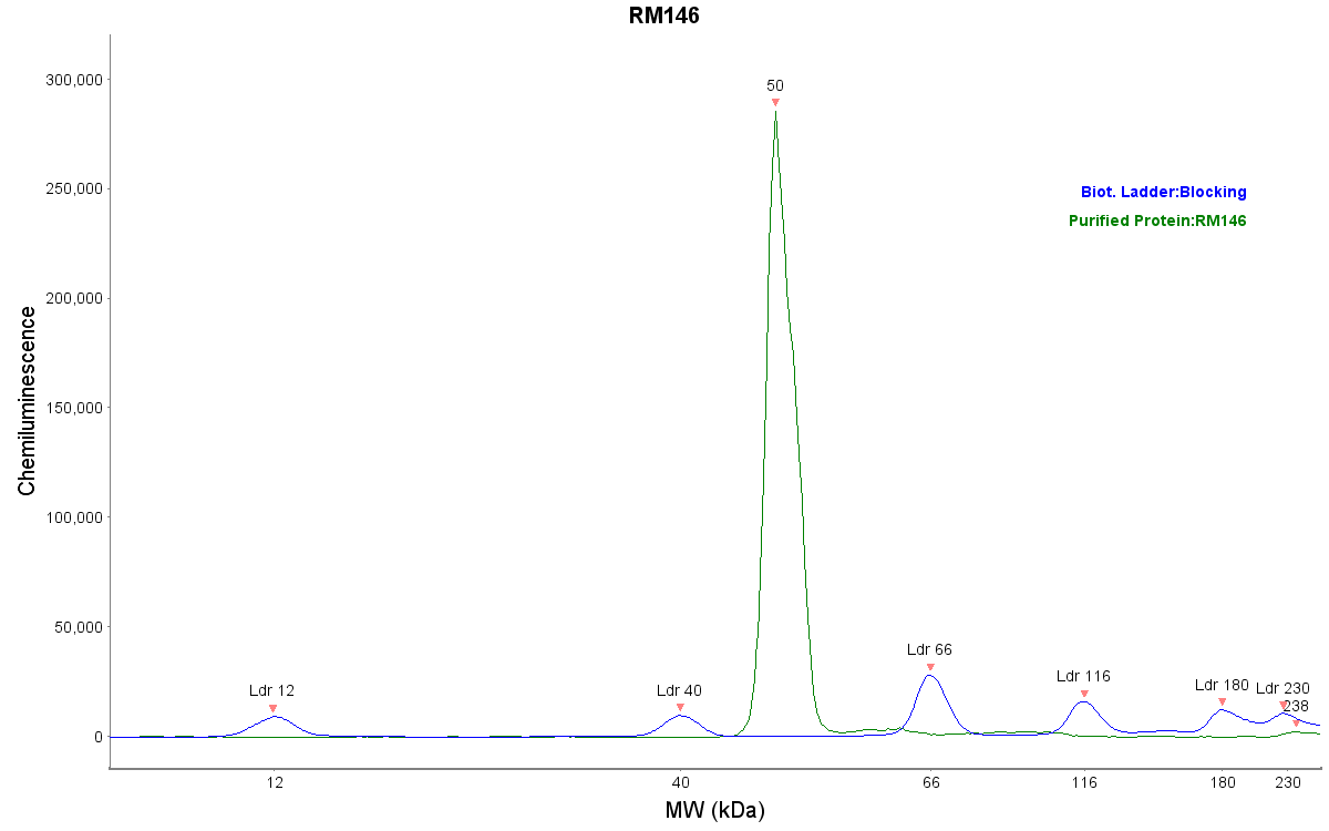
Simple Western: His Tag Antibody (RM146) [NBP2-61482]
Simple Western: His Tag Antibody (RM146) [NBP2-61482] - Human purified protein. Simple Western image submitted by a verified customer review.Applications for His Tag Antibody (RM146)
Application
Recommended Usage
Flow Cytometry
0.5 ug/mL - 2 ug/mL
Immunocytochemistry/ Immunofluorescence
0.5 ug/mL - 2 ug/mL
Immunohistochemistry
0.1 ug/mL - 1 ug/mL
Immunoprecipitation
0.5 ug/ml - 2 ug/ml
Simple Western
reported from a verified customer review.
Western Blot
0.1 ug/ml - 1 ug/ml
Application Notes
His Tag Antibody (RM146) validated for Simple Western from a verified customer review.
See Simple Western Antibody Database for Simple Western validation: Tested in Human purified protein, separated by Size
See Simple Western Antibody Database for Simple Western validation: Tested in Human purified protein, separated by Size
Reviewed Applications
Read 1 review rated 5 using NBP2-61482 in the following applications:
Flow Cytometry Panel Builder
Bio-Techne Knows Flow Cytometry
Save time and reduce costly mistakes by quickly finding compatible reagents using the Panel Builder Tool.
Advanced Features
- Spectra Viewer - Custom analysis of spectra from multiple fluorochromes
- Spillover Popups - Visualize the spectra of individual fluorochromes
- Antigen Density Selector - Match fluorochrome brightness with antigen density

Formulation, Preparation, and Storage
Purification
Protein A purified
Formulation
50% Glycerol/PBS, 1% BSA
Preservative
0.09% Sodium Azide
Concentration
1 mg/ml
Shipping
The product is shipped with polar packs. Upon receipt, store it immediately at the temperature recommended below.
Stability & Storage
Store at -20C. Avoid freeze-thaw cycles.
Background: His Tag
References
1. Malhotra, A. (2009). Tagging for protein expression. Methods in Enzymology, Guide to Protein Purification, 2nd Edition, 463, 239-258. https://doi.org/10.1016/s0076-6879(09)63016-0
2. Terpe, K. (2003). Overview of tag protein fusions: from molecular and biochemical fundamentals to commercial systems. Applied Microbiology and Biotechnology, 60(5), 523-533. https://doi.org/10.1007/s00253-002-1158-6
3. Booth, W. T., Schlachter, C. R., Pote, S., Ussin, N., Mank, N. J., Klapper, V.,... Chruszcz, M. (2018). Impact of an N-terminal polyhistidine tag on protein thermal stability. ACS Omega, 3(1), 760-768. https://doi.org/10.1021/acsomega.7b01598
Long Name
Histidine Tag
Alternate Names
polyHistidine
Additional His Tag Products
Product Documents for His Tag Antibody (RM146)
Certificate of Analysis
To download a Certificate of Analysis, please enter a lot or batch number in the search box below.
Product Specific Notices for His Tag Antibody (RM146)
This product is for research use only and is not approved for use in humans or in clinical diagnosis. Primary Antibodies are guaranteed for 1 year from date of receipt.
Citations for His Tag Antibody (RM146)
 Powered by Bioz
Customer Reviews for His Tag Antibody (RM146) (1)
5 out of 5
1 Customer Rating
Have you used His Tag Antibody (RM146)?
Submit a review and receive an Amazon gift card!
$25/€18/£15/$25CAN/¥2500 Yen for a review with an image
$10/€7/£6/$10CAN/¥1110 Yen for a review without an image
Submit a review
Customer Images
Showing
1
-
1 of
1 review
Showing All
Filter By:
-
Application: Simple WesternSample Tested: Purified proteinSpecies: HumanVerified Customer | Posted 12/04/2018

There are no reviews that match your criteria.
Protocols
Find general support by application which include: protocols, troubleshooting, illustrated assays, videos and webinars.
- 7-Amino Actinomycin D (7-AAD) Cell Viability Flow Cytometry Protocol
- Antigen Retrieval Protocol (PIER)
- Antigen Retrieval for Frozen Sections Protocol
- Appropriate Fixation of IHC/ICC Samples
- Cellular Response to Hypoxia Protocols
- Chromogenic IHC Staining of Formalin-Fixed Paraffin-Embedded (FFPE) Tissue Protocol
- Chromogenic Immunohistochemistry Staining of Frozen Tissue
- ClariTSA™ Fluorophore Kits
- Detection & Visualization of Antibody Binding
- Extracellular Membrane Flow Cytometry Protocol
- Flow Cytometry Protocol for Cell Surface Markers
- Flow Cytometry Protocol for Staining Membrane Associated Proteins
- Flow Cytometry Staining Protocols
- Flow Cytometry Troubleshooting Guide
- Fluorescent IHC Staining of Frozen Tissue Protocol
- Graphic Protocol for Heat-induced Epitope Retrieval
- Graphic Protocol for the Preparation and Fluorescent IHC Staining of Frozen Tissue Sections
- Graphic Protocol for the Preparation and Fluorescent IHC Staining of Paraffin-embedded Tissue Sections
- Graphic Protocol for the Preparation of Gelatin-coated Slides for Histological Tissue Sections
- ICC Cell Smear Protocol for Suspension Cells
- ICC Immunocytochemistry Protocol Videos
- ICC for Adherent Cells
- IHC Sample Preparation (Frozen sections vs Paraffin)
- Immunocytochemistry (ICC) Protocol
- Immunocytochemistry Troubleshooting
- Immunofluorescence of Organoids Embedded in Cultrex Basement Membrane Extract
- Immunofluorescent IHC Staining of Formalin-Fixed Paraffin-Embedded (FFPE) Tissue Protocol
- Immunohistochemistry (IHC) and Immunocytochemistry (ICC) Protocols
- Immunohistochemistry Frozen Troubleshooting
- Immunohistochemistry Paraffin Troubleshooting
- Immunoprecipitation Protocol
- Intracellular Flow Cytometry Protocol Using Alcohol (Methanol)
- Intracellular Flow Cytometry Protocol Using Detergents
- Intracellular Nuclear Staining Flow Cytometry Protocol Using Detergents
- Intracellular Staining Flow Cytometry Protocol Using Alcohol Permeabilization
- Intracellular Staining Flow Cytometry Protocol Using Detergents to Permeabilize Cells
- Preparing Samples for IHC/ICC Experiments
- Preventing Non-Specific Staining (Non-Specific Binding)
- Primary Antibody Selection & Optimization
- Propidium Iodide Cell Viability Flow Cytometry Protocol
- Protocol for Heat-Induced Epitope Retrieval (HIER)
- Protocol for Liperfluo
- Protocol for Making a 4% Formaldehyde Solution in PBS
- Protocol for VisUCyte™ HRP Polymer Detection Reagent
- Protocol for the Characterization of Human Th22 Cells
- Protocol for the Characterization of Human Th9 Cells
- Protocol for the Fluorescent ICC Staining of Cell Smears - Graphic
- Protocol for the Fluorescent ICC Staining of Cultured Cells on Coverslips - Graphic
- Protocol for the Preparation & Fixation of Cells on Coverslips
- Protocol for the Preparation and Chromogenic IHC Staining of Frozen Tissue Sections
- Protocol for the Preparation and Chromogenic IHC Staining of Frozen Tissue Sections - Graphic
- Protocol for the Preparation and Chromogenic IHC Staining of Paraffin-embedded Tissue Sections
- Protocol for the Preparation and Chromogenic IHC Staining of Paraffin-embedded Tissue Sections - Graphic
- Protocol for the Preparation and Fluorescent ICC Staining of Cells on Coverslips
- Protocol for the Preparation and Fluorescent ICC Staining of Non-adherent Cells
- Protocol for the Preparation and Fluorescent ICC Staining of Stem Cells on Coverslips
- Protocol for the Preparation and Fluorescent IHC Staining of Frozen Tissue Sections
- Protocol for the Preparation and Fluorescent IHC Staining of Paraffin-embedded Tissue Sections
- Protocol for the Preparation of Gelatin-coated Slides for Histological Tissue Sections
- Protocol for the Preparation of a Cell Smear for Non-adherent Cell ICC - Graphic
- Protocol: Annexin V and PI Staining by Flow Cytometry
- Protocol: Annexin V and PI Staining for Apoptosis by Flow Cytometry
- R&D Systems Quality Control Western Blot Protocol
- TUNEL and Active Caspase-3 Detection by IHC/ICC Protocol
- The Importance of IHC/ICC Controls
- Troubleshooting Guide: Fluorokine Flow Cytometry Kits
- Troubleshooting Guide: Immunohistochemistry
- Troubleshooting Guide: Western Blot Figures
- Western Blot Conditions
- Western Blot Protocol
- Western Blot Protocol for Cell Lysates
- Western Blot Troubleshooting
- Western Blot Troubleshooting Guide
- View all Protocols, Troubleshooting, Illustrated assays and Webinars
Loading...