Histone H2AX [p Ser139] Antibody (9F3) - BSA Free
Novus Biologicals | Catalog # NBP1-19255
Loading...
Key Product Details
Validated by
Biological Validation
Species Reactivity
Validated:
Human, Mouse, Rat, Porcine, Bovine, Canine, Chicken, Guinea Pig, Hamster, Primate, Rabbit, Sheep
Cited:
Human, Mouse
Applications
Validated:
Western Blot, Immunocytochemistry, Immunocytochemistry/ Immunofluorescence
Cited:
Western Blot, Immunocytochemistry, Immunocytochemistry/ Immunofluorescence
Label
Unconjugated
Antibody Source
Monoclonal Mouse IgG1 kappa Clone # 9F3
Format
BSA Free
Loading...
Product Specifications
Immunogen
This Histone H2AX [p Ser139] Antibody (9F3) was developed against a synthetic phospho-peptide derived from the sequence of human Histone H2AX phosphorylated at Ser139.
Reactivity Notes
Please note that this antibody is reactive to Mouse and derived from the same host, Mouse. Mouse-On-Mouse blocking reagent may be needed for IHC and ICC experiments to reduce high background signal. You can find these reagents under catalog numbers PK-2200-NB and MP-2400-NB. Please contact Technical Support if you have any questions.
Modification
p Ser139
Marker
DNA Double-strand break marker
Specificity
Specific to the portion of Human Histone H2AX phosphorylated at Ser139.
Clonality
Monoclonal
Host
Mouse
Isotype
IgG1 kappa
Theoretical MW
15 kDa.
Disclaimer note: The observed molecular weight of the protein may vary from the listed predicted molecular weight due to post translational modifications, post translation cleavages, relative charges, and other experimental factors.
Disclaimer note: The observed molecular weight of the protein may vary from the listed predicted molecular weight due to post translational modifications, post translation cleavages, relative charges, and other experimental factors.
Description
Licensed to Novus Biologicals LLC under U.S. Patent Nos. 6,362,317 and 6,884,873.
Scientific Data Images for Histone H2AX [p Ser139] Antibody (9F3) - BSA Free
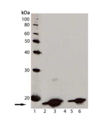
Western Blot: Histone H2AX [p Ser139] Antibody (9F3) [NBP1-19255] - Analysis of Histone H2AX [p Ser139] antibody (9F3) Jurkat cell lysate; Lane 3: Jurkat cell lysate treated with staurosporine; Lane 4: 3T3 cell lysate; Lane 5: CHO-K1 cell lysate; Lane 6: Rat-2 cell lysate. Bands appear at an observed molecular weight of ~15 kDa.
Applications for Histone H2AX [p Ser139] Antibody (9F3) - BSA Free
Application
Recommended Usage
Western Blot
1:1000
Application Notes
Use in Immunocytochemistry reported in scientific literature (PMID:33088425).
Formulation, Preparation, and Storage
Purification
Protein G purified
Formulation
PBS (pH 7.2) and 50% Glycerol
Format
BSA Free
Preservative
0.09% Sodium Azide
Concentration
Please see the vial label for concentration. If unlisted please contact technical services.
Shipping
The product is shipped with polar packs. Upon receipt, store it immediately at the temperature recommended below.
Stability & Storage
Store at -20C. Avoid freeze-thaw cycles.
Background: Histone H2AX
References
1. Palla, V. V., Karaolanis, G., Katafigiotis, I., Anastasiou, I., Patapis, P., Dimitroulis, D., & Perrea, D. (2017). gamma-H2AX: Can it be established as a classical cancer prognostic factor?. Tumour biology : the journal of the International Society for Oncodevelopmental Biology and Medicine. https://doi.org/10.1177/1010428317695931
2. Kuo, L. J., & Yang, L. X. (2008). Gamma-H2AX - a novel biomarker for DNA double-strand breaks. In vivo (Athens, Greece).
3. Kinner, A., Wu, W., Staudt, C., & Iliakis, G. (2008). Gamma-H2AX in recognition and signaling of DNA double-strand breaks in the context of chromatin. Nucleic acids research. https://doi.org/10.1093/nar/gkn550
4. Redon, C. E., Weyemi, U., Parekh, P. R., Huang, D., Burrell, A. S., & Bonner, W. M. (2012). gamma-H2AX and other histone post-translational modifications in the clinic. Biochimica et biophysica acta. https://doi.org/10.1016/j.bbagrm.2012.02.021
5. H2AX: Uniprot (P16104)
Alternate Names
H2AFX
Gene Symbol
H2AX
UniProt
Additional Histone H2AX Products
Product Documents for Histone H2AX [p Ser139] Antibody (9F3) - BSA Free
Certificate of Analysis
To download a Certificate of Analysis, please enter a lot or batch number in the search box below.
Product Specific Notices for Histone H2AX [p Ser139] Antibody (9F3) - BSA Free
This product is for research use only and is not approved for use in humans or in clinical diagnosis. Primary Antibodies are guaranteed for 1 year from date of receipt.
Related Research Areas
Citations for Histone H2AX [p Ser139] Antibody (9F3) - BSA Free
 Powered by Bioz
Customer Reviews for Histone H2AX [p Ser139] Antibody (9F3) - BSA Free
There are currently no reviews for this product. Be the first to review Histone H2AX [p Ser139] Antibody (9F3) - BSA Free and earn rewards!
Have you used Histone H2AX [p Ser139] Antibody (9F3) - BSA Free?
Submit a review and receive an Amazon gift card!
$25/€18/£15/$25CAN/¥2500 Yen for a review with an image
$10/€7/£6/$10CAN/¥1110 Yen for a review without an image
Submit a review
Protocols
Find general support by application which include: protocols, troubleshooting, illustrated assays, videos and webinars.
- Appropriate Fixation of IHC/ICC Samples
- Cellular Response to Hypoxia Protocols
- ClariTSA™ Fluorophore Kits
- Detection & Visualization of Antibody Binding
- ICC Cell Smear Protocol for Suspension Cells
- ICC Immunocytochemistry Protocol Videos
- ICC for Adherent Cells
- Immunocytochemistry (ICC) Protocol
- Immunocytochemistry Troubleshooting
- Immunofluorescence of Organoids Embedded in Cultrex Basement Membrane Extract
- Immunohistochemistry (IHC) and Immunocytochemistry (ICC) Protocols
- Preparing Samples for IHC/ICC Experiments
- Preventing Non-Specific Staining (Non-Specific Binding)
- Primary Antibody Selection & Optimization
- Protocol for VisUCyte™ HRP Polymer Detection Reagent
- Protocol for the Fluorescent ICC Staining of Cell Smears - Graphic
- Protocol for the Fluorescent ICC Staining of Cultured Cells on Coverslips - Graphic
- Protocol for the Preparation and Fluorescent ICC Staining of Cells on Coverslips
- Protocol for the Preparation and Fluorescent ICC Staining of Non-adherent Cells
- Protocol for the Preparation and Fluorescent ICC Staining of Stem Cells on Coverslips
- Protocol for the Preparation of a Cell Smear for Non-adherent Cell ICC - Graphic
- R&D Systems Quality Control Western Blot Protocol
- TUNEL and Active Caspase-3 Detection by IHC/ICC Protocol
- The Importance of IHC/ICC Controls
- Troubleshooting Guide: Western Blot Figures
- Western Blot Conditions
- Western Blot Protocol
- Western Blot Protocol for Cell Lysates
- Western Blot Troubleshooting
- Western Blot Troubleshooting Guide
- View all Protocols, Troubleshooting, Illustrated assays and Webinars
Loading...