Histone H4 [ac Lys12, ac Lys16, ac Lys8, ac Lys5] Antibody
Novus Biologicals | Catalog # NBP2-16848
![Western Blot: Histone H4 [ac Lys12, ac Lys16, ac Lys8, ac Lys5] Antibody [NBP2-16848]](https://resources.rndsystems.com/images/products/Histone-H4-[ac-Lys12--ac-Lys16--ac-Lys8--ac-Lys5]-Antibody-Western-Blot-NBP2-16848-img0015.jpg "Western Blot: Histone H4 [ac Lys12, ac Lys16, ac Lys8, ac Lys5] Antibody [NBP2-16848]")
Loading...
Key Product Details
Species Reactivity
Validated:
Human, Mouse, Rat
Cited:
Human, Mouse
Predicted:
Xenopus (100%). Backed by our 100% Guarantee.
Applications
Validated:
Immunohistochemistry, Immunohistochemistry-Paraffin, Western Blot, Immunocytochemistry/ Immunofluorescence, Simple Western, Dot Blot
Cited:
Western Blot
Label
Unconjugated
Antibody Source
Polyclonal Rabbit IgG
Loading...
Product Specifications
Immunogen
Carrier-protein conjugated synthetic peptide surrounding acetyl Lys5/Lys8/Lys12/Lys16 of human Histone H4. The exact sequence is proprietary.
Reactivity Notes
Immunogen displays the following percentage of sequence identity for non-tested species: Xenopus laevis (100%).
Modification
ac Lys12, ac Lys16, ac Lys8, ac Lys5
Localization
Nucleus
Clonality
Polyclonal
Host
Rabbit
Isotype
IgG
Theoretical MW
11 kDa.
Disclaimer note: The observed molecular weight of the protein may vary from the listed predicted molecular weight due to post translational modifications, post translation cleavages, relative charges, and other experimental factors.
Disclaimer note: The observed molecular weight of the protein may vary from the listed predicted molecular weight due to post translational modifications, post translation cleavages, relative charges, and other experimental factors.
Scientific Data Images for Histone H4 [ac Lys12, ac Lys16, ac Lys8, ac Lys5] Antibody
Western Blot: Histone H4 [ac Lys12, ac Lys16, ac Lys8, ac Lys5] Antibody [NBP2-16848]
Histone-H4-[ac-Lys12--ac-Lys16--ac-Lys8--ac-Lys5]-Antibody-Western-Blot-NBP2-16848-img0015.jpg![Immunohistochemistry-Paraffin: Histone H4 [ac Lys12, ac Lys16, ac Lys8, ac Lys5] Antibody [NBP2-16848]](https://resources.rndsystems.com/images/products/Histone-H4-Antibody-Immunohistochemistry-Paraffin-NBP2-16848-img0010.jpg "Immunohistochemistry-Paraffin: Histone H4 [ac Lys12, ac Lys16, ac Lys8, ac Lys5] Antibody [NBP2-16848]")
Immunohistochemistry-Paraffin: Histone H4 [ac Lys12, ac Lys16, ac Lys8, ac Lys5] Antibody [NBP2-16848]
Immunohistochemistry-Paraffin: Histone H4 Antibody [NBP2-16848] - Human breast cancer, using antibody at 1:500 dilution. Antigen Retrieval: TrilogyTM (EDTA based) buffer, 15min![Western Blot: Histone H4 [ac Lys12, ac Lys16, ac Lys8, ac Lys5] Antibody [NBP2-16848]](https://resources.rndsystems.com/images/products/Histone-H4-Antibody-Western-Blot-NBP2-16848-img0008.jpg "Western Blot: Histone H4 [ac Lys12, ac Lys16, ac Lys8, ac Lys5] Antibody [NBP2-16848]")
Western Blot: Histone H4 [ac Lys12, ac Lys16, ac Lys8, ac Lys5] Antibody [NBP2-16848]
Western Blot: Histone H4 Antibody [NBP2-16848] - WB for total histone in MCF-7 and HCT116 cells. Image from verified customer review.![Western Blot: Histone H4 [ac Lys12, ac Lys16, ac Lys8, ac Lys5] Antibody [NBP2-16848]](https://resources.rndsystems.com/images/products/Histone-H4-Antibody-Western-Blot-NBP2-16848-img0012.jpg "Western Blot: Histone H4 [ac Lys12, ac Lys16, ac Lys8, ac Lys5] Antibody [NBP2-16848]")
Western Blot: Histone H4 [ac Lys12, ac Lys16, ac Lys8, ac Lys5] Antibody [NBP2-16848]
Western Blot: Histone H4 Antibody [NBP2-16848] - Sample (30 ug of whole cell lysate) A: NIH-3T3 B: BCL-1 15% SDS PAGE diluted at 1:10000![Western Blot: Histone H4 [ac Lys12, ac Lys16, ac Lys8, ac Lys5] Antibody [NBP2-16848]](https://resources.rndsystems.com/images/products/Histone-H4-Antibody-Western-Blot-NBP2-16848-img0013.jpg "Western Blot: Histone H4 [ac Lys12, ac Lys16, ac Lys8, ac Lys5] Antibody [NBP2-16848]")
Western Blot: Histone H4 [ac Lys12, ac Lys16, ac Lys8, ac Lys5] Antibody [NBP2-16848]
Western Blot: Histone H4 Antibody [NBP2-16848] - Sample (30 ug of whole cell lysate) A: HeLa 15% SDS PAGE diluted at 1:10000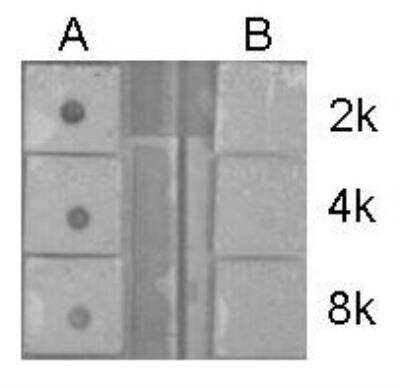
Dot Blot: Histone H4 Antibody [NBP2-16848] - Peptide samples (0.1 ug) were spotted onto positively charged nylon membrane and blotted with Histone H4 (acetyl Lys6, Lys9, Lys13, Lys17) antibody Histone H4 at different dilution as indicated. A: Peptide samples of Histone H4 (acetyl Lys6, Lys9, Lys13, Lys17). B: Peptide samples of Histone H4
![Histone H4 [ac Lys12, ac Lys16, ac Lys8, ac Lys5] Antibody](https://resources.rndsystems.com/images/products/nbp2-16848_rabbit-polyclonal-histone-h4-ac-lys12-ac-lys16-ac-lys8-ac-lys5-antibody-21020242345480.jpg "Western Blot: Histone H4 [ac Lys12, ac Lys16, ac Lys8, ac Lys5] Antibody [NBP2-16848] -")
Western Blot: Histone H4 [ac Lys12, ac Lys16, ac Lys8, ac Lys5] Antibody [NBP2-16848] -
Reduced histone levels in senescent cells is induced in vitro by different means, & in vivo from aged donors. (a) Phase-contrast images of primary fibroblasts induced into senescence by chronic gamma -radiation, oncogene over-expression or exhaustive replication (replicative senescence), & DNA damage from a single acute 4 Gy X-ray dose at an early time-point (1 hour) as a control for DNA damage without senescence. Scale bars 200 µm. (b) Western immunoblot analyses of histones in fibroblasts described in (a). (c) Histone levels in dermal fibroblasts isolated from human neonatal (age 0, donors a & b) & adult donors. Image collected & cropped by CiteAb from the following publication (https://pubmed.ncbi.nlm.nih.gov/32042076), licensed under a CC-BY license. Not internally tested by Novus Biologicals.![Histone H4 [ac Lys12, ac Lys16, ac Lys8, ac Lys5] Antibody](https://resources.rndsystems.com/images/products/nbp2-16848_rabbit-polyclonal-histone-h4-ac-lys12-ac-lys16-ac-lys8-ac-lys5-antibody-210202423452418.jpg "Western Blot: Histone H4 [ac Lys12, ac Lys16, ac Lys8, ac Lys5] Antibody [NBP2-16848] -")
Western Blot: Histone H4 [ac Lys12, ac Lys16, ac Lys8, ac Lys5] Antibody [NBP2-16848] -
Chronic gamma -radiation reduces histone levels. (a) Western blot analyses of histone H2AX & gamma H2AX in primary fibroblasts exposed to various dose-rates of chronic gamma -radiation for 7 days. Cells irradiated with a single acute dose of 4 Gy X-ray were included as control. (b) Effect of chronic gamma -irradiation on H2AX levels in three different isogenic primary cell types from a different donor to that used in (a) at the same dose rates as in (a). (c) Immunoblots of other histones in chronically irradiated primary fibroblasts. (d) All significant histone level changes detected by SILAC LC-MS/MS protein analyses of samples from primary fibroblasts exposed or mock-exposed to chronic gamma -radiation for 7 days. Image collected & cropped by CiteAb from the following publication (https://pubmed.ncbi.nlm.nih.gov/32042076), licensed under a CC-BY license. Not internally tested by Novus Biologicals.![Histone H4 [ac Lys12, ac Lys16, ac Lys8, ac Lys5] Antibody](https://resources.rndsystems.com/images/products/nbp2-16848_rabbit-polyclonal-histone-h4-ac-lys12-ac-lys16-ac-lys8-ac-lys5-antibody-210202423454852.jpg "Western Blot: Histone H4 [ac Lys12, ac Lys16, ac Lys8, ac Lys5] Antibody [NBP2-16848] -")
Western Blot: Histone H4 [ac Lys12, ac Lys16, ac Lys8, ac Lys5] Antibody [NBP2-16848] -
Reduced histone levels in senescent cells is induced in vitro by different means, & in vivo from aged donors. (a) Phase-contrast images of primary fibroblasts induced into senescence by chronic gamma -radiation, oncogene over-expression or exhaustive replication (replicative senescence), & DNA damage from a single acute 4 Gy X-ray dose at an early time-point (1 hour) as a control for DNA damage without senescence. Scale bars 200 µm. (b) Western immunoblot analyses of histones in fibroblasts described in (a). (c) Histone levels in dermal fibroblasts isolated from human neonatal (age 0, donors a & b) & adult donors. Image collected & cropped by CiteAb from the following publication (https://pubmed.ncbi.nlm.nih.gov/32042076), licensed under a CC-BY license. Not internally tested by Novus Biologicals.![Histone H4 [ac Lys12, ac Lys16, ac Lys8, ac Lys5] Antibody](https://resources.rndsystems.com/images/products/nbp2-16848_rabbit-polyclonal-histone-h4-ac-lys12-ac-lys16-ac-lys8-ac-lys5-antibody-91020242021955.jpg "Western Blot: Histone H4 [ac Lys12, ac Lys16, ac Lys8, ac Lys5] Antibody [NBP2-16848] -")
Western Blot: Histone H4 [ac Lys12, ac Lys16, ac Lys8, ac Lys5] Antibody [NBP2-16848] -
Western Blot: Histone H4 [ac Lys12, ac Lys16, ac Lys8, ac Lys5] Antibody [NBP2-16848] - Sample (30 ug of whole cell lysate)A: HeLa
15% SDS PAGE
NBP2-16848 diluted at 1:10000
![Histone H4 [ac Lys12, ac Lys16, ac Lys8, ac Lys5] Antibody](https://resources.rndsystems.com/images/products/nbp2-16848_rabbit-polyclonal-histone-h4-ac-lys12-ac-lys16-ac-lys8-ac-lys5-antibody-immunocytochemistry-immunofluorescence-26112025184230.jpg "Immunocytochemistry/ Immunofluorescence: Histone H4 [ac Lys12, ac Lys16, ac Lys8, ac Lys5] Antibody [NBP2-16848] -")
Immunocytochemistry/ Immunofluorescence: Histone H4 [ac Lys12, ac Lys16, ac Lys8, ac Lys5] Antibody [NBP2-16848] -
Histone H4K5K8K12K16ac (acetyl Lys5/Lys8/Lys12/Lys16) antibody detects Histone H4K5K8K12K16ac (acetyl Lys5/Lys8/Lys12/Lys16) protein at nucleus by immunofluorescent analysis.Sample: HeLa cells were fixed in 4% paraformaldehyde at RT for 15 min.
Green: Histone H4K5K8K12K16ac (acetyl Lys5/Lys8/Lys12/Lys16) protein stained by Histone H4K5K8K12K16ac (acetyl Lys5/Lys8/Lys12/Lys16) antibody (NBP2-16848) diluted at 1:500.
Red: phalloidin, a cytoskeleton marker, diluted at 1:200.
Scale bar = 10 um.
Applications for Histone H4 [ac Lys12, ac Lys16, ac Lys8, ac Lys5] Antibody
Application
Recommended Usage
Dot Blot
Assay dependent
Immunocytochemistry/ Immunofluorescence
1:100-1:1000
Immunohistochemistry-Paraffin
1:100-1:1000
Western Blot
1:5000-1:20000
Application Notes
See Simple Western Antibody Database for Simple Western validation: Tested in Brain, separated by Size, apparent MW was 11 kDa
Reviewed Applications
Read 1 review rated 5 using NBP2-16848 in the following applications:
Formulation, Preparation, and Storage
Purification
Antigen Affinity-purified
Formulation
PBS, 1% BSA, 20% Glycerol
Preservative
0.01% Thimerosal
Concentration
Concentrations vary lot to lot. See vial label for concentration. If unlisted please contact technical services.
Shipping
The product is shipped with polar packs. Upon receipt, store it immediately at the temperature recommended below.
Stability & Storage
Aliquot and store at -20C or -80C. Avoid freeze-thaw cycles.
Background: Histone H4
Alternate Names
H4, H4/M, H4FM, H4M, HIST1H4I, HIST4H4, Histone Cluster 1 H4, Histone Cluster 1 H4i, Histone Cluster 4 H4
Gene Symbol
H4C16
Additional Histone H4 Products
Product Documents for Histone H4 [ac Lys12, ac Lys16, ac Lys8, ac Lys5] Antibody
Certificate of Analysis
To download a Certificate of Analysis, please enter a lot or batch number in the search box below.
Product Specific Notices for Histone H4 [ac Lys12, ac Lys16, ac Lys8, ac Lys5] Antibody
This product is for research use only and is not approved for use in humans or in clinical diagnosis. Primary Antibodies are guaranteed for 1 year from date of receipt.
⚠ WARNING: This product can expose you to chemicals including mercury, which is known to the State of California to cause reproductive toxicity with developmental effects. For more information go to www.P65Warnings.ca.gov.Related Research Areas
Citations for Histone H4 [ac Lys12, ac Lys16, ac Lys8, ac Lys5] Antibody
 Powered by Bioz
Customer Reviews for Histone H4 [ac Lys12, ac Lys16, ac Lys8, ac Lys5] Antibody (1)
5 out of 5
1 Customer Rating
Have you used Histone H4 [ac Lys12, ac Lys16, ac Lys8, ac Lys5] Antibody?
Submit a review and receive an Amazon gift card!
$25/€18/£15/$25CAN/¥2500 Yen for a review with an image
$10/€7/£6/$10CAN/¥1110 Yen for a review without an image
Submit a review
Customer Images
![Histone H4 [ac Lys12, ac Lys16, ac Lys8, ac Lys5] Antibody NBP2-16848](https://resources.rndsystems.com/images/reviews/Western-Blot_Histone-H4-Antibody-(NBP2-16848)-(01-ml)_NBP2-16848_9036.jpg)
Showing
1
-
1 of
1 review
Showing All
Filter By:
-
Application: Western BlotSample Tested: MCF-7 and HCT116 cellsSpecies: HumanVerified Customer | Posted 07/25/2014WB for total hsitoen in MCF-7 and HCT116 cells
![Histone H4 [ac Lys12, ac Lys16, ac Lys8, ac Lys5] Antibody NBP2-16848](data:image/png;base64,R0lGODlhAQABAAD/ACwAAAAAAQABAAACADs=)
There are no reviews that match your criteria.
Protocols
Find general support by application which include: protocols, troubleshooting, illustrated assays, videos and webinars.
- Antigen Retrieval Protocol (PIER)
- Antigen Retrieval for Frozen Sections Protocol
- Appropriate Fixation of IHC/ICC Samples
- Cellular Response to Hypoxia Protocols
- Chromogenic IHC Staining of Formalin-Fixed Paraffin-Embedded (FFPE) Tissue Protocol
- Chromogenic Immunohistochemistry Staining of Frozen Tissue
- ClariTSA™ Fluorophore Kits
- Detection & Visualization of Antibody Binding
- Fluorescent IHC Staining of Frozen Tissue Protocol
- Graphic Protocol for Heat-induced Epitope Retrieval
- Graphic Protocol for the Preparation and Fluorescent IHC Staining of Frozen Tissue Sections
- Graphic Protocol for the Preparation and Fluorescent IHC Staining of Paraffin-embedded Tissue Sections
- Graphic Protocol for the Preparation of Gelatin-coated Slides for Histological Tissue Sections
- ICC Cell Smear Protocol for Suspension Cells
- ICC Immunocytochemistry Protocol Videos
- ICC for Adherent Cells
- IHC Sample Preparation (Frozen sections vs Paraffin)
- Immunocytochemistry (ICC) Protocol
- Immunocytochemistry Troubleshooting
- Immunofluorescence of Organoids Embedded in Cultrex Basement Membrane Extract
- Immunofluorescent IHC Staining of Formalin-Fixed Paraffin-Embedded (FFPE) Tissue Protocol
- Immunohistochemistry (IHC) and Immunocytochemistry (ICC) Protocols
- Immunohistochemistry Frozen Troubleshooting
- Immunohistochemistry Paraffin Troubleshooting
- Preparing Samples for IHC/ICC Experiments
- Preventing Non-Specific Staining (Non-Specific Binding)
- Primary Antibody Selection & Optimization
- Protocol for Heat-Induced Epitope Retrieval (HIER)
- Protocol for Making a 4% Formaldehyde Solution in PBS
- Protocol for VisUCyte™ HRP Polymer Detection Reagent
- Protocol for the Fluorescent ICC Staining of Cell Smears - Graphic
- Protocol for the Fluorescent ICC Staining of Cultured Cells on Coverslips - Graphic
- Protocol for the Preparation & Fixation of Cells on Coverslips
- Protocol for the Preparation and Chromogenic IHC Staining of Frozen Tissue Sections
- Protocol for the Preparation and Chromogenic IHC Staining of Frozen Tissue Sections - Graphic
- Protocol for the Preparation and Chromogenic IHC Staining of Paraffin-embedded Tissue Sections
- Protocol for the Preparation and Chromogenic IHC Staining of Paraffin-embedded Tissue Sections - Graphic
- Protocol for the Preparation and Fluorescent ICC Staining of Cells on Coverslips
- Protocol for the Preparation and Fluorescent ICC Staining of Non-adherent Cells
- Protocol for the Preparation and Fluorescent ICC Staining of Stem Cells on Coverslips
- Protocol for the Preparation and Fluorescent IHC Staining of Frozen Tissue Sections
- Protocol for the Preparation and Fluorescent IHC Staining of Paraffin-embedded Tissue Sections
- Protocol for the Preparation of Gelatin-coated Slides for Histological Tissue Sections
- Protocol for the Preparation of a Cell Smear for Non-adherent Cell ICC - Graphic
- R&D Systems Quality Control Western Blot Protocol
- TUNEL and Active Caspase-3 Detection by IHC/ICC Protocol
- The Importance of IHC/ICC Controls
- Troubleshooting Guide: Immunohistochemistry
- Troubleshooting Guide: Western Blot Figures
- Western Blot Conditions
- Western Blot Protocol
- Western Blot Protocol for Cell Lysates
- Western Blot Troubleshooting
- Western Blot Troubleshooting Guide
- View all Protocols, Troubleshooting, Illustrated assays and Webinars
Loading...
Associated Pathways
