Histone H4 [Trimethyl Lys20] Antibody - BSA Free
Novus Biologicals | Catalog # NBP2-59259
Loading...
Key Product Details
Species Reactivity
Human, Mouse
Applications
Western Blot, ELISA, Immunocytochemistry/ Immunofluorescence, Chromatin Immunoprecipitation (ChIP), Chromatin Immunoprecipitation Sequencing, Dot Blot
Label
Unconjugated
Antibody Source
Polyclonal Rabbit IgG
Format
BSA Free
Loading...
Product Specifications
Immunogen
The exact immunogen is propietary information.
Modification
Trimethyl Lys20
Clonality
Polyclonal
Host
Rabbit
Isotype
IgG
Scientific Data Images for Histone H4 [Trimethyl Lys20] Antibody - BSA Free
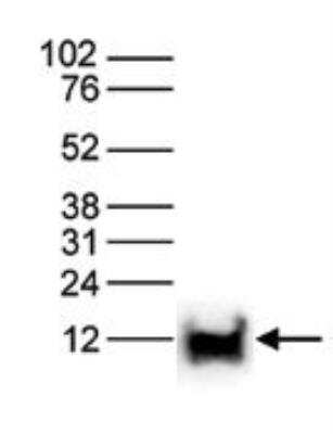
Western Blot: Histone H4 [Trimethyl Lys20] Antibody [NBP2-59259] - Histone extracts of HeLa cells (15 ug) were analysed by Western blot using Histone H4 [Trimethyl Lys20] Antibody diluted 1:1,000 in TBS-Tween containing 5% skimmed milk. The position of the protein of interest is indicated on the right; the marker (in kDa) is shown on the left.
![Immunocytochemistry/ Immunofluorescence: Histone H4 [Trimethyl Lys20] Antibody [NBP2-59259]](https://resources.rndsystems.com/images/products/Histone-H4-[Trimethyl-Lys20]-Antibody-Immunocytochemistry-Immunofluorescence-NBP2-59259-img0005.jpg "Immunocytochemistry/ Immunofluorescence: Histone H4 [Trimethyl Lys20] Antibody [NBP2-59259]")
Immunocytochemistry/ Immunofluorescence: Histone H4 [Trimethyl Lys20] Antibody [NBP2-59259]
Immunocytochemistry/Immunofluorescence: Histone H4 [Trimethyl Lys20] Antibody [NBP2-59259] - (Top): Human osteosarcoma (U2OS) cells were stained with and DAPI. Cells were fixed with ice cold methanol for 10' and blocked with PBS/TX-100 containing 5% normal goat serum. Figure 6A: cells were immunofluorescently labeled with (left) diluted 1:300 in blocking solution followed by an anti-rabbit antibody conjugated to Alexa568 or with DAPI (right), which specifically labels DNA. (Bottom): Staining of the cells with the after incubation of the antibody with blocking peptide, concentration: 5 ng/ul.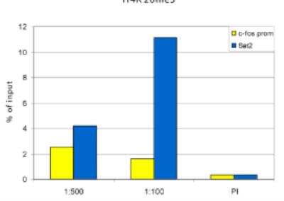
Chromatin Immunoprecipitation: Histone H4 [Trimethyl Lys20] Antibody [NBP2-59259] - ChIP assays were performed using undifferentiated human teratocarcinoma cells (NCCIT), the antibody against H4K20me3 and optimized PCR primer sets for qPCR. Sheared chromatin from 10,000 cells was used per ChIP experiment. The antibody was tested at two different dilutions of 1:500 and 1:100. The pre-immune serum (PI) was used as a negative control. Quantitative PCR was performed using primer sets for the satellite repeat Sat2 as a positive control and for the promoter of the house keeping gene c-fos, as a negative control. Figure 1 shows the recovery, expressed as a % of input (the relative amount of immunoprecipitated DNA compared to input DNA after qPCR analysis). These results are in accordance with the observation that H4K20me3 is preferably present at heterochromatin.
![Western Blot: Histone H4 [Trimethyl Lys20] Antibody [NBP2-59259]](https://resources.rndsystems.com/images/products/Histone-H4-[Trimethyl-Lys20]-Antibody-Western-Blot-NBP2-59259-img0002.jpg "Western Blot: Histone H4 [Trimethyl Lys20] Antibody [NBP2-59259]")
Western Blot: Histone H4 [Trimethyl Lys20] Antibody [NBP2-59259]
Western Blot: Histone H4 [Trimethyl Lys20] Antibody [NBP2-59259] - Histone (acid) extracts of NB4 cells were analysed by Western blot using the Diagenode antibody against H4K20me3, diluted 1:750 in TBS-Tween containing 5% skimmed milk. The location of the protein of interest is indicated on the right.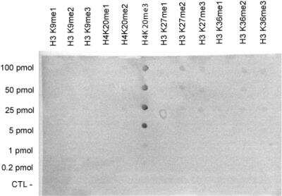
Dot Blot: Histone H4 [Trimethyl Lys20] Antibody [NBP2-59259] - Dot Blot was used to check the specificity of the antibody against H4K20me3 with peptides containing other modifications of histone H3 and H4. Other histone modifications include monoand dimethylation of the same lysine and mono-, di- and trimethylation of lysines 9, 27 and 36 of H3. One hundred to 0.2 pmol of peptide containing the respective histone modification were spotted on a membrane. The antibody was used at a dilution of 1:1,000. Figure shows a high specificity of the antibody for the modification of interest.
![ELISA: Histone H4 [Trimethyl Lys20] Antibody [NBP2-59259]](https://resources.rndsystems.com/images/products/Histone-H4-[Trimethyl-Lys20]-Antibody-ELISA-NBP2-59259-img0003.jpg "ELISA: Histone H4 [Trimethyl Lys20] Antibody [NBP2-59259]")
ELISA: Histone H4 [Trimethyl Lys20] Antibody [NBP2-59259]
ELISA: Histone H4 [Trimethyl Lys20] Antibody [NBP2-59259] - To determine the titer, an ELISA was performed using a serial dilution of the Diagenode antibody directed against H4K20me3. The antigen used was a peptide containing the histone modification of interest. By plotting the absorbance against the antibody dilution, the titer of the antibody was estimated to be 1:700.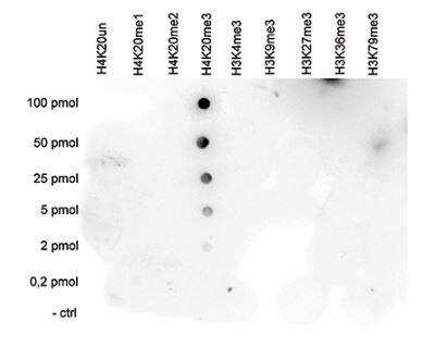
Dot Blot: Histone H4 [Trimethyl Lys20] Antibody [NBP2-59259] - A Dot Blot analysis was performed to test the cross reactivity of Histone H4 [Trimethyl Lys20] Antibody with peptides containing other histone modifications and the unmodified H4K20. One hundred to 0.2 pmol of the respective peptides were spotted on a membrane. The antibody was used at a dilution of 1:20,000. Figure shows a high specificity of the antibody for the modification of interest
Applications for Histone H4 [Trimethyl Lys20] Antibody - BSA Free
Application
Recommended Usage
Chromatin Immunoprecipitation (ChIP)
1:100
Dot Blot
1:1000
ELISA
1:50
Western Blot
1:750
Formulation, Preparation, and Storage
Purification
Peptide affinity purified
Formulation
PBS
Format
BSA Free
Preservative
0.05% Sodium Azide and 0.05% ProClin 300
Concentration
Please see the vial label for concentration. If unlisted please contact technical services.
Shipping
The product is shipped with polar packs. Upon receipt, store it immediately at the temperature recommended below.
Stability & Storage
Store at -20C. Avoid freeze-thaw cycles.
Background: Histone H4
Alternate Names
H4, H4/M, H4FM, H4M, HIST1H4I, HIST4H4, Histone Cluster 1 H4, Histone Cluster 1 H4i, Histone Cluster 4 H4, H4K20Me3
Gene Symbol
H4C16
Additional Histone H4 Products
Product Documents for Histone H4 [Trimethyl Lys20] Antibody - BSA Free
Certificate of Analysis
To download a Certificate of Analysis, please enter a lot or batch number in the search box below.
Product Specific Notices for Histone H4 [Trimethyl Lys20] Antibody - BSA Free
This product is for research use only and is not approved for use in humans or in clinical diagnosis. Primary Antibodies are guaranteed for 1 year from date of receipt.
Related Research Areas
Customer Reviews for Histone H4 [Trimethyl Lys20] Antibody - BSA Free
There are currently no reviews for this product. Be the first to review Histone H4 [Trimethyl Lys20] Antibody - BSA Free and earn rewards!
Have you used Histone H4 [Trimethyl Lys20] Antibody - BSA Free?
Submit a review and receive an Amazon gift card!
$25/€18/£15/$25CAN/¥2500 Yen for a review with an image
$10/€7/£6/$10CAN/¥1110 Yen for a review without an image
Submit a review
Protocols
Find general support by application which include: protocols, troubleshooting, illustrated assays, videos and webinars.
- Appropriate Fixation of IHC/ICC Samples
- Cellular Response to Hypoxia Protocols
- ChIP Protocol Video
- Chromatin Immunoprecipitation (ChIP) Protocol
- Chromatin Immunoprecipitation Protocol
- ClariTSA™ Fluorophore Kits
- Detection & Visualization of Antibody Binding
- ELISA Sample Preparation & Collection Guide
- ELISA Troubleshooting Guide
- How to Run an R&D Systems DuoSet ELISA
- How to Run an R&D Systems Quantikine ELISA
- How to Run an R&D Systems Quantikine™ QuicKit™ ELISA
- ICC Cell Smear Protocol for Suspension Cells
- ICC Immunocytochemistry Protocol Videos
- ICC for Adherent Cells
- Immunocytochemistry (ICC) Protocol
- Immunocytochemistry Troubleshooting
- Immunofluorescence of Organoids Embedded in Cultrex Basement Membrane Extract
- Immunohistochemistry (IHC) and Immunocytochemistry (ICC) Protocols
- Preparing Samples for IHC/ICC Experiments
- Preventing Non-Specific Staining (Non-Specific Binding)
- Primary Antibody Selection & Optimization
- Protocol for VisUCyte™ HRP Polymer Detection Reagent
- Protocol for the Fluorescent ICC Staining of Cell Smears - Graphic
- Protocol for the Fluorescent ICC Staining of Cultured Cells on Coverslips - Graphic
- Protocol for the Preparation and Fluorescent ICC Staining of Cells on Coverslips
- Protocol for the Preparation and Fluorescent ICC Staining of Non-adherent Cells
- Protocol for the Preparation and Fluorescent ICC Staining of Stem Cells on Coverslips
- Protocol for the Preparation of a Cell Smear for Non-adherent Cell ICC - Graphic
- Quantikine HS ELISA Kit Assay Principle, Alkaline Phosphatase
- Quantikine HS ELISA Kit Principle, Streptavidin-HRP Polymer
- R&D Systems Quality Control Western Blot Protocol
- Sandwich ELISA (Colorimetric) – Biotin/Streptavidin Detection Protocol
- Sandwich ELISA (Colorimetric) – Direct Detection Protocol
- TUNEL and Active Caspase-3 Detection by IHC/ICC Protocol
- The Importance of IHC/ICC Controls
- Troubleshooting Guide: ELISA
- Troubleshooting Guide: Western Blot Figures
- Western Blot Conditions
- Western Blot Protocol
- Western Blot Protocol for Cell Lysates
- Western Blot Troubleshooting
- Western Blot Troubleshooting Guide
- View all Protocols, Troubleshooting, Illustrated assays and Webinars
Loading...
Associated Pathways
