HMG-CoA Reductase/HMGCR Antibody (JF0981)
Novus Biologicals | Catalog # NBP2-66888
Recombinant Monoclonal Antibody
![Western Blot: HMG-CoA Reductase/HMGCR Antibody (JF0981) [NBP2-66888]](https://resources.rndsystems.com/images/products/HMG-CoA-Reductase-HMGCR-Antibody-JF0981-Western-Blot-NBP2-66888-img0003.jpg "Western Blot: HMG-CoA Reductase/HMGCR Antibody (JF0981) [NBP2-66888]")
Loading...
Key Product Details
Species Reactivity
Validated:
Human, Mouse, Rat
Cited:
Human, Mouse
Applications
Validated:
Immunohistochemistry-Paraffin, Western Blot, Simple Western, Immunoprecipitation
Cited:
Western Blot, Simple Western
Label
Unconjugated
Antibody Source
Recombinant Monoclonal Rabbit IgG Clone # JF0981 expressed in HEK293
Loading...
Product Specifications
Immunogen
Synthetic peptide within Human HMG-CoA Reductase/HMGCR aa 438-489 / 888. (SwissProt: P04035 Human; SwissProt: Q01237 Mouse; SwissProt: P51639 Rat)
Localization
Endoplasmic reticulum membrane.
Clonality
Monoclonal
Host
Rabbit
Isotype
IgG
Scientific Data Images for HMG-CoA Reductase/HMGCR Antibody (JF0981)
Western Blot: HMG-CoA Reductase/HMGCR Antibody (JF0981) [NBP2-66888]
Western Blot: HMG-CoA Reductase/HMGCR Antibody (JF0981) [NBP2-66888] - Analysis of HMGCR on different lysates. Proteins were transferred to a PVDF membrane and blocked with 5% BSA in PBS for 1 hour at room temperature. The primary antibody (ET1702-41, 1/500) was used in 5% BSA at room temperature for 2 hours. Goat Anti-Rabbit IgG - HRP Secondary Antibody (HA1001) at 1:5,000 dilution was used for 1 hour at room temperature.Positive control: Lane 1: Hela cell lysateLane 2: THP-1 cell lysate![Simple Western: HMG-CoA Reductase/HMGCR Antibody (JF0981) [NBP2-66888]](https://resources.rndsystems.com/images/products/HMG-CoA-Reductase-HMGCR-Antibody-JF0981-Simple-Western-NBP2-66888-img0002.jpg "Simple Western: HMG-CoA Reductase/HMGCR Antibody (JF0981) [NBP2-66888]")
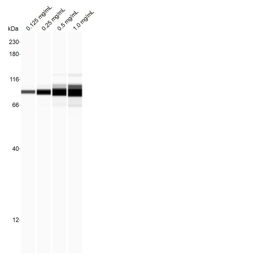
Simple Western: HMG-CoA Reductase/HMGCR Antibody (JF0981) [NBP2-66888]
Simple Western: HMG-CoA Reductase/HMGCR Antibody (JF0981) [NBP2-66888] - SVG-A whole cell lysate. Antibody at 1:25 dilution. Image submitted by a verified customer review.Applications for HMG-CoA Reductase/HMGCR Antibody (JF0981)
Application
Recommended Usage
Immunohistochemistry-Paraffin
1:100
Simple Western
1:25
Western Blot
1:500-1:2000
Application Notes
HMG-CoA Reductase/HMGCR antibody validated for Simple Western from a verified customer review. IP: Use at an assay dependent concentration.
See Simple Western Antibody Database for Simple Western validation: Tested in SVG-A whole cell lysate, separated by Size, antibody dilution of 1:25
See Simple Western Antibody Database for Simple Western validation: Tested in SVG-A whole cell lysate, separated by Size, antibody dilution of 1:25
Reviewed Applications
Read 1 review rated 5 using NBP2-66888 in the following applications:
Formulation, Preparation, and Storage
Purification
Protein A purified
Formulation
TBS (pH7.4), 0.05% BSA, 40% Glycerol
Preservative
0.05% Sodium Azide
Concentration
1 mg/ml
Shipping
The product is shipped with polar packs. Upon receipt, store it immediately at the temperature recommended below.
Stability & Storage
Store at 4C short term. Aliquot and store at -20C long term. Avoid freeze-thaw cycles.
Background: HMG-CoA Reductase/HMGCR
Long Name
3-Hydroxy-3-Methylglutaryl-CoA Reductase
Alternate Names
HMGCR, Hydroxymethylglutaryl-CoA Reductase, LDLCQ3
Gene Symbol
HMGCR
Additional HMG-CoA Reductase/HMGCR Products
Product Documents for HMG-CoA Reductase/HMGCR Antibody (JF0981)
Certificate of Analysis
To download a Certificate of Analysis, please enter a lot or batch number in the search box below.
Product Specific Notices for HMG-CoA Reductase/HMGCR Antibody (JF0981)
This product is for research use only and is not approved for use in humans or in clinical diagnosis. Primary Antibodies are guaranteed for 1 year from date of receipt.
Related Research Areas
Citations for HMG-CoA Reductase/HMGCR Antibody (JF0981)
 Powered by Bioz
Customer Reviews for HMG-CoA Reductase/HMGCR Antibody (JF0981) (1)
5 out of 5
1 Customer Rating
Have you used HMG-CoA Reductase/HMGCR Antibody (JF0981)?
Submit a review and receive an Amazon gift card!
$25/€18/£15/$25CAN/¥2500 Yen for a review with an image
$10/€7/£6/$10CAN/¥1110 Yen for a review without an image
Submit a review
Customer Images
Showing
1
-
1 of
1 review
Showing All
Filter By:
-
Application: Simple WesternSample Tested: SVG-A whole cell lysateSpecies: HumanVerified Customer | Posted 07/03/2018SVG-A whole cell lysate run at the indicated concentration. Antibody was tested at 1:25 dil.

There are no reviews that match your criteria.
Protocols
Find general support by application which include: protocols, troubleshooting, illustrated assays, videos and webinars.
- Antigen Retrieval Protocol (PIER)
- Antigen Retrieval for Frozen Sections Protocol
- Appropriate Fixation of IHC/ICC Samples
- Cellular Response to Hypoxia Protocols
- Chromogenic IHC Staining of Formalin-Fixed Paraffin-Embedded (FFPE) Tissue Protocol
- Chromogenic Immunohistochemistry Staining of Frozen Tissue
- ClariTSA™ Fluorophore Kits
- Detection & Visualization of Antibody Binding
- Fluorescent IHC Staining of Frozen Tissue Protocol
- Graphic Protocol for Heat-induced Epitope Retrieval
- Graphic Protocol for the Preparation and Fluorescent IHC Staining of Frozen Tissue Sections
- Graphic Protocol for the Preparation and Fluorescent IHC Staining of Paraffin-embedded Tissue Sections
- Graphic Protocol for the Preparation of Gelatin-coated Slides for Histological Tissue Sections
- IHC Sample Preparation (Frozen sections vs Paraffin)
- Immunofluorescent IHC Staining of Formalin-Fixed Paraffin-Embedded (FFPE) Tissue Protocol
- Immunohistochemistry (IHC) and Immunocytochemistry (ICC) Protocols
- Immunohistochemistry Frozen Troubleshooting
- Immunohistochemistry Paraffin Troubleshooting
- Immunoprecipitation Protocol
- Preparing Samples for IHC/ICC Experiments
- Preventing Non-Specific Staining (Non-Specific Binding)
- Primary Antibody Selection & Optimization
- Protocol for Heat-Induced Epitope Retrieval (HIER)
- Protocol for Making a 4% Formaldehyde Solution in PBS
- Protocol for VisUCyte™ HRP Polymer Detection Reagent
- Protocol for the Preparation & Fixation of Cells on Coverslips
- Protocol for the Preparation and Chromogenic IHC Staining of Frozen Tissue Sections
- Protocol for the Preparation and Chromogenic IHC Staining of Frozen Tissue Sections - Graphic
- Protocol for the Preparation and Chromogenic IHC Staining of Paraffin-embedded Tissue Sections
- Protocol for the Preparation and Chromogenic IHC Staining of Paraffin-embedded Tissue Sections - Graphic
- Protocol for the Preparation and Fluorescent IHC Staining of Frozen Tissue Sections
- Protocol for the Preparation and Fluorescent IHC Staining of Paraffin-embedded Tissue Sections
- Protocol for the Preparation of Gelatin-coated Slides for Histological Tissue Sections
- R&D Systems Quality Control Western Blot Protocol
- TUNEL and Active Caspase-3 Detection by IHC/ICC Protocol
- The Importance of IHC/ICC Controls
- Troubleshooting Guide: Immunohistochemistry
- Troubleshooting Guide: Western Blot Figures
- Western Blot Conditions
- Western Blot Protocol
- Western Blot Protocol for Cell Lysates
- Western Blot Troubleshooting
- Western Blot Troubleshooting Guide
- View all Protocols, Troubleshooting, Illustrated assays and Webinars
Loading...