Key Product Details
Species Reactivity
Validated:
Cited:
Applications
Validated:
Cited:
Label
Antibody Source
Product Specifications
Immunogen
Localization
Specificity
Clonality
Host
Isotype
Theoretical MW
Disclaimer note: The observed molecular weight of the protein may vary from the listed predicted molecular weight due to post translational modifications, post translation cleavages, relative charges, and other experimental factors.
Description
Antibody with azide - store at 2 to 8C. Antibody without azide - store at -20 to -80 C.
Scientific Data Images for HSP27 Antibody (G3.1)
![Western Blot: HSP27 Antibody (G3.1) [NBP2-32972]](https://resources.rndsystems.com/images/products/HSP27-Antibody-G3-1-Western-Blot-NBP2-32972-img0008.jpg "Western Blot: HSP27 Antibody (G3.1) [NBP2-32972]")
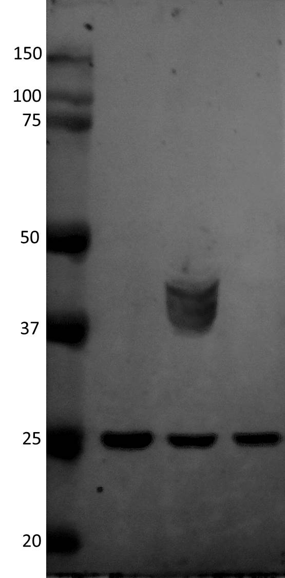
Western Blot: HSP27 Antibody (G3.1) [NBP2-32972]
Western Blot: HSP27 Antibody (G3.1) [NBP2-32972] - Western Blot Analysis of HeLa cell lysate using HSP27 Antibody (G3.1).![Immunocytochemistry/ Immunofluorescence: HSP27 Antibody (G3.1) [NBP2-32972]](https://resources.rndsystems.com/images/products/HSP27-Antibody-G3-1-Immunocytochemistry-Immunofluorescence-NBP2-32972-img0007.jpg "Immunocytochemistry/ Immunofluorescence: HSP27 Antibody (G3.1) [NBP2-32972]")
Immunocytochemistry/ Immunofluorescence: HSP27 Antibody (G3.1) [NBP2-32972]
Immunocytochemistry/Immunofluorescence: HSP27 Antibody (G3.1) [NBP2-32972] - Immunofluorescence Analysis of PFA-fixed MCF-7 cells labeling HSP27 with followed by Goat anti-Mouse IgG-CF488 (Green). The nuclear counterstain is Red Dot (Red)![Immunohistochemistry-Paraffin: HSP27 Antibody (G3.1) [NBP2-32972]](https://resources.rndsystems.com/images/products/HSP27-Antibody-G3-1-Immunohistochemistry-Paraffin-NBP2-32972-img0004.jpg "Immunohistochemistry-Paraffin: HSP27 Antibody (G3.1) [NBP2-32972]")
Immunohistochemistry-Paraffin: HSP27 Antibody (G3.1) [NBP2-32972]
Immunohistochemistry-Paraffin: HSP27 Antibody (G3.1) [NBP2-32972] - Formalin-fixed. paraffin-embedded human Breast Carcinome stained with HSP27 Monoclonal Antibody (G3.1)![Flow Cytometry: HSP27 Antibody (G3.1) [NBP2-32972]](https://resources.rndsystems.com/images/products/HSP27-Antibody-G3-1-Flow-Cytometry-NBP2-32972-img0006.jpg "Flow Cytometry: HSP27 Antibody (G3.1) [NBP2-32972]")
Flow Cytometry: HSP27 Antibody (G3.1) [NBP2-32972]
Flow Cytometry: HSP27 Antibody (G3.1) [NBP2-32972] - Flow Cytometric Analysis of PFA-fixed MCF-7 cells using HSP27 Antibody (G3.1)followed by Goat anti- Mouse- IgG-CF488 (Blue); Isotype Control (Red).![Immunohistochemistry-Paraffin: HSP27 Antibody (G3.1) [NBP2-32972]](https://resources.rndsystems.com/images/products/HSP27-Antibody-G3-1-Immunohistochemistry-Paraffin-NBP2-32972-img0003.jpg "Immunohistochemistry-Paraffin: HSP27 Antibody (G3.1) [NBP2-32972]")
Immunohistochemistry-Paraffin: HSP27 Antibody (G3.1) [NBP2-32972]
Immunohistochemistry-Paraffin: HSP27 Antibody (G3.1) [NBP2-32972] - Formalin-fixed. paraffim-embedded human prostate Carcinoma stained with HSP27 Monoclonal Antibody (G3.1) [NBP2-32972] -")
Western Blot: HSP27 Antibody (G3.1) [NBP2-32972] -
Hsp90 inhibition reduces cell viability in a concentration-dependent manner and increases cytosolic Hsp70 and Hsp27 levels. (A, B) Toxicity assay of B16F10 (A) and LS174T (B) WT and LDH−/− cells treated with NVP-AUY922 (0, 5, 10, 50, 100 nM) for 24 h. (C, D) Representative immunoblot showing intracellular Hsp70, Hsp27 and beta -Actin levels in B16F10 (C) and LS174T (D) cells upon treatment with NVP-AUY922 (100 nM) for 24 h. Quantification of the heat shock protein (HSP) levels are shown in the adjacent bar chart (**p ≤ 0.01, ***p ≤ 0.001). Image collected and cropped by CiteAb from the following open publication (https://pubmed.ncbi.nlm.nih.gov/35463341), licensed under a CC-BY license. Not internally tested by Novus Biologicals.Applications for HSP27 Antibody (G3.1)
Flow Cytometry
Immunocytochemistry/ Immunofluorescence
Immunohistochemistry-Paraffin
Western Blot
Optimal dilution for a specific application should be determined.
Reviewed Applications
Read 1 review rated 4 using NBP2-32972 in the following applications:
Flow Cytometry Panel Builder
Bio-Techne Knows Flow Cytometry
Save time and reduce costly mistakes by quickly finding compatible reagents using the Panel Builder Tool.
Advanced Features
- Spectra Viewer - Custom analysis of spectra from multiple fluorochromes
- Spillover Popups - Visualize the spectra of individual fluorochromes
- Antigen Density Selector - Match fluorochrome brightness with antigen density

Formulation, Preparation, and Storage
Purification
Formulation
Preservative
Concentration
Shipping
Stability & Storage
Background: HSP27
Additional HSP27 Products
Product Documents for HSP27 Antibody (G3.1)
Certificate of Analysis
To download a Certificate of Analysis, please enter a lot or batch number in the search box below.
Product Specific Notices for HSP27 Antibody (G3.1)
This product is for research use only and is not approved for use in humans or in clinical diagnosis. Primary Antibodies are guaranteed for 1 year from date of receipt.
Related Research Areas
Citations for HSP27 Antibody (G3.1)
 Powered by Bioz
Customer Reviews for HSP27 Antibody (G3.1) (1)
Have you used HSP27 Antibody (G3.1)?
Submit a review and receive an Amazon gift card!
$25/€18/£15/$25CAN/¥2500 Yen for a review with an image
$10/€7/£6/$10CAN/¥1110 Yen for a review without an image
Submit a review
Customer Images
-
Application: Western BlotSample Tested: SW480 (colon)Species: HumanVerified Customer | Posted 12/24/2016Marker, SW480 (colon), HT29 (colon), Colo320 (colon) (25ug protein per well). HT29 gave a non-specific set of bands at around 40kDaPrimary mouse anti HSP27 antibody diluted 1:1000 in TBS. Membrane incubation for 2 hours at RT. Secondary IRDye 800CW goat anti mouse antibody diluted 1:10000 in TBS. Membrane incubation for 1 hour at RT. Detection by Li-Cor Odyssey

There are no reviews that match your criteria.
Protocols
Find general support by application which include: protocols, troubleshooting, illustrated assays, videos and webinars.
- 7-Amino Actinomycin D (7-AAD) Cell Viability Flow Cytometry Protocol
- Antigen Retrieval Protocol (PIER)
- Antigen Retrieval for Frozen Sections Protocol
- Appropriate Fixation of IHC/ICC Samples
- Cellular Response to Hypoxia Protocols
- Chromogenic IHC Staining of Formalin-Fixed Paraffin-Embedded (FFPE) Tissue Protocol
- Chromogenic Immunohistochemistry Staining of Frozen Tissue
- ClariTSA™ Fluorophore Kits
- Detection & Visualization of Antibody Binding
- Extracellular Membrane Flow Cytometry Protocol
- Flow Cytometry Protocol for Cell Surface Markers
- Flow Cytometry Protocol for Staining Membrane Associated Proteins
- Flow Cytometry Staining Protocols
- Flow Cytometry Troubleshooting Guide
- Fluorescent IHC Staining of Frozen Tissue Protocol
- Graphic Protocol for Heat-induced Epitope Retrieval
- Graphic Protocol for the Preparation and Fluorescent IHC Staining of Frozen Tissue Sections
- Graphic Protocol for the Preparation and Fluorescent IHC Staining of Paraffin-embedded Tissue Sections
- Graphic Protocol for the Preparation of Gelatin-coated Slides for Histological Tissue Sections
- ICC Cell Smear Protocol for Suspension Cells
- ICC Immunocytochemistry Protocol Videos
- ICC for Adherent Cells
- IHC Sample Preparation (Frozen sections vs Paraffin)
- Immunocytochemistry (ICC) Protocol
- Immunocytochemistry Troubleshooting
- Immunofluorescence of Organoids Embedded in Cultrex Basement Membrane Extract
- Immunofluorescent IHC Staining of Formalin-Fixed Paraffin-Embedded (FFPE) Tissue Protocol
- Immunohistochemistry (IHC) and Immunocytochemistry (ICC) Protocols
- Immunohistochemistry Frozen Troubleshooting
- Immunohistochemistry Paraffin Troubleshooting
- Intracellular Flow Cytometry Protocol Using Alcohol (Methanol)
- Intracellular Flow Cytometry Protocol Using Detergents
- Intracellular Nuclear Staining Flow Cytometry Protocol Using Detergents
- Intracellular Staining Flow Cytometry Protocol Using Alcohol Permeabilization
- Intracellular Staining Flow Cytometry Protocol Using Detergents to Permeabilize Cells
- Preparing Samples for IHC/ICC Experiments
- Preventing Non-Specific Staining (Non-Specific Binding)
- Primary Antibody Selection & Optimization
- Propidium Iodide Cell Viability Flow Cytometry Protocol
- Protocol for Heat-Induced Epitope Retrieval (HIER)
- Protocol for Liperfluo
- Protocol for Making a 4% Formaldehyde Solution in PBS
- Protocol for VisUCyte™ HRP Polymer Detection Reagent
- Protocol for the Characterization of Human Th22 Cells
- Protocol for the Characterization of Human Th9 Cells
- Protocol for the Fluorescent ICC Staining of Cell Smears - Graphic
- Protocol for the Fluorescent ICC Staining of Cultured Cells on Coverslips - Graphic
- Protocol for the Preparation & Fixation of Cells on Coverslips
- Protocol for the Preparation and Chromogenic IHC Staining of Frozen Tissue Sections
- Protocol for the Preparation and Chromogenic IHC Staining of Frozen Tissue Sections - Graphic
- Protocol for the Preparation and Chromogenic IHC Staining of Paraffin-embedded Tissue Sections
- Protocol for the Preparation and Chromogenic IHC Staining of Paraffin-embedded Tissue Sections - Graphic
- Protocol for the Preparation and Fluorescent ICC Staining of Cells on Coverslips
- Protocol for the Preparation and Fluorescent ICC Staining of Non-adherent Cells
- Protocol for the Preparation and Fluorescent ICC Staining of Stem Cells on Coverslips
- Protocol for the Preparation and Fluorescent IHC Staining of Frozen Tissue Sections
- Protocol for the Preparation and Fluorescent IHC Staining of Paraffin-embedded Tissue Sections
- Protocol for the Preparation of Gelatin-coated Slides for Histological Tissue Sections
- Protocol for the Preparation of a Cell Smear for Non-adherent Cell ICC - Graphic
- Protocol: Annexin V and PI Staining by Flow Cytometry
- Protocol: Annexin V and PI Staining for Apoptosis by Flow Cytometry
- R&D Systems Quality Control Western Blot Protocol
- TUNEL and Active Caspase-3 Detection by IHC/ICC Protocol
- The Importance of IHC/ICC Controls
- Troubleshooting Guide: Fluorokine Flow Cytometry Kits
- Troubleshooting Guide: Immunohistochemistry
- Troubleshooting Guide: Western Blot Figures
- Western Blot Conditions
- Western Blot Protocol
- Western Blot Protocol for Cell Lysates
- Western Blot Troubleshooting
- Western Blot Troubleshooting Guide
- View all Protocols, Troubleshooting, Illustrated assays and Webinars
FAQs for HSP27 Antibody (G3.1)
-
Q: What is the concentration of the existing lot of NBP2-32972-0.1ml
A: The concentration for product NBP2-32972 is 0.2mg/ml.


