Hsp47 Antibody (M16.10A1) - BSA Free
Novus Biologicals | Catalog # NBP1-97491
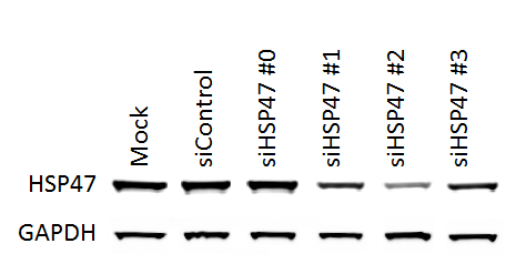
![Western Blot: Hsp47 Antibody (M16.10A1) [NBP1-97491]](https://resources.rndsystems.com/images/products/Hsp47-Antibody-M16-10A1-Western-Blot-NBP1-97491-img0006.jpg "Western Blot: Hsp47 Antibody (M16.10A1) [NBP1-97491]")
Key Product Details
Species Reactivity
Validated:
Human, Mouse, Rat, Porcine, Bovine, Canine, Chicken, Guinea Pig, Hamster, Monkey, Rabbit, Sheep
Cited:
Human, Mouse, Rat
Applications
Validated:
Immunohistochemistry, Immunohistochemistry-Paraffin, Immunohistochemistry-Frozen, Western Blot, Flow Cytometry, Immunocytochemistry/ Immunofluorescence, Simple Western, Immunoprecipitation, Knockdown Validated
Cited:
Immunohistochemistry-Paraffin, Western Blot, Flow Cytometry, Immunocytochemistry/ Immunofluorescence, Immunoprecipitation, IF/IHC
Label
Unconjugated
Antibody Source
Monoclonal Mouse IgG2B Clone # M16.10A1
Format
BSA Free
Loading...
Product Specifications
Immunogen
Native rat HSP47.
Reactivity Notes
Recognizes Sheep HSP47. Please note that this antibody is reactive to Mouse and derived from the same host, Mouse. Mouse-On-Mouse blocking reagent may be needed for IHC and ICC experiments to reduce high background signal. You can find these reagents under catalog numbers PK-2200-NB and MP-2400-NB. Please contact Technical Support if you have any questions.
Clonality
Monoclonal
Host
Mouse
Isotype
IgG2B
Scientific Data Images for Hsp47 Antibody (M16.10A1) - BSA Free
Western Blot: Hsp47 Antibody (M16.10A1) [NBP1-97491]
Western Blot: Hsp47 Antibody (M16.10A1) [NBP1-97491] - analysis of Hsp47 in NIH/3T3 cell lysate (20ug/lane) using anti-Hsp47 antibody. 5x10^4 NIH/3T3 cells were seeded in 6-well plate and transfected with 50nM of HSP47 siRNA the following day. Total protein was harvested 72h after transfection. Image from verified customer review.![Immunocytochemistry/ Immunofluorescence: Hsp47 Antibody (M16.10A1) [NBP1-97491]](https://resources.rndsystems.com/images/products/Hsp47-Antibody-M16-10A1-Immunocytochemistry-Immunofluorescence-NBP1-97491-img0007.jpg "Immunocytochemistry/ Immunofluorescence: Hsp47 Antibody (M16.10A1) [NBP1-97491]")
Immunocytochemistry/ Immunofluorescence: Hsp47 Antibody (M16.10A1) [NBP1-97491]
Hsp47-Antibody-M16-10A1-Immunocytochemistry-Immunofluorescence-NBP1-97491-img0007.jpg![Immunohistochemistry-Frozen: Hsp47 Antibody (M16.10A1) [NBP1-97491]](https://resources.rndsystems.com/images/products/Hsp47-Antibody-M16-10A1-Immunohistochemistry-Frozen-NBP1-97491-img0004.jpg "Immunohistochemistry-Frozen: Hsp47 Antibody (M16.10A1) [NBP1-97491]")
Immunohistochemistry-Frozen: Hsp47 Antibody (M16.10A1) [NBP1-97491]
Immunohistochemistry-Frozen: Hsp47 Antibody (M16.10A1) [NBP1-97491] - Hsp47 antibody in guinea pig skin (subdermis). Image from verified customer review.![Western Blot: Hsp47 Antibody (M16.10A1) [NBP1-97491]](https://resources.rndsystems.com/images/products/Hsp47-Antibody-M16-10A1-Western-Blot-NBP1-97491-img0003.jpg "Western Blot: Hsp47 Antibody (M16.10A1) [NBP1-97491]")
Western Blot: Hsp47 Antibody (M16.10A1) [NBP1-97491]
Western Blot: Hsp47 Antibody (M16.10A1) [NBP1-97491] - Analysis of HSP47, mAb (M16.10A1): Lane 1: Rat recombinant HSP47; Lane 2: Rat-2 (heat shocked); Lane 3: L929 (heat shocked); Lane 4: 3T3 (heat shocked); Lane 5: CHO (heat shocked); Lane 6: HeLa (heat shocked).![Immunohistochemistry-Paraffin: Hsp47 Antibody (M16.10A1) [NBP1-97491]](https://resources.rndsystems.com/images/products/Hsp47-Antibody-M16-10A1-Immunohistochemistry-Paraffin-NBP1-97491-img0001.jpg "Immunohistochemistry-Paraffin: Hsp47 Antibody (M16.10A1) [NBP1-97491]")
Immunohistochemistry-Paraffin: Hsp47 Antibody (M16.10A1) [NBP1-97491]
Immunohistochemistry-Paraffin: Hsp47 Antibody (M16.10A1) [NBP1-97491] - Analysis of paraffin-embedded tissue section of rat carotid artery 14 days after balloon-injury to the artery, stained using Hsp47 (Colligin) mAb (M16.10A1).![Immunohistochemistry-Paraffin: Hsp47 Antibody (M16.10A1) [NBP1-97491]](https://resources.rndsystems.com/images/products/Hsp47-Antibody-M16-10A1-Immunohistochemistry-Paraffin-NBP1-97491-img0002.jpg "Immunohistochemistry-Paraffin: Hsp47 Antibody (M16.10A1) [NBP1-97491]")
Immunohistochemistry-Paraffin: Hsp47 Antibody (M16.10A1) [NBP1-97491]
Immunohistochemistry-Paraffin: Hsp47 Antibody (M16.10A1) [NBP1-97491] - Analysis of human breast cancer tissue immunohistochemically stained using Hsp47 mAb (M16.10A1).![Simple Western: Hsp47 Antibody (M16.10A1) [NBP1-97491]](https://resources.rndsystems.com/images/products/Hsp47-Antibody-M16-10A1-Simple-Western-NBP1-97491-img0005.jpg "Simple Western: Hsp47 Antibody (M16.10A1) [NBP1-97491]")
Simple Western: Hsp47 Antibody (M16.10A1) [NBP1-97491]
Simple Western: Hsp47 Antibody (M16.10A1) [NBP1-97491] - Simple Western lane view shows a specific band for Hsp47 in 0.5 mg/ml of NIH-3T3 lysate. This experiment was performed under reducing conditions using the 12-230 kDa separation system. [NBP1-97491] -")
Immunocytochemistry/ Immunofluorescence: Hsp47 Antibody (M16.10A1) [NBP1-97491] -
Immunocytochemistry/ Immunofluorescence: Hsp47 Antibody (M16.10A1) [NBP1-97491] - WNT & BMP signaling pathways play crucial roles in regeneration. a–d Real-time PCR for WNT & BMP signals, including Wnt7b (a), Wnt10a (b), Lef1 (c), & Bmp2 (d) in response to stretch (day 1 & day 7) & release of stretch (day 9); n = 6 for each group. e Schematic of hair cycle activator & inhibitor in response to strain alteration. f Dual immunostaining for K15 & beta -catenin revealed nuclear staining of beta -catenin in hair follicles at day 9 & day 14 when stretch was released. Scale bar = 50 μm. g Dual immunostaining for K15 & phospho-Smad1/5 (pSmad1/5) revealed the nuclear staining of pSmad1/5 in hair stem cells at day 7 when skin was stretched (arrow). Scale bar = 50 μm. h, i Immunostaining (h) & confocal microscope (i) revealed co-localization of HSP47 & BMP-2 signals at day 7 when skin was stretched. Scale bar = 100 μm (h) or 10 μm (i). j, k Immunostaining (j) & confocal microscope (k) revealed co-localization of Perilipin-1 & BMP-2 signals at day 7 when skin was stretched. Scale bar = 100 μm (j) or 10 μm (k). Statistical significance was determined using ANOVA followed by a Bonferroni post hoc test. Data are presented as means ± SEM. *p < 0.05. **p < 0.01. ***p < 0.001. Source data are provided as a Source Data file. *Autofluorescence of hair shafts in g, h Image collected & cropped by CiteAb from the following publication (https://pubmed.ncbi.nlm.nih.gov/30944305), licensed under a CC-BY license. Not internally tested by Novus Biologicals. [NBP1-97491] -")
Immunocytochemistry/ Immunofluorescence: Hsp47 Antibody (M16.10A1) [NBP1-97491] -
Immunocytochemistry/ Immunofluorescence: Hsp47 Antibody (M16.10A1) [NBP1-97491] - WNT & BMP signaling pathways play crucial roles in regeneration. a–d Real-time PCR for WNT & BMP signals, including Wnt7b (a), Wnt10a (b), Lef1 (c), & Bmp2 (d) in response to stretch (day 1 & day 7) & release of stretch (day 9); n = 6 for each group. e Schematic of hair cycle activator & inhibitor in response to strain alteration. f Dual immunostaining for K15 & beta -catenin revealed nuclear staining of beta -catenin in hair follicles at day 9 & day 14 when stretch was released. Scale bar = 50 μm. g Dual immunostaining for K15 & phospho-Smad1/5 (pSmad1/5) revealed the nuclear staining of pSmad1/5 in hair stem cells at day 7 when skin was stretched (arrow). Scale bar = 50 μm. h, i Immunostaining (h) & confocal microscope (i) revealed co-localization of HSP47 & BMP-2 signals at day 7 when skin was stretched. Scale bar = 100 μm (h) or 10 μm (i). j, k Immunostaining (j) & confocal microscope (k) revealed co-localization of Perilipin-1 & BMP-2 signals at day 7 when skin was stretched. Scale bar = 100 μm (j) or 10 μm (k). Statistical significance was determined using ANOVA followed by a Bonferroni post hoc test. Data are presented as means ± SEM. *p < 0.05. **p < 0.01. ***p < 0.001. Source data are provided as a Source Data file. *Autofluorescence of hair shafts in g, h Image collected & cropped by CiteAb from the following publication (https://pubmed.ncbi.nlm.nih.gov/30944305), licensed under a CC-BY license. Not internally tested by Novus Biologicals. - BSA Free [NBP1-97491] -")
Western Blot: Hsp47 Antibody (M16.10A1) - BSA Free [NBP1-97491] -
Biodistribution and HSP47 silencing activity of LNPs in fibrotic mice.a Ex vivo fluorescence imaging and signal quantification of major organs from PBS, AA-T3A-C12 LNP/Cy5-siRNA or MC3 LNP/Cy5-siRNA treated fibrotic mice (representative dataset from n = 3/group). b Scheme of CCl4 and LNP treatment. Mice received intraperitoneal (i.p.) injections of 20% CCl4 (0.7 μl/g) in corn oil twice a week for 4 weeks. LNPs were intravenously (i.v.) administered at a siRNA dose of 5 μg/mouse twice weekly for 2 weeks. c Body weight changes of mice over time during the experiment (n = 5/group). d Body weight at the end of the experiment (n = 5/group). e IF staining of HSP47 in liver sections (representative dataset from n = 5/group). Arrows indicate central veins. Quantitative analysis was performed using ImageJ software (n = 5/group). Scale bar: 100 μm. f Western blot analysis of HSP47 expression in liver lysates (representative dataset from n = 3/group). GAPDH was used as an internal control. Representative images for two sets of mouse liver samples are shown. Quantitative analysis was performed using ImageJ software (n = 3/group). Data are presented as mean +/- SD. G1, healthy mice; G2, PBS-treated fibrotic mice; G3, AA-T3A-C12/siGFP LNP-treated fibrotic mice; G4, AA-T3A-C12/siHSP47 LNP-treated fibrotic mice; G5, MC3/siHSP47 LNP-treated fibrotic mice. ns, not significant; *p < 0.05; **p < 0.01; ***p < 0.001. a, d, e one-way ANOVA with Tukey’s correction. Source data are provided as a Source Data file. Image collected and cropped by CiteAb from the following open publication (https://pubmed.ncbi.nlm.nih.gov/36650129), licensed under a CC-BY license. Not internally tested by Novus Biologicals. - BSA Free [NBP1-97491] -")
Western Blot: Hsp47 Antibody (M16.10A1) - BSA Free [NBP1-97491] -
AA-T3A-C12 LNP-mediated GFP and HSP47 knockdown in activated fibroblasts.a GFP knockdown using AA-T3A-C12/siGFP LNP (n = 3/group). Activated 3T3-GFP fibroblasts were treated with AA-T3A-C12/siGFP LNP at the indicated dose for 24 or 48 h. b Flow cytometry analysis of GFP expression after AA-T3A-C12/siGFP LNP treatment for 48 h (representative dataset from n = 3/group). c Immunofluorescence (IF) staining of HSP47 in LNP-treated activated 3T3 fibroblasts. Scale bar: 20 μm. d and e Western blot analysis of HSP47 expression in LNP-treated activated 3T3 fibroblasts and primary HSCs (representative dataset from n = 3/group). GAPDH was used as an internal control. Quantitative analysis was performed using ImageJ software. Data are presented as mean +/- SD (n = 3). **p < 0.01; ***p < 0.001. b, d, e one-way ANOVA with Tukey’s correction. Source data are provided as a Source Data file. Image collected and cropped by CiteAb from the following open publication (https://pubmed.ncbi.nlm.nih.gov/36650129), licensed under a CC-BY license. Not internally tested by Novus Biologicals. - BSA Free [NBP1-97491] -")
Western Blot: Hsp47 Antibody (M16.10A1) - BSA Free [NBP1-97491] -
AA-T3A-C12 LNP-mediated GFP and HSP47 knockdown in activated fibroblasts.a GFP knockdown using AA-T3A-C12/siGFP LNP (n = 3/group). Activated 3T3-GFP fibroblasts were treated with AA-T3A-C12/siGFP LNP at the indicated dose for 24 or 48 h. b Flow cytometry analysis of GFP expression after AA-T3A-C12/siGFP LNP treatment for 48 h (representative dataset from n = 3/group). c Immunofluorescence (IF) staining of HSP47 in LNP-treated activated 3T3 fibroblasts. Scale bar: 20 μm. d and e Western blot analysis of HSP47 expression in LNP-treated activated 3T3 fibroblasts and primary HSCs (representative dataset from n = 3/group). GAPDH was used as an internal control. Quantitative analysis was performed using ImageJ software. Data are presented as mean +/- SD (n = 3). **p < 0.01; ***p < 0.001. b, d, e one-way ANOVA with Tukey’s correction. Source data are provided as a Source Data file. Image collected and cropped by CiteAb from the following open publication (https://pubmed.ncbi.nlm.nih.gov/36650129), licensed under a CC-BY license. Not internally tested by Novus Biologicals.Applications for Hsp47 Antibody (M16.10A1) - BSA Free
Application
Recommended Usage
Flow Cytometry
1:10-1:1000
Immunocytochemistry/ Immunofluorescence
1:10-1:500
Immunohistochemistry
1:10-1:500
Immunohistochemistry-Frozen
1:10-1:500
Immunohistochemistry-Paraffin
1:50
Immunoprecipitation
1:10-1:500
Simple Western
1:100
Western Blot
1:1000
Application Notes
This antibody is useful in IHC and detects a band of ~47kDa by WB. Immunoprecipitation, Flow Cytometry and ICC/IF were reported in scientific literature.
In Simple Western only 10 - 15 uL of the recommended dilution is used per data point.
See Simple Western Antibody Database for Simple Western validation: Tested in NIH-3T3 lysate, separated by Size, antibody dilution of 1:100, apparent MW was 57 kDa.
In Simple Western only 10 - 15 uL of the recommended dilution is used per data point.
See Simple Western Antibody Database for Simple Western validation: Tested in NIH-3T3 lysate, separated by Size, antibody dilution of 1:100, apparent MW was 57 kDa.
Reviewed Applications
Read 2 reviews rated 4 using NBP1-97491 in the following applications:
Flow Cytometry Panel Builder
Bio-Techne Knows Flow Cytometry
Save time and reduce costly mistakes by quickly finding compatible reagents using the Panel Builder Tool.
Advanced Features
- Spectra Viewer - Custom analysis of spectra from multiple fluorochromes
- Spillover Popups - Visualize the spectra of individual fluorochromes
- Antigen Density Selector - Match fluorochrome brightness with antigen density

Formulation, Preparation, and Storage
Purification
Protein G purified
Formulation
PBS (pH 7.2) and 50% Glycerol
Format
BSA Free
Preservative
0.09% Sodium Azide
Concentration
1.0 mg/ml
Shipping
The product is shipped with polar packs. Upon receipt, store it immediately at the temperature recommended below.
Stability & Storage
Store at -20C. Avoid freeze-thaw cycles.
Background: HSP47
Long Name
Heat Shock Protein 47
Alternate Names
AsTP3, BERF-1, CBP1, CBP2, Colligin, gp46, OI10, PPROM, RA-A47, Serpin H1, SERPINH1
Gene Symbol
SERPINH1
Additional HSP47 Products
Product Documents for Hsp47 Antibody (M16.10A1) - BSA Free
Certificate of Analysis
To download a Certificate of Analysis, please enter a lot or batch number in the search box below.
Product Specific Notices for Hsp47 Antibody (M16.10A1) - BSA Free
This product is for research use only and is not approved for use in humans or in clinical diagnosis. Primary Antibodies are guaranteed for 1 year from date of receipt.
Related Research Areas
Citations for Hsp47 Antibody (M16.10A1) - BSA Free
 Powered by Bioz
Customer Reviews for Hsp47 Antibody (M16.10A1) - BSA Free (2)
4 out of 5
2 Customer Ratings
Have you used Hsp47 Antibody (M16.10A1) - BSA Free?
Submit a review and receive an Amazon gift card!
$25/€18/£15/$25CAN/¥2500 Yen for a review with an image
$10/€7/£6/$10CAN/¥1110 Yen for a review without an image
Submit a review
Customer Images
-(005-mg)_NBP1-97491_6676.bmp)
Showing
1
-
2 of
2 reviews
Showing All
Filter By:
-
Application: Western BlotSample Tested: NIH3T3 Whole Cell Lysate, 20ug loaded per laneSpecies: MouseVerified Customer | Posted 01/07/2015HSP47 Knockdown

-
Application: Immunohistochemistry-FrozenSample Tested: Guinea pig skinSpecies: OtherVerified Customer | Posted 03/28/2014Hsp47 antibody in guinea pig skin (subdermis)
There are no reviews that match your criteria.
Protocols
Find general support by application which include: protocols, troubleshooting, illustrated assays, videos and webinars.
- 7-Amino Actinomycin D (7-AAD) Cell Viability Flow Cytometry Protocol
- Antigen Retrieval Protocol (PIER)
- Antigen Retrieval for Frozen Sections Protocol
- Appropriate Fixation of IHC/ICC Samples
- Cellular Response to Hypoxia Protocols
- Chromogenic IHC Staining of Formalin-Fixed Paraffin-Embedded (FFPE) Tissue Protocol
- Chromogenic Immunohistochemistry Staining of Frozen Tissue
- ClariTSA™ Fluorophore Kits
- Detection & Visualization of Antibody Binding
- Extracellular Membrane Flow Cytometry Protocol
- Flow Cytometry Protocol for Cell Surface Markers
- Flow Cytometry Protocol for Staining Membrane Associated Proteins
- Flow Cytometry Staining Protocols
- Flow Cytometry Troubleshooting Guide
- Fluorescent IHC Staining of Frozen Tissue Protocol
- Graphic Protocol for Heat-induced Epitope Retrieval
- Graphic Protocol for the Preparation and Fluorescent IHC Staining of Frozen Tissue Sections
- Graphic Protocol for the Preparation and Fluorescent IHC Staining of Paraffin-embedded Tissue Sections
- Graphic Protocol for the Preparation of Gelatin-coated Slides for Histological Tissue Sections
- ICC Cell Smear Protocol for Suspension Cells
- ICC Immunocytochemistry Protocol Videos
- ICC for Adherent Cells
- IHC Sample Preparation (Frozen sections vs Paraffin)
- Immunocytochemistry (ICC) Protocol
- Immunocytochemistry Troubleshooting
- Immunofluorescence of Organoids Embedded in Cultrex Basement Membrane Extract
- Immunofluorescent IHC Staining of Formalin-Fixed Paraffin-Embedded (FFPE) Tissue Protocol
- Immunohistochemistry (IHC) and Immunocytochemistry (ICC) Protocols
- Immunohistochemistry Frozen Troubleshooting
- Immunohistochemistry Paraffin Troubleshooting
- Immunoprecipitation Protocol
- Intracellular Flow Cytometry Protocol Using Alcohol (Methanol)
- Intracellular Flow Cytometry Protocol Using Detergents
- Intracellular Nuclear Staining Flow Cytometry Protocol Using Detergents
- Intracellular Staining Flow Cytometry Protocol Using Alcohol Permeabilization
- Intracellular Staining Flow Cytometry Protocol Using Detergents to Permeabilize Cells
- Preparing Samples for IHC/ICC Experiments
- Preventing Non-Specific Staining (Non-Specific Binding)
- Primary Antibody Selection & Optimization
- Propidium Iodide Cell Viability Flow Cytometry Protocol
- Protocol for Heat-Induced Epitope Retrieval (HIER)
- Protocol for Liperfluo
- Protocol for Making a 4% Formaldehyde Solution in PBS
- Protocol for VisUCyte™ HRP Polymer Detection Reagent
- Protocol for the Characterization of Human Th22 Cells
- Protocol for the Characterization of Human Th9 Cells
- Protocol for the Fluorescent ICC Staining of Cell Smears - Graphic
- Protocol for the Fluorescent ICC Staining of Cultured Cells on Coverslips - Graphic
- Protocol for the Preparation & Fixation of Cells on Coverslips
- Protocol for the Preparation and Chromogenic IHC Staining of Frozen Tissue Sections
- Protocol for the Preparation and Chromogenic IHC Staining of Frozen Tissue Sections - Graphic
- Protocol for the Preparation and Chromogenic IHC Staining of Paraffin-embedded Tissue Sections
- Protocol for the Preparation and Chromogenic IHC Staining of Paraffin-embedded Tissue Sections - Graphic
- Protocol for the Preparation and Fluorescent ICC Staining of Cells on Coverslips
- Protocol for the Preparation and Fluorescent ICC Staining of Non-adherent Cells
- Protocol for the Preparation and Fluorescent ICC Staining of Stem Cells on Coverslips
- Protocol for the Preparation and Fluorescent IHC Staining of Frozen Tissue Sections
- Protocol for the Preparation and Fluorescent IHC Staining of Paraffin-embedded Tissue Sections
- Protocol for the Preparation of Gelatin-coated Slides for Histological Tissue Sections
- Protocol for the Preparation of a Cell Smear for Non-adherent Cell ICC - Graphic
- Protocol: Annexin V and PI Staining by Flow Cytometry
- Protocol: Annexin V and PI Staining for Apoptosis by Flow Cytometry
- R&D Systems Quality Control Western Blot Protocol
- TUNEL and Active Caspase-3 Detection by IHC/ICC Protocol
- The Importance of IHC/ICC Controls
- Troubleshooting Guide: Fluorokine Flow Cytometry Kits
- Troubleshooting Guide: Immunohistochemistry
- Troubleshooting Guide: Western Blot Figures
- Western Blot Conditions
- Western Blot Protocol
- Western Blot Protocol for Cell Lysates
- Western Blot Troubleshooting
- Western Blot Troubleshooting Guide
- View all Protocols, Troubleshooting, Illustrated assays and Webinars
Loading...