HSP70/HSPA1A Antibody (BB70) - BSA Free
Novus Biologicals | Catalog # NB110-96427
![Western Blot: HSP70/HSPA1A Antibody (BB70) [NB110-96427]](https://resources.rndsystems.com/images/products/HSP70-HSPA1A-Antibody-BB70-Western-Blot-NB110-96427-img0016.jpg "Western Blot: HSP70/HSPA1A Antibody (BB70) [NB110-96427]")
Key Product Details
Species Reactivity
Validated:
Human, Mouse, Rat, Porcine, Bovine, Canine, Chicken, Drosophila, Fish, Guinea Pig, Hamster, Rabbit, Sheep, Xenopus, Yeast
Cited:
Canine
Applications
Validated:
Immunohistochemistry, Immunohistochemistry-Paraffin, Western Blot, Immunocytochemistry/ Immunofluorescence, Immunoprecipitation
Cited:
Immunohistochemistry, IF/IHC
Label
Unconjugated
Antibody Source
Monoclonal Mouse IgG2A Clone # BB70
Format
BSA Free
Loading...
Product Specifications
Immunogen
Chicken HSP70/HSP90 complex
Localization
Cytoplasm
Specificity
Detects approx 72 (HSP) and approx 73kDa (HSC).
Clonality
Monoclonal
Host
Mouse
Isotype
IgG2A
Theoretical MW
70 kDa.
Disclaimer note: The observed molecular weight of the protein may vary from the listed predicted molecular weight due to post translational modifications, post translation cleavages, relative charges, and other experimental factors.
Disclaimer note: The observed molecular weight of the protein may vary from the listed predicted molecular weight due to post translational modifications, post translation cleavages, relative charges, and other experimental factors.
Scientific Data Images for HSP70/HSPA1A Antibody (BB70) - BSA Free
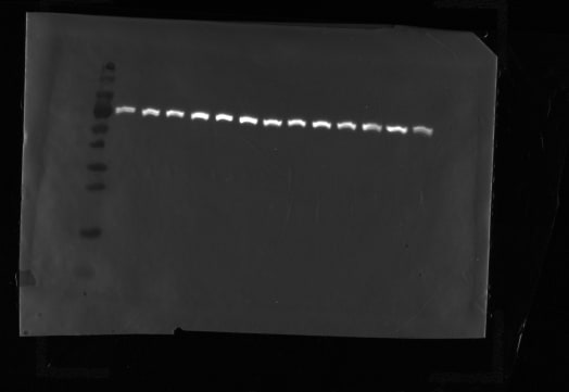
Western Blot: HSP70/HSPA1A Antibody (BB70) [NB110-96427]
Western Blot: HSP70/HSPA1A Antibody (BB70) [NB110-96427] - Western Blot analysis of Human Cervical cancer cell line (HeLa) lysate showing detection of Hsp70 protein using Mouse Anti-Hsp70 Monoclonal Antibody, Clone BB70 (NB110-96427). Primary Antibody: Mouse Anti-Hsp70 Monoclonal Antibody (NB110-96427) at 1:1000. Secondary Antibody: HRP Goat Anti-Rat.![Immunohistochemistry: HSP70/HSPA1A Antibody (BB70) [NB110-96427]](https://resources.rndsystems.com/images/products/HSP70-HSPA1A-Antibody-BB70-Immunohistochemistry-NB110-96427-img0018.jpg "Immunohistochemistry: HSP70/HSPA1A Antibody (BB70) [NB110-96427]")
Immunohistochemistry: HSP70/HSPA1A Antibody (BB70) [NB110-96427]
Immunohistochemistry: HSP70/HSPA1A Antibody (BB70) [NB110-96427] - Immunohistochemistry analysis using Mouse Anti-Hsp70 Monoclonal Antibody, Clone BB70 (NB110-96427). Tissue: colon carcinoma. Species: Human. Fixation: Formalin. Primary Antibody: Mouse Anti-Hsp70 Monoclonal Antibody (NB110-96427) at 1:10000 for 12 hours at 4C. Secondary Antibody: Biotin Goat Anti-Mouse at 1:2000 for 1 hour at RT. Counterstain: Mayer Hematoxylin (purple/blue) nuclear stain at 200 l for 2 minutes at RT. Localization: Inflammatory cells. Magnification: 40x. HSP70/HSPA1A cells stained brown. This image was produced using an amplifying IHC wash buffer. The antibody has therefore been diluted more than is recommended for other applications..![Western Blot: HSP70/HSPA1A Antibody (BB70) [NB110-96427]](https://resources.rndsystems.com/images/products/HSP70-HSPA1A-Antibody-BB70-Western-Blot-NB110-96427-img0013.jpg "Western Blot: HSP70/HSPA1A Antibody (BB70) [NB110-96427]")
Western Blot: HSP70/HSPA1A Antibody (BB70) [NB110-96427]
Western Blot: HSP70/HSPA1A Antibody (BB70) [NB110-96427] - analysis of Bovine MDBK cell lysates showing detection of Hsp70 protein using Mouse Anti-Hsp70 Monoclonal Antibody, Clone BB70. Primary Antibody: Mouse Anti-Hsp70 Monoclonal Antibody at 1:1000.![Immunohistochemistry: HSP70/HSPA1A Antibody (BB70) [NB110-96427]](https://resources.rndsystems.com/images/products/HSP70-HSPA1A-Antibody-BB70-Immunohistochemistry-NB110-96427-img0008.jpg "Immunohistochemistry: HSP70/HSPA1A Antibody (BB70) [NB110-96427]")
Immunohistochemistry: HSP70/HSPA1A Antibody (BB70) [NB110-96427]
Immunohistochemistry: HSP70/HSPA1A Antibody (BB70) [NB110-96427] - Detection in paraffin embedded liver sections and nuclear sections of rat hepatocytes by staining with BB70. First pictures in series show two hours after exposure to stress, the second shows the control.![Immunohistochemistry-Paraffin: HSP70/HSPA1A Antibody (BB70) [NB110-96427]](https://resources.rndsystems.com/images/products/HSP70-HSPA1A-Antibody-BB70-Immunohistochemistry-Paraffin-NB110-96427-img0015.jpg "Immunohistochemistry-Paraffin: HSP70/HSPA1A Antibody (BB70) [NB110-96427]")
Immunohistochemistry-Paraffin: HSP70/HSPA1A Antibody (BB70) [NB110-96427]
Immunohistochemistry-Paraffin: HSP70/HSPA1A Antibody (BB70) [NB110-96427] - Tissue: hepatocytes. Species: Rat. Fixation: Paraffin Embedded. Primary Antibody: Mouse Anti-Hsp70 Monoclonal Antibody at 1:200. Courtesy of: G. Matic, University of Belgrade, Serbia.![Immunohistochemistry: HSP70/HSPA1A Antibody (BB70) [NB110-96427]](https://resources.rndsystems.com/images/products/HSP70-HSPA1A-Antibody-BB70-Immunohistochemistry-NB110-96427-img0017.jpg "Immunohistochemistry: HSP70/HSPA1A Antibody (BB70) [NB110-96427]")
Immunohistochemistry: HSP70/HSPA1A Antibody (BB70) [NB110-96427]
Immunohistochemistry: HSP70/HSPA1A Antibody (BB70) [NB110-96427] - Immunohistochemistry analysis using Mouse Anti-Hsp70 Monoclonal Antibody, Clone BB70 (NB110-96427). Tissue: inflamed colon. Species: Mouse. Fixation: Formalin. Primary Antibody: Mouse Anti-Hsp70 Monoclonal Antibody (NB110-96427) at 1:10000 for 12 hours at 4C. Secondary Antibody: Biotin Goat Anti-Mouse at 1:2000 for 1 hour at RT. Counterstain: Mayer Hematoxylin (purple/blue) nuclear stain at 200 l for 2 minutes at RT. Localization: Inflammatory cells. Magnification: 40x. Inflammatory cells. HSP70/HSPA1A stained brown. This image was produced using an amplifying IHC wash buffer. The antibody has therefore been diluted more than is recommended for other applications.. - BSA Free [NB110-96427] -")
Western Blot: HSP70/HSPA1A Antibody (BB70) - BSA Free [NB110-96427] -
Effects of 7-d dirual HS and ZEN treatment on glycolytic skeletal muscle. (A) After 7 d of HS, relative protein abundance of MDA- and 4-HNE-modified proteins was similar between all groups. (B) Antioxidant enzymes relative protein abundance were assessed via western blotting. Treatment with ZEN decreased protein abundance in GPX1; however, all other antioxidant enzymes were similar between groups (n = 6–7/group). (C) Relative protein abundance of select HSPs following environmental HS and ZEN exposure. Ponceau S Stain (PonS) was used as a loading control. Values represent the mean +/- SEM. Groups were compared using a 2 × 2 ANOVA; a main effect of environment (P < 0.05) and/or a main effect of zearalenone (ZEN) (P < 0.05) is indicated. Similar groups are indicated by the same letter where appropriate, with differences determined by a Newman–Keuls post hoc test. Image collected and cropped by CiteAb from the following open publication (https://pubmed.ncbi.nlm.nih.gov/35908787), licensed under a CC-BY license. Not internally tested by Novus Biologicals.Applications for HSP70/HSPA1A Antibody (BB70) - BSA Free
Application
Recommended Usage
Immunocytochemistry/ Immunofluorescence
1:200
Immunohistochemistry
1:200
Immunohistochemistry-Paraffin
1:10-1:500
Western Blot
1:1000
Application Notes
1 ug/ml of HSP70/HSC70 Antibody was sufficient for detection of HSP70 and HSC70 in 20 ug of heat shocked HeLa cell lysate by colorimetric immunoblot analysis using Goat anti-mouse IgG:HRP as the secondary Antibody.
Reviewed Applications
Read 1 review rated 5 using NB110-96427 in the following applications:
Formulation, Preparation, and Storage
Purification
Protein G purified
Formulation
PBS (pH 7.2), 50% Glycerol
Format
BSA Free
Preservative
0.09% Sodium Azide
Concentration
1 mg/ml
Shipping
The product is shipped with polar packs. Upon receipt, store it immediately at the temperature recommended below.
Stability & Storage
Store at 4C short term. Aliquot and store at -20C long term. Avoid freeze-thaw cycles.
Background: HSP70/HSPA1A
Long Name
Heat Shock Protein 70
Alternate Names
HSP70-1A, HSP72, HSPA1A
Gene Symbol
HSPA1B
UniProt
Additional HSP70/HSPA1A Products
Product Documents for HSP70/HSPA1A Antibody (BB70) - BSA Free
Certificate of Analysis
To download a Certificate of Analysis, please enter a lot or batch number in the search box below.
Product Specific Notices for HSP70/HSPA1A Antibody (BB70) - BSA Free
This product is for research use only and is not approved for use in humans or in clinical diagnosis. Primary Antibodies are guaranteed for 1 year from date of receipt.
Related Research Areas
Citations for HSP70/HSPA1A Antibody (BB70) - BSA Free
 Powered by Bioz
Customer Reviews for HSP70/HSPA1A Antibody (BB70) - BSA Free (1)
5 out of 5
1 Customer Rating
Have you used HSP70/HSPA1A Antibody (BB70) - BSA Free?
Submit a review and receive an Amazon gift card!
$25/€18/£15/$25CAN/¥2500 Yen for a review with an image
$10/€7/£6/$10CAN/¥1110 Yen for a review without an image
Submit a review
Customer Images
Showing
1
-
1 of
1 review
Showing All
Filter By:
-
Application: Western BlotSample Tested: Skeletal muscle tissueSpecies: PigVerified Customer | Posted 06/30/2017Analysis of pig skeletal muscle lysates showing detection of Hsp70 protein. Load: 50 ug protein. Block: 5% milk TBST for 1 hour at RT. Primary Antibody: 1:5000 for o/n 4˚C. Secondary Antibody: Horse Anti-Mouse IgG: HRP for 1 minute at RT.

There are no reviews that match your criteria.
Protocols
Find general support by application which include: protocols, troubleshooting, illustrated assays, videos and webinars.
- Antigen Retrieval Protocol (PIER)
- Antigen Retrieval for Frozen Sections Protocol
- Appropriate Fixation of IHC/ICC Samples
- Cellular Response to Hypoxia Protocols
- Chromogenic IHC Staining of Formalin-Fixed Paraffin-Embedded (FFPE) Tissue Protocol
- Chromogenic Immunohistochemistry Staining of Frozen Tissue
- ClariTSA™ Fluorophore Kits
- Detection & Visualization of Antibody Binding
- Fluorescent IHC Staining of Frozen Tissue Protocol
- Graphic Protocol for Heat-induced Epitope Retrieval
- Graphic Protocol for the Preparation and Fluorescent IHC Staining of Frozen Tissue Sections
- Graphic Protocol for the Preparation and Fluorescent IHC Staining of Paraffin-embedded Tissue Sections
- Graphic Protocol for the Preparation of Gelatin-coated Slides for Histological Tissue Sections
- ICC Cell Smear Protocol for Suspension Cells
- ICC Immunocytochemistry Protocol Videos
- ICC for Adherent Cells
- IHC Sample Preparation (Frozen sections vs Paraffin)
- Immunocytochemistry (ICC) Protocol
- Immunocytochemistry Troubleshooting
- Immunofluorescence of Organoids Embedded in Cultrex Basement Membrane Extract
- Immunofluorescent IHC Staining of Formalin-Fixed Paraffin-Embedded (FFPE) Tissue Protocol
- Immunohistochemistry (IHC) and Immunocytochemistry (ICC) Protocols
- Immunohistochemistry Frozen Troubleshooting
- Immunohistochemistry Paraffin Troubleshooting
- Immunoprecipitation Protocol
- Preparing Samples for IHC/ICC Experiments
- Preventing Non-Specific Staining (Non-Specific Binding)
- Primary Antibody Selection & Optimization
- Protocol for Heat-Induced Epitope Retrieval (HIER)
- Protocol for Making a 4% Formaldehyde Solution in PBS
- Protocol for VisUCyte™ HRP Polymer Detection Reagent
- Protocol for the Fluorescent ICC Staining of Cell Smears - Graphic
- Protocol for the Fluorescent ICC Staining of Cultured Cells on Coverslips - Graphic
- Protocol for the Preparation & Fixation of Cells on Coverslips
- Protocol for the Preparation and Chromogenic IHC Staining of Frozen Tissue Sections
- Protocol for the Preparation and Chromogenic IHC Staining of Frozen Tissue Sections - Graphic
- Protocol for the Preparation and Chromogenic IHC Staining of Paraffin-embedded Tissue Sections
- Protocol for the Preparation and Chromogenic IHC Staining of Paraffin-embedded Tissue Sections - Graphic
- Protocol for the Preparation and Fluorescent ICC Staining of Cells on Coverslips
- Protocol for the Preparation and Fluorescent ICC Staining of Non-adherent Cells
- Protocol for the Preparation and Fluorescent ICC Staining of Stem Cells on Coverslips
- Protocol for the Preparation and Fluorescent IHC Staining of Frozen Tissue Sections
- Protocol for the Preparation and Fluorescent IHC Staining of Paraffin-embedded Tissue Sections
- Protocol for the Preparation of Gelatin-coated Slides for Histological Tissue Sections
- Protocol for the Preparation of a Cell Smear for Non-adherent Cell ICC - Graphic
- R&D Systems Quality Control Western Blot Protocol
- TUNEL and Active Caspase-3 Detection by IHC/ICC Protocol
- The Importance of IHC/ICC Controls
- Troubleshooting Guide: Immunohistochemistry
- Troubleshooting Guide: Western Blot Figures
- Western Blot Conditions
- Western Blot Protocol
- Western Blot Protocol for Cell Lysates
- Western Blot Troubleshooting
- Western Blot Troubleshooting Guide
- View all Protocols, Troubleshooting, Illustrated assays and Webinars
Loading...
Associated Pathways
