![Western Blot: HSP90 Antibody (3B6) [NB120-5455]](https://resources.rndsystems.com/images/products/HSP90-Antibody-3B6-Western-Blot-NB120-5455-img0013.jpg "Western Blot: HSP90 Antibody (3B6) [NB120-5455]")
Loading...
Key Product Details
Species Reactivity
Validated:
Human, Mouse, Rat
Cited:
Human
Applications
Validated:
Immunohistochemistry, Immunohistochemistry-Paraffin, Western Blot, Immunocytochemistry/ Immunofluorescence
Cited:
Western Blot
Label
Unconjugated
Antibody Source
Monoclonal Mouse IgG2B Clone # 3B6
Loading...
Product Specifications
Immunogen
Purified mouse HSP90.
Reactivity Notes
Please note that this antibody is reactive to Mouse and derived from the same host, Mouse. Additional Mouse on Mouse blocking steps may be required for IHC and ICC experiments. Please contact Technical Support for more information.
Localization
Cytoplasmic
Specificity
Detects heat shock protein 90 kDa (Hsp 90) from human, mouse and rat tissues. This does not detect chicken Hsp 90.
Clonality
Monoclonal
Host
Mouse
Isotype
IgG2B
Scientific Data Images for HSP90 Antibody (3B6)
Western Blot: HSP90 Antibody (3B6) [NB120-5455]
Western Blot: HSP90 Antibody (3B6) [NB120-5455] - Analysis of 50ug of various whole cell lysates and 15ul of PageRuler Prestained Protein Ladder onto a 4-20% Tris-HCl polyacrylamide gel.![Immunocytochemistry/ Immunofluorescence: HSP90 Antibody (3B6) [NB120-5455]](https://resources.rndsystems.com/images/products/HSP90-Antibody-3B6-Immunocytochemistry-Immunofluorescence-NB120-5455-img0008.jpg "Immunocytochemistry/ Immunofluorescence: HSP90 Antibody (3B6) [NB120-5455]")
Immunocytochemistry/ Immunofluorescence: HSP90 Antibody (3B6) [NB120-5455]
Immunocytochemistry/Immunofluorescence: HSP90 Antibody (3B6) [NB120-5455] - Heat Shock Protein 90 staining (green), F-Actin staining with Phalloidin (red) and nuclei with DAPI (blue) is shown. Cells were grown on chamber slides and fixed with formaldehyde prior to staining. Cells were probed without (control) or with or an antibody recognizing Heat Shock Protein 90 at a dilution of 1:100-1:200 over night at 4C, washed with PBS and incubated with a DyLight-488 conjugated.![Immunohistochemistry-Paraffin: HSP90 Antibody (3B6) [NB120-5455]](https://resources.rndsystems.com/images/products/HSP90-Antibody-3B6-Immunohistochemistry-Paraffin-NB120-5455-img0012.jpg "Immunohistochemistry-Paraffin: HSP90 Antibody (3B6) [NB120-5455]")
Immunohistochemistry-Paraffin: HSP90 Antibody (3B6) [NB120-5455]
Immunohistochemistry-Paraffin: HSP90 Antibody (3B6) [NB120-5455] - Both normal and cancer biopsies of deparaffinized Human breast carcinoma tissues.![Immunocytochemistry/ Immunofluorescence: HSP90 Antibody (3B6) [NB120-5455]](https://resources.rndsystems.com/images/products/HSP90-Antibody-3B6-Immunocytochemistry-Immunofluorescence-NB120-5455-img0005.jpg "Immunocytochemistry/ Immunofluorescence: HSP90 Antibody (3B6) [NB120-5455]")
Immunocytochemistry/ Immunofluorescence: HSP90 Antibody (3B6) [NB120-5455]
Immunocytochemistry/Immunofluorescence: HSP90 Antibody (3B6) [NB120-5455] - Formalin fixed cells were permeabilized with 0.1% Triton X-100 in TBS for 10 minutes at room temperature and blocked with 1% Blocker BSA for 15 minutes at room temperature. Cells were probed without (left panel) or with (right panel) an HSP90 monoclonal antibody at a dilution of 1:100 for at least 1 hour at room temperature, washed with PBS, and incubated with DyLight 488 goat anti-mouse IgG secondary antibody at a dilution of 1:400 for 30 minutes at room temperature. F-Actin (red) was stained with DyLight 554 Phalloidin and nuclei (blue) were stained with Hoechst 33342 dye.![Immunocytochemistry/ Immunofluorescence: HSP90 Antibody (3B6) [NB120-5455]](https://resources.rndsystems.com/images/products/HSP90-Antibody-3B6-Immunocytochemistry-Immunofluorescence-NB120-5455-img0006.jpg "Immunocytochemistry/ Immunofluorescence: HSP90 Antibody (3B6) [NB120-5455]")
Immunocytochemistry/ Immunofluorescence: HSP90 Antibody (3B6) [NB120-5455]
Immunocytochemistry/Immunofluorescence: HSP90 Antibody (3B6) [NB120-5455] - Heat Shock Protein 90 staining (green), F-Actin staining with Phalloidin (red) and nuclei with DAPI (blue) is shown. Cells were grown on chamber slides and fixed with formaldehyde prior to staining. Cells were probed without (control) or with or an antibody recognizing Heat Shock Protein 90 at a dilution of 1:100-1:200 over night at 4C, washed with PBS and incubated with a DyLight-488 conjugated.![Immunocytochemistry/ Immunofluorescence: HSP90 Antibody (3B6) [NB120-5455]](https://resources.rndsystems.com/images/products/HSP90-Antibody-3B6-Immunocytochemistry-Immunofluorescence-NB120-5455-img0007.jpg "Immunocytochemistry/ Immunofluorescence: HSP90 Antibody (3B6) [NB120-5455]")
Immunocytochemistry/ Immunofluorescence: HSP90 Antibody (3B6) [NB120-5455]
Immunocytochemistry/Immunofluorescence: HSP90 Antibody (3B6) [NB120-5455] - Heat Shock Protein 90 staining (green), F-Actin staining with Phalloidin (red) and nuclei with DAPI (blue) is shown. Cells were grown on chamber slides and fixed with formaldehyde prior to staining. Cells were probed without (control) or with or an antibody recognizing Heat Shock Protein 90 at a dilution of 1:100-1:200 over night at 4C, washed with PBS and incubated with a DyLight-488 conjugated.![Immunohistochemistry-Paraffin: HSP90 Antibody (3B6) [NB120-5455]](https://resources.rndsystems.com/images/products/HSP90-Antibody-3B6-Immunohistochemistry-Paraffin-NB120-5455-img0009.jpg "Immunohistochemistry-Paraffin: HSP90 Antibody (3B6) [NB120-5455]")
Immunohistochemistry-Paraffin: HSP90 Antibody (3B6) [NB120-5455]
Immunohistochemistry-Paraffin: HSP90 Antibody (3B6) [NB120-5455] - Staining of human colon cancer tissue sections.![Immunohistochemistry-Paraffin: HSP90 Antibody (3B6) [NB120-5455]](https://resources.rndsystems.com/images/products/HSP90-Antibody-3B6-Immunohistochemistry-Paraffin-NB120-5455-img0010.jpg "Immunohistochemistry-Paraffin: HSP90 Antibody (3B6) [NB120-5455]")
Immunohistochemistry-Paraffin: HSP90 Antibody (3B6) [NB120-5455]
Immunohistochemistry-Paraffin: HSP90 Antibody (3B6) [NB120-5455] - Both normal and cancer biopsies of deparaffinized Human tonsil tissues.![Immunohistochemistry-Paraffin: HSP90 Antibody (3B6) [NB120-5455]](https://resources.rndsystems.com/images/products/HSP90-Antibody-3B6-Immunohistochemistry-Paraffin-NB120-5455-img0011.jpg "Immunohistochemistry-Paraffin: HSP90 Antibody (3B6) [NB120-5455]")
Immunohistochemistry-Paraffin: HSP90 Antibody (3B6) [NB120-5455]
Immunohistochemistry-Paraffin: HSP90 Antibody (3B6) [NB120-5455] - Both normal and cancer biopsies of deparaffinized Human kidney tissues.Applications for HSP90 Antibody (3B6)
Application
Recommended Usage
Immunocytochemistry/ Immunofluorescence
1:100 - 1:200
Immunohistochemistry
1:20
Immunohistochemistry-Paraffin
1:20
Western Blot
1:500
Application Notes
WB: Detects an approx. 90 kDa protein representing Hsp 90 from MCF7 cell extract. ab5455 does not immunoprecipitate Hsp 90 in any form (free or complexed).
Reviewed Applications
Read 1 review rated 5 using NB120-5455 in the following applications:
Formulation, Preparation, and Storage
Purification
Unpurified
Formulation
Ascites diluted with PBS
Preservative
0.05% Sodium Azide
Concentration
This product is unpurified. The exact concentration of antibody is not quantifiable.
Shipping
The product is shipped with polar packs. Upon receipt, store it immediately at the temperature recommended below.
Stability & Storage
Store at -20C. Avoid freeze-thaw cycles.
Background: HSP90
Long Name
Heat Shock Protein 90
Alternate Names
FLJ31884, Heat shock 86 kDa, heat shock 90kD protein 1, alpha, heat shock 90kD protein 1, alpha-like 4, heat shock 90kD protein, alpha-like 4, heat shock 90kDa protein 1, alpha, heat shock protein 90kDa alpha (cytosolic), class A member 1, heat shock protein HSP 90-alpha, Hsp89, Hsp90, HSP90AHSP86, HSP90N, HSPC1HSP 86, HSPCAHSP89A, HSPCAL1, HSPCAL4, HSPN, LAP2, Renal carcinoma antigen NY-REN-38
Gene Symbol
HSP90AA1
Additional HSP90 Products
Product Documents for HSP90 Antibody (3B6)
Certificate of Analysis
To download a Certificate of Analysis, please enter a lot or batch number in the search box below.
Product Specific Notices for HSP90 Antibody (3B6)
This product is for research use only and is not approved for use in humans or in clinical diagnosis. Primary Antibodies are guaranteed for 1 year from date of receipt.
Related Research Areas
Citations for HSP90 Antibody (3B6)
 Powered by Bioz
Customer Reviews for HSP90 Antibody (3B6) (1)
5 out of 5
1 Customer Rating
Have you used HSP90 Antibody (3B6)?
Submit a review and receive an Amazon gift card!
$25/€18/£15/$25CAN/¥2500 Yen for a review with an image
$10/€7/£6/$10CAN/¥1110 Yen for a review without an image
Submit a review
Customer Images
Showing
1
-
1 of
1 review
Showing All
Filter By:
-
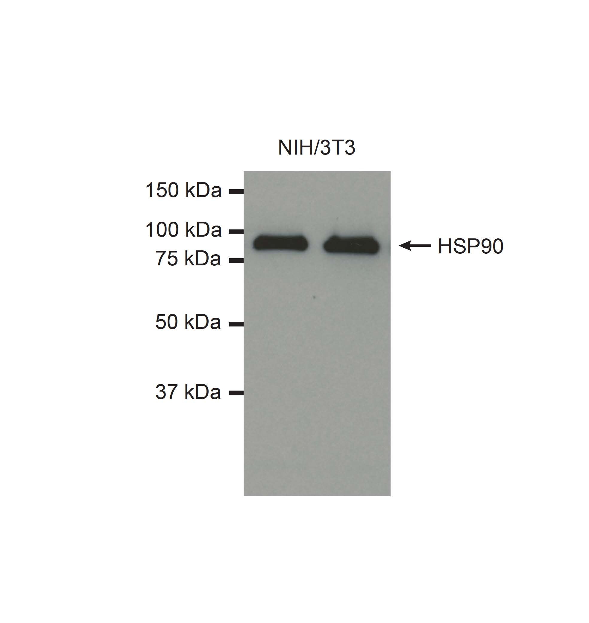
Application: Western BlotSample Tested: NIH/3T3 whole cell lysateSpecies: MouseVerified Customer | Posted 06/03/2015Western blot for HSP90 in NIH/3T3 cell lysates (30ug per lane) using NB120-5455.

There are no reviews that match your criteria.
Protocols
Find general support by application which include: protocols, troubleshooting, illustrated assays, videos and webinars.
- Antigen Retrieval Protocol (PIER)
- Antigen Retrieval for Frozen Sections Protocol
- Appropriate Fixation of IHC/ICC Samples
- Cellular Response to Hypoxia Protocols
- Chromogenic IHC Staining of Formalin-Fixed Paraffin-Embedded (FFPE) Tissue Protocol
- Chromogenic Immunohistochemistry Staining of Frozen Tissue
- ClariTSA™ Fluorophore Kits
- Detection & Visualization of Antibody Binding
- Fluorescent IHC Staining of Frozen Tissue Protocol
- Graphic Protocol for Heat-induced Epitope Retrieval
- Graphic Protocol for the Preparation and Fluorescent IHC Staining of Frozen Tissue Sections
- Graphic Protocol for the Preparation and Fluorescent IHC Staining of Paraffin-embedded Tissue Sections
- Graphic Protocol for the Preparation of Gelatin-coated Slides for Histological Tissue Sections
- ICC Cell Smear Protocol for Suspension Cells
- ICC Immunocytochemistry Protocol Videos
- ICC for Adherent Cells
- IHC Sample Preparation (Frozen sections vs Paraffin)
- Immunocytochemistry (ICC) Protocol
- Immunocytochemistry Troubleshooting
- Immunofluorescence of Organoids Embedded in Cultrex Basement Membrane Extract
- Immunofluorescent IHC Staining of Formalin-Fixed Paraffin-Embedded (FFPE) Tissue Protocol
- Immunohistochemistry (IHC) and Immunocytochemistry (ICC) Protocols
- Immunohistochemistry Frozen Troubleshooting
- Immunohistochemistry Paraffin Troubleshooting
- Preparing Samples for IHC/ICC Experiments
- Preventing Non-Specific Staining (Non-Specific Binding)
- Primary Antibody Selection & Optimization
- Protocol for Heat-Induced Epitope Retrieval (HIER)
- Protocol for Making a 4% Formaldehyde Solution in PBS
- Protocol for VisUCyte™ HRP Polymer Detection Reagent
- Protocol for the Fluorescent ICC Staining of Cell Smears - Graphic
- Protocol for the Fluorescent ICC Staining of Cultured Cells on Coverslips - Graphic
- Protocol for the Preparation & Fixation of Cells on Coverslips
- Protocol for the Preparation and Chromogenic IHC Staining of Frozen Tissue Sections
- Protocol for the Preparation and Chromogenic IHC Staining of Frozen Tissue Sections - Graphic
- Protocol for the Preparation and Chromogenic IHC Staining of Paraffin-embedded Tissue Sections
- Protocol for the Preparation and Chromogenic IHC Staining of Paraffin-embedded Tissue Sections - Graphic
- Protocol for the Preparation and Fluorescent ICC Staining of Cells on Coverslips
- Protocol for the Preparation and Fluorescent ICC Staining of Non-adherent Cells
- Protocol for the Preparation and Fluorescent ICC Staining of Stem Cells on Coverslips
- Protocol for the Preparation and Fluorescent IHC Staining of Frozen Tissue Sections
- Protocol for the Preparation and Fluorescent IHC Staining of Paraffin-embedded Tissue Sections
- Protocol for the Preparation of Gelatin-coated Slides for Histological Tissue Sections
- Protocol for the Preparation of a Cell Smear for Non-adherent Cell ICC - Graphic
- R&D Systems Quality Control Western Blot Protocol
- TUNEL and Active Caspase-3 Detection by IHC/ICC Protocol
- The Importance of IHC/ICC Controls
- Troubleshooting Guide: Immunohistochemistry
- Troubleshooting Guide: Western Blot Figures
- Western Blot Conditions
- Western Blot Protocol
- Western Blot Protocol for Cell Lysates
- Western Blot Troubleshooting
- Western Blot Troubleshooting Guide
- View all Protocols, Troubleshooting, Illustrated assays and Webinars
Loading...
Associated Pathways
