Four distinct genes encode alkaline phosphatases (APs) in humans (1). The ALPL gene encodes the liver/bone/kidney isozyme, also known as the tissue-nonspecific AP (TNAP). In comparison, ALPI, ALPP and ALPPL2 encode intestinal, placental and placental-like or germ cell APs, respectively. The serum levels of human APs are useful tumor markers (2). There are many mutations in the ALPL gene, leading to different forms of hypophosphatasia, characterized by poorly mineralized cartilage and bones (3). The native ALPL is a glycosylated homodimer attached to the membrane through a GPI-anchor. The C-terminal pro peptide (residues 503‑524) is not present in the mature form.
Human Alkaline Phosphatase/ALPL Antibody (388828)
R&D Systems | Catalog # MAB29091

Key Product Details
Species Reactivity
Validated:
Human
Cited:
Mouse
Applications
Validated:
Immunocytochemistry
Cited:
Immunohistochemistry, Immunohistochemistry-Paraffin, Immunocytochemistry
Label
Unconjugated
Antibody Source
Monoclonal Rat IgG2A Clone # 388828
Loading...
Product Specifications
Immunogen
E. coli-derived recombinant human Alkaline Phosphatase/ALPL
Leu18-Ser502
Accession # P05186
Leu18-Ser502
Accession # P05186
Specificity
Detects human Alkaline Phosphatase/ALPL in direct ELISAs. In direct ELISAs, 80-100%
cross-reactivity with recombinant mouse Alkaline Phosphatase/ALPL is observed
and no cross-reactivity with recombinant human (rh) Alkaline Phosphatase/ALPP
or rhAlkaline Phosphatase/ALPI is observed.
Clonality
Monoclonal
Host
Rat
Isotype
IgG2A
Scientific Data Images for Human Alkaline Phosphatase/ALPL Antibody (388828)
Alkaline Phosphatase/ALPL in BG01V Human Embryonic Stem Cells.
Alkaline Phosphatase/ALPL was detected in immersion fixed BG01V human embryonic stem cells using Rat Anti-Human Alkaline Phosphatase/ALPL Monoclonal Antibody (Catalog # MAB29091) at 10 µg/mL for 3 hours at room temperature. Cells were stained using the NorthernLights™ 557-conjugated Anti-Rat IgG Secondary Antibody (red; Catalog # NL013) and counterstained with DAPI (blue). Specific staining was localized to cell surfaces. View our protocol for Fluorescent ICC Staining of Cells on Coverslips.Applications for Human Alkaline Phosphatase/ALPL Antibody (388828)
Application
Recommended Usage
Immunocytochemistry
8-25 µg/mL
Sample: Immersion fixed BG01V human embryonic stem cells
Sample: Immersion fixed BG01V human embryonic stem cells
Reviewed Applications
Read 3 reviews rated 5 using MAB29091 in the following applications:
Formulation, Preparation, and Storage
Purification
Protein A or G purified from hybridoma culture supernatant
Reconstitution
Sterile PBS to a final concentration of 0.5 mg/mL. For liquid material, refer to CoA for concentration.
Loading...
Formulation
Lyophilized from a 0.2 μm filtered solution in PBS with Trehalose. *Small pack size (SP) is supplied either lyophilized or as a 0.2 µm filtered solution in PBS.
Shipping
Lyophilized product is shipped at ambient temperature. Liquid small pack size (-SP) is shipped with polar packs. Upon receipt, store immediately at the temperature recommended below.
Stability & Storage
Use a manual defrost freezer and avoid repeated freeze-thaw cycles.
- 12 months from date of receipt, -20 to -70 °C as supplied.
- 1 month, 2 to 8 °C under sterile conditions after reconstitution.
- 6 months, -20 to -70 °C under sterile conditions after reconstitution.
Calculators
Background: Alkaline Phosphatase/ALPL
References
- Le Du, M-H. and J.L. Millan (2002) J. Biol. Chem. 277:49808.
- Millan, J.L. and W.H. Fishman (1995) Crit. Rev. Clin. Lab. Sci. 32:1.
- Di Mauro, S. et al. (2002) J. Bone Miner. Res. 17:1383.
Long Name
Alkaline Phosphatase Liver
Alternate Names
Akp2, AP-TNAP, HOPS, TNAP, TNSALP
Gene Symbol
ALPL
UniProt
Additional Alkaline Phosphatase/ALPL Products
Product Documents for Human Alkaline Phosphatase/ALPL Antibody (388828)
Certificate of Analysis
To download a Certificate of Analysis, please enter a lot or batch number in the search box below.
Note: Certificate of Analysis not available for kit components.
Product Specific Notices for Human Alkaline Phosphatase/ALPL Antibody (388828)
For research use only
Related Research Areas
Citations for Human Alkaline Phosphatase/ALPL Antibody (388828)
 Powered by Bioz
Customer Reviews for Human Alkaline Phosphatase/ALPL Antibody (388828) (3)
5 out of 5
3 Customer Ratings
Have you used Human Alkaline Phosphatase/ALPL Antibody (388828)?
Submit a review and receive an Amazon gift card!
$25/€18/£15/$25CAN/¥2500 Yen for a review with an image
$10/€7/£6/$10CAN/¥1110 Yen for a review without an image
Submit a review
Customer Images
Showing
1
-
3 of
3 reviews
Showing All
Filter By:
-
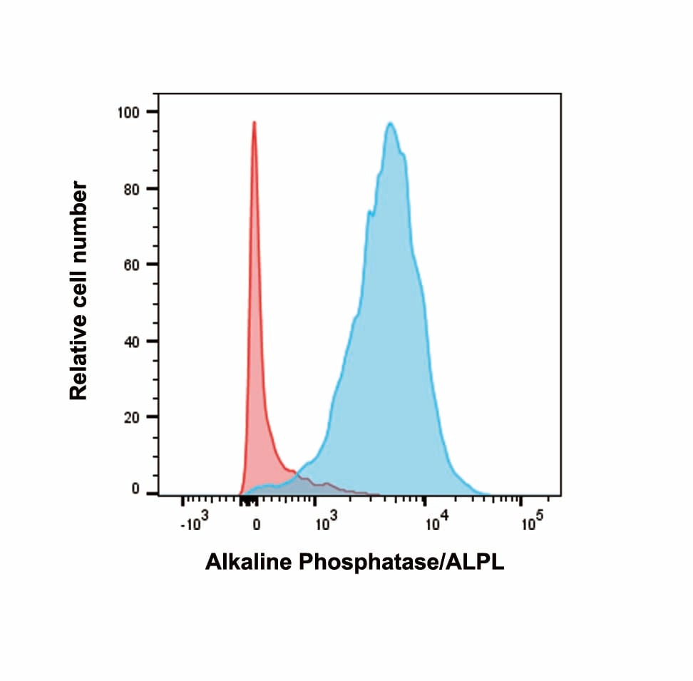
Application: Flow CytometrySample Tested: activated mouse CD8 T cellSpecies: MouseVerified Customer | Posted 02/16/2020

-
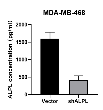
Application: ELISASample Tested: Cell Culture MediaSpecies: HumanVerified Customer | Posted 02/16/2020
-
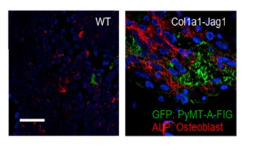
Application: Immunocytochemistry/ImmunofluorescenceSample Tested: bone marrowSpecies: MouseVerified Customer | Posted 10/29/2018Antibody staining was performed on mouse bone marrow sections. Please refer to PMID 29232552 for this staining, experimental details, and other data using this antibody.
There are no reviews that match your criteria.
Protocols
Find general support by application which include: protocols, troubleshooting, illustrated assays, videos and webinars.
- Appropriate Fixation of IHC/ICC Samples
- Cellular Response to Hypoxia Protocols
- ClariTSA™ Fluorophore Kits
- Detection & Visualization of Antibody Binding
- ICC Cell Smear Protocol for Suspension Cells
- ICC Immunocytochemistry Protocol Videos
- ICC for Adherent Cells
- Immunocytochemistry (ICC) Protocol
- Immunocytochemistry Troubleshooting
- Immunofluorescence of Organoids Embedded in Cultrex Basement Membrane Extract
- Immunohistochemistry (IHC) and Immunocytochemistry (ICC) Protocols
- Preparing Samples for IHC/ICC Experiments
- Preventing Non-Specific Staining (Non-Specific Binding)
- Primary Antibody Selection & Optimization
- Protocol for VisUCyte™ HRP Polymer Detection Reagent
- Protocol for the Fluorescent ICC Staining of Cell Smears - Graphic
- Protocol for the Fluorescent ICC Staining of Cultured Cells on Coverslips - Graphic
- Protocol for the Preparation and Fluorescent ICC Staining of Cells on Coverslips
- Protocol for the Preparation and Fluorescent ICC Staining of Non-adherent Cells
- Protocol for the Preparation and Fluorescent ICC Staining of Stem Cells on Coverslips
- Protocol for the Preparation of a Cell Smear for Non-adherent Cell ICC - Graphic
- TUNEL and Active Caspase-3 Detection by IHC/ICC Protocol
- The Importance of IHC/ICC Controls
- View all Protocols, Troubleshooting, Illustrated assays and Webinars
Loading...