Human Angiopoietin-4 (Ang-4) (1), alternatively named Ang-3 (2), is a secreted glycoprotein belonging to the angiopoietin family. It has the characteristic structural motifs of angiopoietins including the coiled-coiled domain near the amino-terminus and a fibrinogen-like domain at the C-terminus. Human Ang-4 cDNA encodes a 503 amino acid (aa) precursor protein with a 23 aa signal peptide. It shares 45%, 47%, and 54% aa sequence identity with human Ang-1, human Ang-2, and mouse Ang‑3, respectively. Although the sequence homology is much higher between the human and mouse counterparts for Ang-1 (97%) and Ang-2 (85%), mouse Ang‑3 is believed to be an ortholog of human Ang-4 based on chromosomal localization studies (2). Human Ang-4 is highly expressed in lung and in cultured human umbilical vein endothelial cells (HUVECs). In contrast, mouse Ang-3 is expressed multiple mouse tissues. Human Ang-4 is an agonist that can bind and activate Tie-2, a receptor tyrosine kinase with immunoglobulin and epidermal growth factor homology domains expressed primarily on endothelial cells and early hematopoietic cells (2, 3). Mouse Ang-3 has been reported to be a Tie-2 antagonist. It is likely that mouse Ang-3, like Ang-2, may exert agonist or antagonist activities depending on the cell context (1, 3, 4).
Human Angiopoietin-4 Antibody (156215)
R&D Systems | Catalog # MAB964

Key Product Details
Species Reactivity
Validated:
Human
Cited:
Xenograft
Applications
Validated:
Immunohistochemistry, Western Blot
Cited:
Simple Western
Label
Unconjugated
Antibody Source
Monoclonal Mouse IgG1 Clone # 156215
Loading...
Product Specifications
Immunogen
Mouse myeloma cell line NS0-derived recombinant human Angiopoietin-4
Met1-Ile503
Accession # Q9Y264
Met1-Ile503
Accession # Q9Y264
Specificity
Detects human Angiopoietin-4 in direct ELISAs and Western blots. In direct ELISAs, this antibody does not cross-react with recombinant human (rh) Ang‑1, rhAng‑2, rhAng-X, or rhANGPTL3.
Clonality
Monoclonal
Host
Mouse
Isotype
IgG1
Scientific Data Images for Human Angiopoietin-4 Antibody (156215)
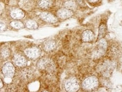
Angiopoietin‑4 in Human Kidney.
Angiopoietin-4 was detected in immersion fixed paraffin-embedded sections of human kidney using Human Angiopoietin-4 Monoclonal Antibody (Catalog # MAB964) at 15 µg/mL overnight at 4 °C. Before incubation with the primary antibody, tissue was subjected to heat-induced epitope retrieval using Antigen Retrieval Reagent-Basic (Catalog # CTS013). Tissue was stained using the Anti-Mouse HRP-DAB Cell & Tissue Staining Kit (brown; Catalog # CTS002) and counterstained with hematoxylin (blue). View our protocol for Chromogenic IHC Staining of Paraffin-embedded Tissue Sections.Applications for Human Angiopoietin-4 Antibody (156215)
Application
Recommended Usage
Immunohistochemistry
8-25 µg/mL
Sample: Immersion fixed paraffin-embedded sections of human kidney
Sample: Immersion fixed paraffin-embedded sections of human kidney
Western Blot
1 µg/mL
Sample: Recombinant Human Angiopoietin‑4 (Catalog # 964-AN)
Sample: Recombinant Human Angiopoietin‑4 (Catalog # 964-AN)
Reviewed Applications
Read 1 review rated 5 using MAB964 in the following applications:
Formulation, Preparation, and Storage
Purification
Protein A or G purified from hybridoma culture supernatant
Reconstitution
Reconstitute at 0.5 mg/mL in sterile PBS. For liquid material, refer to CoA for concentration.
Loading...
Formulation
Lyophilized from a 0.2 μm filtered solution in PBS with Trehalose. *Small pack size (SP) is supplied either lyophilized or as a 0.2 µm filtered solution in PBS.
Shipping
Lyophilized product is shipped at ambient temperature. Liquid small pack size (-SP) is shipped with polar packs. Upon receipt, store immediately at the temperature recommended below.
Stability & Storage
Use a manual defrost freezer and avoid repeated freeze-thaw cycles.
- 12 months from date of receipt, -20 to -70 °C as supplied.
- 1 month, 2 to 8 °C under sterile conditions after reconstitution.
- 6 months, -20 to -70 °C under sterile conditions after reconstitution.
Calculators
Background: Angiopoietin-4
References
- Valenzuela, D.M. et al. (1999) Proc. Natl. Acad. Sci. USA 96:1904.
- Nishimura, M. et al. (1999) FEBS Lett. 448:254.
- Jones, N. et al. (2001) Nat. Rev. Mol. Cell Biol. 2:257.
- Teichert-Kuliszewska, K. et al. (2001) Cardiovasc. Res. 49:659.
Alternate Names
ANGPT4
Entrez Gene IDs
51378 (Human)
Gene Symbol
ANGPT4
UniProt
Additional Angiopoietin-4 Products
Product Documents for Human Angiopoietin-4 Antibody (156215)
Certificate of Analysis
To download a Certificate of Analysis, please enter a lot or batch number in the search box below.
Note: Certificate of Analysis not available for kit components.
Product Specific Notices for Human Angiopoietin-4 Antibody (156215)
For research use only
Related Research Areas
Citations for Human Angiopoietin-4 Antibody (156215)
 Powered by Bioz
Customer Reviews for Human Angiopoietin-4 Antibody (156215) (1)
5 out of 5
1 Customer Rating
Have you used Human Angiopoietin-4 Antibody (156215)?
Submit a review and receive an Amazon gift card!
$25/€18/£15/$25CAN/¥2500 Yen for a review with an image
$10/€7/£6/$10CAN/¥1110 Yen for a review without an image
Submit a review
Customer Images
Showing
1
-
1 of
1 review
Showing All
Filter By:
-
Application: ImmunohistochemistrySample Tested: Kidney tissueSpecies: HumanVerified Customer | Posted 05/30/2022

There are no reviews that match your criteria.
Protocols
Find general support by application which include: protocols, troubleshooting, illustrated assays, videos and webinars.
- Antigen Retrieval Protocol (PIER)
- Antigen Retrieval for Frozen Sections Protocol
- Appropriate Fixation of IHC/ICC Samples
- Cellular Response to Hypoxia Protocols
- Chromogenic IHC Staining of Formalin-Fixed Paraffin-Embedded (FFPE) Tissue Protocol
- Chromogenic Immunohistochemistry Staining of Frozen Tissue
- ClariTSA™ Fluorophore Kits
- Detection & Visualization of Antibody Binding
- Fluorescent IHC Staining of Frozen Tissue Protocol
- Graphic Protocol for Heat-induced Epitope Retrieval
- Graphic Protocol for the Preparation and Fluorescent IHC Staining of Frozen Tissue Sections
- Graphic Protocol for the Preparation and Fluorescent IHC Staining of Paraffin-embedded Tissue Sections
- Graphic Protocol for the Preparation of Gelatin-coated Slides for Histological Tissue Sections
- IHC Sample Preparation (Frozen sections vs Paraffin)
- Immunofluorescent IHC Staining of Formalin-Fixed Paraffin-Embedded (FFPE) Tissue Protocol
- Immunohistochemistry (IHC) and Immunocytochemistry (ICC) Protocols
- Immunohistochemistry Frozen Troubleshooting
- Immunohistochemistry Paraffin Troubleshooting
- Preparing Samples for IHC/ICC Experiments
- Preventing Non-Specific Staining (Non-Specific Binding)
- Primary Antibody Selection & Optimization
- Protocol for Heat-Induced Epitope Retrieval (HIER)
- Protocol for Making a 4% Formaldehyde Solution in PBS
- Protocol for VisUCyte™ HRP Polymer Detection Reagent
- Protocol for the Preparation & Fixation of Cells on Coverslips
- Protocol for the Preparation and Chromogenic IHC Staining of Frozen Tissue Sections
- Protocol for the Preparation and Chromogenic IHC Staining of Frozen Tissue Sections - Graphic
- Protocol for the Preparation and Chromogenic IHC Staining of Paraffin-embedded Tissue Sections
- Protocol for the Preparation and Chromogenic IHC Staining of Paraffin-embedded Tissue Sections - Graphic
- Protocol for the Preparation and Fluorescent IHC Staining of Frozen Tissue Sections
- Protocol for the Preparation and Fluorescent IHC Staining of Paraffin-embedded Tissue Sections
- Protocol for the Preparation of Gelatin-coated Slides for Histological Tissue Sections
- R&D Systems Quality Control Western Blot Protocol
- TUNEL and Active Caspase-3 Detection by IHC/ICC Protocol
- The Importance of IHC/ICC Controls
- Troubleshooting Guide: Immunohistochemistry
- Troubleshooting Guide: Western Blot Figures
- Western Blot Conditions
- Western Blot Protocol
- Western Blot Protocol for Cell Lysates
- Western Blot Troubleshooting
- Western Blot Troubleshooting Guide
- View all Protocols, Troubleshooting, Illustrated assays and Webinars
Loading...