BAFF (also known as TALL-1, BLyS, and THANK) is a type II transmembrane glycoprotein belonging to the TNF superfamily and has been designated as TNF superfamily member 13B (TNFSF13B). Human BAFF is a 285 amino acid (aa) protein consisting of a 218 aa extracellular domain, a 21 aa transmembrane region and a 46 aa cytoplasmic tail (1, 2). BAFF has the typical structural characteristics of the TNF superfamily ligands. It is a homotrimeric protein having the structurally conserved motif known as TNF homology domain (3, 4). A higher ordered structure composed of a cluster of trimeric units resembling the structure of a viral capsid has also been reported (4). Human BAFF may be shed from the cell surface by proteolytic cleavage between R133 and Ala134 to yield a soluble form of the protein that is detectable in serum (1, 5). Within the TNF superfamily BAFF shares the highest homology (48%) with APRIL (1). BAFF shares with APRIL the ability to bind to BCMA and TACI and also binds specifically to BAFF receptor (BAFF R, also known as BR3 or TNFSFR13C), which is the principal BAFF receptor (6‑8). All three receptors are type III transmembrane proteins that are expressd in B cells. BAFF and APRIL can form active heteromers that bind to TACI (9). BAFF is expressed in peripheral blood mononuclear cells, in spleen and lymph nodes. Its expression in resting monocytes is up-regulated by IFN-alpha, IFN-beta, LPS and IL-10. BAFF provides critical survival signals to a subset of B cells with intermediate maturation status (T2 B cells) during the immune response (10). BAFF also plays an important role in the development of lymphoid tissue and enhances the survival of activated memory B cells (7, 11). Human and mouse BAFF share 86% aa sequence identity (1).
Human BAFF/BLyS/TNFSF13B Antibody
R&D Systems | Catalog # AF124

Key Product Details
Validated by
Biological Validation
Species Reactivity
Validated:
Human
Cited:
Human, Porcine
Applications
Validated:
Immunohistochemistry, Western Blot, Neutralization
Cited:
Immunohistochemistry, Immunohistochemistry-Paraffin, Western Blot, Neutralization, Immunocytochemistry
Label
Unconjugated
Antibody Source
Polyclonal Goat IgG
Loading...
Product Specifications
Immunogen
Mouse myeloma cell line NS0-derived recombinant human BAFF/BLyS/TNFSF13B
Ala134-Leu285
Accession # Q9Y275
Ala134-Leu285
Accession # Q9Y275
Specificity
Detects human BAFF/BLyS/TNFSF13B in direct ELISAs and Western blots. In direct ELISAs, less than 15% cross-reactivity with recombinant murine BAFF and less than 1% cross‑reactivity with recombinant human (rh) APRIL is observed.
Clonality
Polyclonal
Host
Goat
Isotype
IgG
Endotoxin Level
<0.60 EU per 1 μg of the antibody by the LAL method.
Scientific Data Images for Human BAFF/BLyS/TNFSF13B Antibody
Cell Proliferation Induced By BAFF/BLyS/TNFSF13B and Neutralization by Human BAFF/BLyS/TNFSF13B Antibody.
In the presence of Goat F(ab')2 Anti-mouse IgM (1 µg/mL), Recombinant Human BAFF/BLyS/TNFSF13B (Catalog # 2149-BF) stimulates proliferation in mouse B cells in a dose-dependent manner (orange line). Under these conditions, proliferation elicited by Recombinant Human BAFF/BLyS/TNFSF13B (5 ng/mL) is neutralized (green line) by increasing concentrations of Goat Anti-Human BAFF/BLyS/TNFSF13B Antigen Affinity-purified Polyclonal Antibody (Catalog # AF124). The ND50 is typically 3-12 ng/mL.
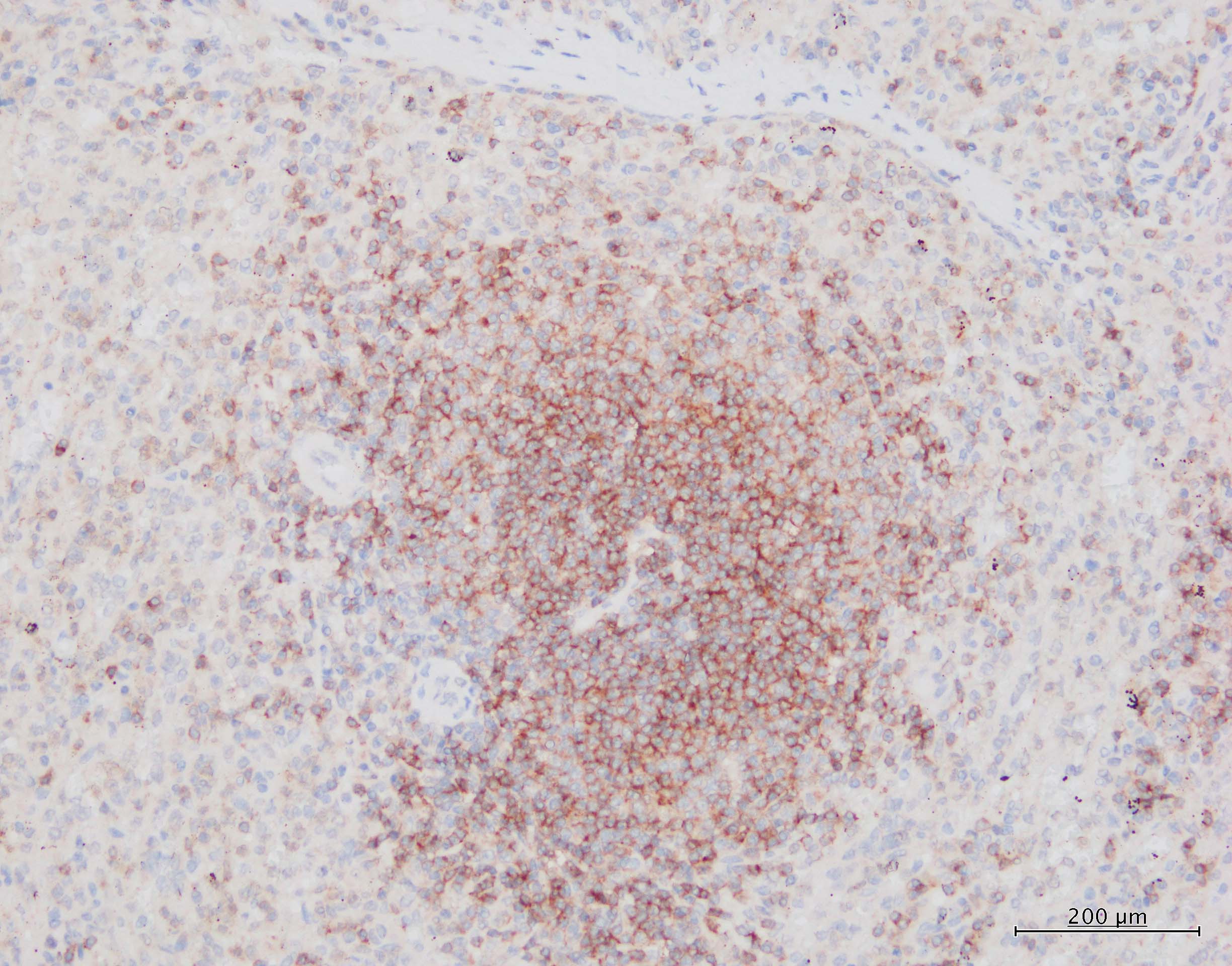
BAFF/BLyS/TNFSF13B in Human Spleen.
BAFF/BLyS/TNFSF13B was detected in formalin fixed paraffin-embedded sections of human spleen using 15 µg/mL Goat Anti-Human BAFF/BLyS/TNFSF13B Antigen Affinity-purified Polyclonal Antibody (Catalog # AF124) overnight at 4 °C. Tissue was stained with the Anti-Goat HRP-DAB Cell & Tissue Staining Kit (brown; Catalog # CTS008) and counterstained with hematoxylin (blue). View our protocol for Chromogenic IHC Staining of Paraffin-embedded Tissue Sections.
Detection of Human BAFF/BLyS/TNFSF13B by Immunocytochemistry/ Immunofluorescence
Role of p65 activation in BLyS up-regulation. (A) HIF-1 alpha and p65 protein levels in MDA-MB-435 in hypoxic conditions for different time points by Western Blotting. (B) CAPE(50 μM)and PX 12 (10 μM) were used to determine the roles of p65 and HIF-1 alpha in the regulation of BLyS expression by Western Blotting. The cells were treated with or without inhibitor in normoxic or hypoxic conditions for 6 h. (C) Effects of CAPE(50 μM)and PX 12 (10 μM) on BLyS promoter activity. Data were average luciferase activities of three independent transfections with ± SD. *, P < 0.05, vs pGL3-Basic/BP. (D) Localization of p65 protein and expression level of BLyS by immunofluorescence. MDA-MB-435 cells were challenged with CAPE (50 μM) for 6 h (original magnification 200 ×). Image collected and cropped by CiteAb from the following open publication (https://jeccr.biomedcentral.com/articles/10.1186/1756-9966-31-31), licensed under a CC-BY license. Not internally tested by R&D Systems.
Detection of Human BAFF/BLyS/TNFSF13B by Immunocytochemistry/ Immunofluorescence
Expressions of BLyS, TACI, BCMA and BAFF-R in human breast cancer cell lines. (A) BLyS and its three receptors in human breast cancer cell lines MDA-MB-435, MDA-MB-231, MDA-MB-468 and B cell line Ramos by immunofluorescence (original magnification 200 ×) and Western Blotting. (B) The mRNA level of BLyS in the three cell lines were detected by real-time PCR under hypoxia for different time points. Data were means of triplicate samples with ± SD; vs normoxia, *, P < 0.05; **, P < 0.01; ***, P < 0.001. (C) BLyS protein level in MDA-MB-435 cells by Western Blotting analysis. Image collected and cropped by CiteAb from the following open publication (https://jeccr.biomedcentral.com/articles/10.1186/1756-9966-31-31), licensed under a CC-BY license. Not internally tested by R&D Systems.
Detection of Human BAFF/BLyS/TNFSF13B by Western Blot
Role of p65 activation in BLyS up-regulation. (A) HIF-1 alpha and p65 protein levels in MDA-MB-435 in hypoxic conditions for different time points by Western Blotting. (B) CAPE(50 μM)and PX 12 (10 μM) were used to determine the roles of p65 and HIF-1 alpha in the regulation of BLyS expression by Western Blotting. The cells were treated with or without inhibitor in normoxic or hypoxic conditions for 6 h. (C) Effects of CAPE(50 μM)and PX 12 (10 μM) on BLyS promoter activity. Data were average luciferase activities of three independent transfections with ± SD. *, P < 0.05, vs pGL3-Basic/BP. (D) Localization of p65 protein and expression level of BLyS by immunofluorescence. MDA-MB-435 cells were challenged with CAPE (50 μM) for 6 h (original magnification 200 ×). Image collected and cropped by CiteAb from the following open publication (https://jeccr.biomedcentral.com/articles/10.1186/1756-9966-31-31), licensed under a CC-BY license. Not internally tested by R&D Systems.Applications for Human BAFF/BLyS/TNFSF13B Antibody
Application
Recommended Usage
Immunohistochemistry
5-15 µg/mL
Sample: Formalin fixed paraffin-embedded sections of human spleen
Sample: Formalin fixed paraffin-embedded sections of human spleen
Western Blot
0.1 µg/mL
Sample: Recombinant Human BAFF/BLyS/TNFSF13B (Catalog # 2149-BF)
Sample: Recombinant Human BAFF/BLyS/TNFSF13B (Catalog # 2149-BF)
Neutralization
Measured by its ability to neutralize BAFF/BLyS/TNFSF13B-induced proliferation in mouse B cells. The Neutralization Dose (ND50) is typically 3-12 ng/mL in the presence of 5 ng/mL Recombinant Human BAFF/BLyS/TNFSF13B and 1 µg/mL Goat F(ab')2 Anti-mouse IgM.
Reviewed Applications
Read 2 reviews rated 5 using AF124 in the following applications:
Formulation, Preparation, and Storage
Purification
Antigen Affinity-purified
Reconstitution
Reconstitute at 0.2 mg/mL in sterile PBS. For liquid material, refer to CoA for concentration.
Loading...
Formulation
Lyophilized from a 0.2 μm filtered solution in PBS with Trehalose. *Small pack size (SP) is supplied either lyophilized or as a 0.2 µm filtered solution in PBS.
Shipping
Lyophilized product is shipped at ambient temperature. Liquid small pack size (-SP) is shipped with polar packs. Upon receipt, store immediately at the temperature recommended below.
Stability & Storage
Use a manual defrost freezer and avoid repeated freeze-thaw cycles.
- 12 months from date of receipt, -20 to -70 °C as supplied.
- 1 month, 2 to 8 °C under sterile conditions after reconstitution.
- 6 months, -20 to -70 °C under sterile conditions after reconstitution.
Calculators
Background: BAFF/BLyS/TNFSF13B
References
- Schneider, P. et al. (1999) J. Exp. Med. 189:1747.
- Mukhopadhyay, A. et al. (1999) J. Biol. Chem. 274:15978.
- Karpusas, M. et al. (2002) J. Mol. Biol. 315:1145.
- Liu, Y. et al. (2002) Cell 108:383.
- Cheema, G.S. et al. (2001) Arthr. Rheum. 44:1313.
- Marsters, S.A. et al. (2000) Curr. Biol. 10:785.
- Thompson, J.S. et al. (2001) Science 293:2108.
- Ng, L.G. et al. (2004) J. Immunol. 173:807.
- Roschke, V. et al. (2002) J. Immunol. 169:4314.
- Batten, M. et al. (2000) J. Exp. Med. 192:1453.
- Avery, D.T. et al. (2003) J. Clin. Invest. 112:286.
Long Name
B cell Activating Factor
Alternate Names
BLyS, CD257, TALL1, THANK, TNFSF13B, ZTNF4
Gene Symbol
TNFSF13B
UniProt
Additional BAFF/BLyS/TNFSF13B Products
Product Documents for Human BAFF/BLyS/TNFSF13B Antibody
Certificate of Analysis
To download a Certificate of Analysis, please enter a lot or batch number in the search box below.
Note: Certificate of Analysis not available for kit components.
Product Specific Notices for Human BAFF/BLyS/TNFSF13B Antibody
For research use only
Citations for Human BAFF/BLyS/TNFSF13B Antibody
 Powered by Bioz
Customer Reviews for Human BAFF/BLyS/TNFSF13B Antibody (2)
5 out of 5
2 Customer Ratings
Have you used Human BAFF/BLyS/TNFSF13B Antibody?
Submit a review and receive an Amazon gift card!
$25/€18/£15/$25CAN/¥2500 Yen for a review with an image
$10/€7/£6/$10CAN/¥1110 Yen for a review without an image
Submit a review
Customer Images
Showing
1
-
2 of
2 reviews
Showing All
Filter By:
-
Application: ELISASample Tested: Recombinant proteinSpecies: HumanVerified Customer | Posted 09/09/2024ELISA signal with AF124I used it to detect rhBAFF after incubation with BCMA in ELISA experiment and it produced a good signal as expected compared to control (no signal).

-
Application: ImmunohistochemistrySample Tested: Spleen tissueSpecies: HumanVerified Customer | Posted 02/18/2022Heat-retrieval in high-pH buffer primary Ab 10ug/mL for 60min ImmPRESS polymer HRP and DAB
There are no reviews that match your criteria.
Protocols
Find general support by application which include: protocols, troubleshooting, illustrated assays, videos and webinars.
- Antigen Retrieval Protocol (PIER)
- Antigen Retrieval for Frozen Sections Protocol
- Appropriate Fixation of IHC/ICC Samples
- Cellular Response to Hypoxia Protocols
- Chromogenic IHC Staining of Formalin-Fixed Paraffin-Embedded (FFPE) Tissue Protocol
- Chromogenic Immunohistochemistry Staining of Frozen Tissue
- ClariTSA™ Fluorophore Kits
- Detection & Visualization of Antibody Binding
- Fluorescent IHC Staining of Frozen Tissue Protocol
- Graphic Protocol for Heat-induced Epitope Retrieval
- Graphic Protocol for the Preparation and Fluorescent IHC Staining of Frozen Tissue Sections
- Graphic Protocol for the Preparation and Fluorescent IHC Staining of Paraffin-embedded Tissue Sections
- Graphic Protocol for the Preparation of Gelatin-coated Slides for Histological Tissue Sections
- IHC Sample Preparation (Frozen sections vs Paraffin)
- Immunofluorescent IHC Staining of Formalin-Fixed Paraffin-Embedded (FFPE) Tissue Protocol
- Immunohistochemistry (IHC) and Immunocytochemistry (ICC) Protocols
- Immunohistochemistry Frozen Troubleshooting
- Immunohistochemistry Paraffin Troubleshooting
- Preparing Samples for IHC/ICC Experiments
- Preventing Non-Specific Staining (Non-Specific Binding)
- Primary Antibody Selection & Optimization
- Protocol for Heat-Induced Epitope Retrieval (HIER)
- Protocol for Making a 4% Formaldehyde Solution in PBS
- Protocol for VisUCyte™ HRP Polymer Detection Reagent
- Protocol for the Preparation & Fixation of Cells on Coverslips
- Protocol for the Preparation and Chromogenic IHC Staining of Frozen Tissue Sections
- Protocol for the Preparation and Chromogenic IHC Staining of Frozen Tissue Sections - Graphic
- Protocol for the Preparation and Chromogenic IHC Staining of Paraffin-embedded Tissue Sections
- Protocol for the Preparation and Chromogenic IHC Staining of Paraffin-embedded Tissue Sections - Graphic
- Protocol for the Preparation and Fluorescent IHC Staining of Frozen Tissue Sections
- Protocol for the Preparation and Fluorescent IHC Staining of Paraffin-embedded Tissue Sections
- Protocol for the Preparation of Gelatin-coated Slides for Histological Tissue Sections
- R&D Systems Quality Control Western Blot Protocol
- TUNEL and Active Caspase-3 Detection by IHC/ICC Protocol
- The Importance of IHC/ICC Controls
- Troubleshooting Guide: Immunohistochemistry
- Troubleshooting Guide: Western Blot Figures
- Western Blot Conditions
- Western Blot Protocol
- Western Blot Protocol for Cell Lysates
- Western Blot Troubleshooting
- Western Blot Troubleshooting Guide
- View all Protocols, Troubleshooting, Illustrated assays and Webinars
Loading...
Associated Pathways
