
Carbonic Anhydrase (CA) catalyzes the reversible reaction of CO2 + H2O = HCO3- + H+, which is fundamental to many processes such as respiration, renal tubular acidification and bone resorption (1-3). Topics in the CA meeting (6th International Conference on the CAs, June 20-25, 2003; Slovakia) ranged from use of CAs as markers for tumor and hypoxia in clinic, as nutritional supplement in milk, and as a tool for CO2 removal and mosquito control in industry. CA9, also known as membrane antigen MN and renal cell carcinoma (RCC)-associated antigen G250, is a transmembrane enzyme expressed primarily in carcinoma cells. It is one of the best markers for hypoxia and for RCC (4, 5). rhCA9 corresponds to the extracellular portion of human CA9.
Human Carbonic Anhydrase IX/CA9 Antibody (303123) [FITC]
R&D Systems | Catalog # FAB2188F

Scientific images may be resized, cropped, or adjusted for brightness or contrast for presentation purposes. These updates are limited to presentation and do not affect the underlying scientific interpretation of the data. Where available, additional source data may be provided upon request.
Key Product Details
Species Reactivity
Validated:
Human
Cited:
Human, Mouse
Applications
Validated:
Flow Cytometry
Cited:
Immunohistochemistry, Flow Cytometry
Label
FITC (Excitation = 488 nm, Emission = 515-545 nm)
Antibody Source
Monoclonal Mouse IgG2A Clone # 303123
Loading...
Product Specifications
Immunogen
Mouse myeloma cell line NS0-derived recombinant human Carbonic Anhydrase IX
Pro59-Asp414
Accession # Q16790
Pro59-Asp414
Accession # Q16790
Specificity
Detects human Carbonic Anhydrase IX (CA9) in direct ELISAs. In direct ELISAs, this antibody does not cross-react with recombinant mouse (rm) CA9 or with rhCA1, 2, 3, 4, 5A, 6, 7, 8, 10, 12, 13, or 14.
Clonality
Monoclonal
Host
Mouse
Isotype
IgG2A
Scientific Data Images for Human Carbonic Anhydrase IX/CA9 Antibody (303123) [FITC]
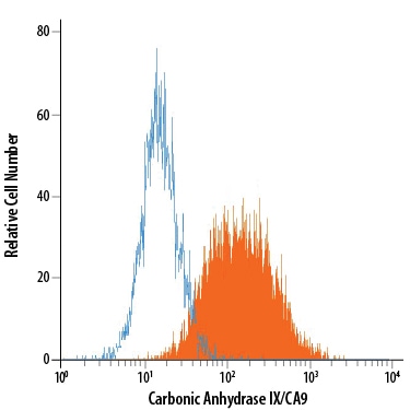
Detection of Carbonic Anhydrase IX/CA9 in U‑87 MG Human Cell Line by Flow Cytometry.
U-87 MG human glioblastoma/ astrocytoma cell line was stained with Mouse Anti-Human Carbonic Anhydrase IX/CA9 Fluorescein-conjugated Monoclonal Antibody (Catalog # FAB2188F, filled histogram) or isotype control antibody (Catalog # IC003F, open histogram). View our protocol for Staining Membrane-associated Proteins.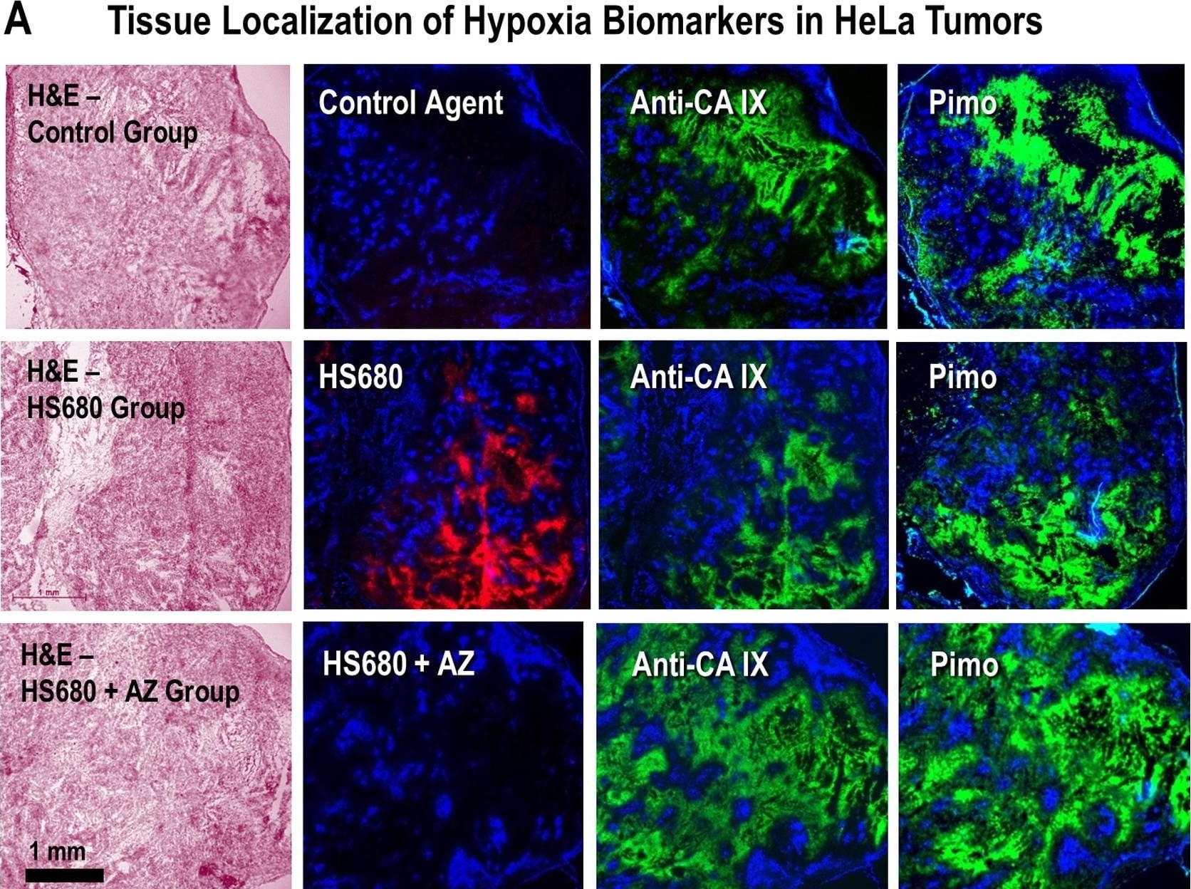
Detection of Carbonic Anhydrase IX/CA9 by Immunohistochemistry
Localization of HS680 and tumor hypoxia markers in HeLa xenografts. A, The tissue staining patterns of the control agent, HS680, and HS680+AZ (red) from the same or adjacent tumor sections as staining with fluorescent CA IX antibody or pimonidazole (green) and the Hoechst perfusion stain (blue). H&E staining of tissue sections that were used for localization are shown on the left side of images. HS680 was specifically localized in regions with low Hoechst staining indicative of low oxygen (less perfused) but positive staining with both the CA IX antibody and Pimonidazole. Preinjection of the mice with unlabeled AZ blocked the binding of HS680 to control levels. B, Co-localization (overlay) of HS680 with CA IX antibody or pimonidazole (hypoxyprobe) is shown on the right side images indicating HS680 was clearly co-localized with both anti-CA IX antibody and hypoxyprobe (pimonidazole). Similar results were obtained using HT-29 tumors (Figure S4A and Figure S4B). Image collected and cropped by CiteAb from the following open publication (https://pubmed.ncbi.nlm.nih.gov/23226406), licensed under a CC-BY license. Not internally tested by R&D Systems.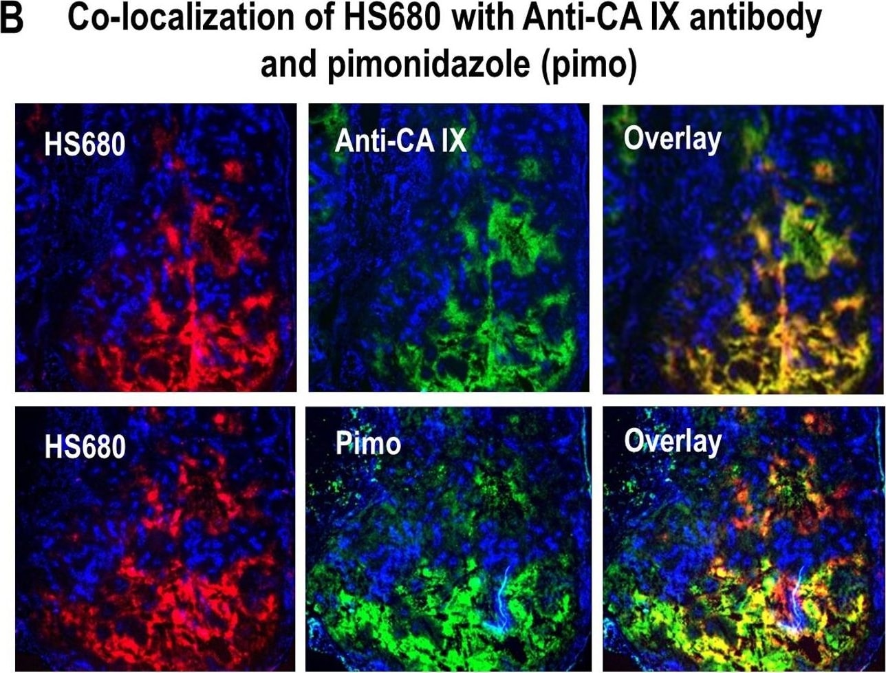
Detection of Carbonic Anhydrase IX/CA9 by Immunohistochemistry
Localization of HS680 and tumor hypoxia markers in HeLa xenografts. A, The tissue staining patterns of the control agent, HS680, and HS680+AZ (red) from the same or adjacent tumor sections as staining with fluorescent CA IX antibody or pimonidazole (green) and the Hoechst perfusion stain (blue). H&E staining of tissue sections that were used for localization are shown on the left side of images. HS680 was specifically localized in regions with low Hoechst staining indicative of low oxygen (less perfused) but positive staining with both the CA IX antibody and Pimonidazole. Preinjection of the mice with unlabeled AZ blocked the binding of HS680 to control levels. B, Co-localization (overlay) of HS680 with CA IX antibody or pimonidazole (hypoxyprobe) is shown on the right side images indicating HS680 was clearly co-localized with both anti-CA IX antibody and hypoxyprobe (pimonidazole). Similar results were obtained using HT-29 tumors (Figure S4A and Figure S4B). Image collected and cropped by CiteAb from the following open publication (https://pubmed.ncbi.nlm.nih.gov/23226406), licensed under a CC-BY license. Not internally tested by R&D Systems.Applications for Human Carbonic Anhydrase IX/CA9 Antibody (303123) [FITC]
Application
Recommended Usage
Flow Cytometry
10 µL/106 cells
Sample: U‑87 MG human glioblastoma/astrocytoma cell line
Sample: U‑87 MG human glioblastoma/astrocytoma cell line
Reviewed Applications
Read 2 reviews rated 5 using FAB2188F in the following applications:
Spectra Viewer
Plan Your Experiments
Use our spectra viewer to interactively plan your experiments, assessing multiplexing options. View the excitation and emission spectra for our fluorescent dye range and other commonly used dyes.
Spectra Viewer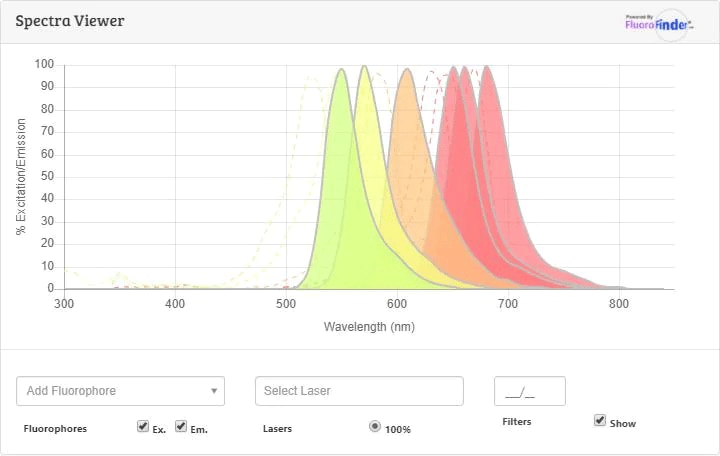
Flow Cytometry Panel Builder
Bio-Techne Knows Flow Cytometry
Save time and reduce costly mistakes by quickly finding compatible reagents using the Panel Builder Tool.
Advanced Features
- Spectra Viewer - Custom analysis of spectra from multiple fluorochromes
- Spillover Popups - Visualize the spectra of individual fluorochromes
- Antigen Density Selector - Match fluorochrome brightness with antigen density

Formulation, Preparation, and Storage
Purification
Protein A or G purified from hybridoma culture supernatant
Formulation
Supplied in a saline solution containing BSA and Sodium Azide.
Shipping
The product is shipped with polar packs. Upon receipt, store it immediately at the temperature recommended below.
Stability & Storage
Protect from light. Do not freeze.
- 12 months from date of receipt, 2 to 8 °C as supplied.
Background: Carbonic Anhydrase IX/CA9
References
- Pastorek, J. et al. (1994) Oncogene 9:2877.
- Opavsky, R. et al. (1996) Genomics 33:480.
- Hewett-Emmett, D. and R.E. Tashian (1996) Mol. Phylogenet. Evol. 5:50.
- Kaluzova, M. et al. (2004) Mol. Cell Biol. 24:5757.
- Mukouyama, H. et al. (2004) Clin. Cancer Res. 10:1421.
Alternate Names
CA9, G250, MN, RCC
Gene Symbol
CA9
UniProt
Additional Carbonic Anhydrase IX/CA9 Products
Product Documents for Human Carbonic Anhydrase IX/CA9 Antibody (303123) [FITC]
Certificate of Analysis
To download a Certificate of Analysis, please enter a lot or batch number in the search box below.
Note: Certificate of Analysis not available for kit components.
Product Specific Notices for Human Carbonic Anhydrase IX/CA9 Antibody (303123) [FITC]
For research use only
Related Research Areas
Citations for Human Carbonic Anhydrase IX/CA9 Antibody (303123) [FITC]
 Powered by Bioz
Customer Reviews for Human Carbonic Anhydrase IX/CA9 Antibody (303123) [FITC] (2)
5 out of 5
2 Customer Ratings
Have you used Human Carbonic Anhydrase IX/CA9 Antibody (303123) [FITC]?
Submit a review and receive an Amazon gift card!
$25/€18/£15/$25CAN/¥2500 for a review with an image
$10/€7/£6/$10CAN/¥1110 for a review without an image
Submit a review
Showing
1
-
2 of
2 reviews
Showing All
Filter By:
-
Application: Flow CytometrySample Tested: Tumour tissueSpecies: HumanVerified Customer | Posted 08/24/2016
-
Application: Flow CytometrySample Tested: 786-O human renal cell adenocarcinoma cell lineSpecies: HumanVerified Customer | Posted 07/12/2016
There are no reviews that match your criteria.
Protocols
Find general support by application which include: protocols, troubleshooting, illustrated assays, videos and webinars.
- 7-Amino Actinomycin D (7-AAD) Cell Viability Flow Cytometry Protocol
- Extracellular Membrane Flow Cytometry Protocol
- Flow Cytometry Protocol for Cell Surface Markers
- Flow Cytometry Protocol for Staining Membrane Associated Proteins
- Flow Cytometry Staining Protocols
- Flow Cytometry Troubleshooting Guide
- Intracellular Flow Cytometry Protocol Using Alcohol (Methanol)
- Intracellular Flow Cytometry Protocol Using Detergents
- Intracellular Nuclear Staining Flow Cytometry Protocol Using Detergents
- Intracellular Staining Flow Cytometry Protocol Using Alcohol Permeabilization
- Intracellular Staining Flow Cytometry Protocol Using Detergents to Permeabilize Cells
- Propidium Iodide Cell Viability Flow Cytometry Protocol
- Protocol for Liperfluo
- Protocol for the Characterization of Human Th22 Cells
- Protocol for the Characterization of Human Th9 Cells
- Protocol: Annexin V and PI Staining by Flow Cytometry
- Protocol: Annexin V and PI Staining for Apoptosis by Flow Cytometry
- Troubleshooting Guide: Fluorokine Flow Cytometry Kits
- View all Protocols, Troubleshooting, Illustrated assays and Webinars
Loading...