CCL27, also known as CTACK (cutaneous T cell-attracting chemokine), ALP, ILC, and ESkine, is a member of the CC family of chemokines (1). Mature human CCL27 is an 88 amino acid (aa) protein that shares 57% aa sequence identity with mouse and rat CCL27 (2). It shares 11%‑35% aa sequence identity with other human CC chemokines. An alternately spliced form of mouse CCL27, known as PESKY, is localized to the nucleus and promotes cellular migration (3). CCL27 is constitutively expressed by keratinocytes and is up‑regulated by inflammatory stimuli and in wounded skin (4‑7). CCL27 binds the chemokine receptor CCR10, glycosaminoglycans in the extracellular matrix, sulfated tyrosine residues on PSGL-1, and determinants on the surface of fibroblasts and endothelial cells (5, 7‑9). CCL27 cooperates with CCL17/TARC in inducing the migration of cutaneous lymphocyte antigen (CLA) positive memory T cells to the skin during inflammation (4, 6, 10‑12). Endothelial cell-bound CCL27 can mediate the adhesion of those cells to CLA+ T cells (6). CCL27 also induces the migration of keratinocyte precursors from bone marrow to the skin, thereby promoting wound healing (7). In humans, serum CCL27 levels are elevated and correlate with disease severity in atopic dermatitis, psoriasis vulgaris, and mycosis fungoides (13‑15).

Key Product Details
Species Reactivity
Validated:
Human
Cited:
Human, Xenograft
Applications
Validated:
Immunohistochemistry, Western Blot, Neutralization
Cited:
Immunohistochemistry-Paraffin, Neutralization, Immunocytochemistry, Granzyme B Activity
Label
Unconjugated
Antibody Source
Monoclonal Mouse IgG1 Clone # 124308
Loading...
Product Specifications
Immunogen
E. coli-derived recombinant human CCL27/CTACK
Phe25-Gly112
Accession # NP_006655
Phe25-Gly112
Accession # NP_006655
Specificity
Detects human CCL27/CTACK in direct ELISAs and Western blots. In direct ELISAs, no cross-reactivity with recombinant
human CCL1, 2, 3, 4, 5, 7, 8, 11, 13, 14, 15, 16, 17, 18, 19, 20, 21, 22, 23, 24,
25, 26, recombinant mouse CCL1, 2, 3, 4, 5, 6, 7, 9/10, 11, 12, 17, 19, 20, 21,
22, 24, 25, or recombinant rat CCL20 is observed.
Clonality
Monoclonal
Host
Mouse
Isotype
IgG1
Endotoxin Level
<0.10 EU per 1 μg of the antibody by the LAL method.
Scientific Data Images for Human CCL27/CTACK Antibody
Chemotaxis Induced by CCL27/CTACK and Neutralization by Human CCL27/CTACK Antibody.
Recombinant Human CCL27/CTACK (Catalog # 376-CT) chemoattracts the BaF3 mouse pro-B cell line transfected with human CCR10 in a dose-dependent manner (orange line). The amount of cells that migrated through to the lower chemotaxis chamber was measured by Resazurin (Catalog # AR002). Chemotaxis elicited by Recombinant Human CCL27/CTACK (1 µg/mL) is neutralized (green line) by increasing concentrations of Mouse Anti-Human CCL27/CTACK Monoclonal Antibody (Catalog # MAB376). The ND50 is typically 0.5-3.0 µg/mL.
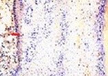
CCL27/CTACK in Human Skin.
CCL27/CTACK was detected in immersion fixed paraffin-embedded sections of human skin using Mouse Anti-Human CCL27/CTACK Monoclonal Antibody (Catalog # MAB376) at 25 µg/mL overnight at 4 °C. Tissue was stained using the Anti-Mouse HRP-DAB Cell & Tissue Staining Kit (brown; Catalog # CTS002) and counterstained with hematoxylin (blue). Specific staining was localized to cytoplasm in keratinocytes. View our protocol for Chromogenic IHC Staining of Paraffin-embedded Tissue Sections.Applications for Human CCL27/CTACK Antibody
Application
Recommended Usage
Immunohistochemistry
8-25 µg/mL
Sample: Immersion fixed paraffin-embedded sections of human skin
Sample: Immersion fixed paraffin-embedded sections of human skin
Western Blot
1 µg/mL
Sample: Recombinant Human CCL27/CTACK (Catalog # 376-CT)
Sample: Recombinant Human CCL27/CTACK (Catalog # 376-CT)
Neutralization
Measured by its ability to neutralize CCL27/CTACK-induced chemotaxis in the BaF3 mouse pro‑B cell line transfected with human CCR10. The Neutralization Dose (ND50) is typically 0.5-3.0 µg/mL in the presence of 1 µg/mL Recombinant Human CCL27/CTACK.
Reviewed Applications
Read 3 reviews rated 5 using MAB376 in the following applications:
Formulation, Preparation, and Storage
Purification
Protein A or G purified from hybridoma culture supernatant
Reconstitution
Reconstitute at 0.5 mg/mL in sterile PBS. For liquid material, refer to CoA for concentration.
Loading...
Formulation
Lyophilized from a 0.2 μm filtered solution in PBS with Trehalose. *Small pack size (SP) is supplied either lyophilized or as a 0.2 µm filtered solution in PBS.
Shipping
Lyophilized product is shipped at ambient temperature. Liquid small pack size (-SP) is shipped with polar packs. Upon receipt, store immediately at the temperature recommended below.
Stability & Storage
Use a manual defrost freezer and avoid repeated freeze-thaw cycles.
- 12 months from date of receipt, -20 to -70 °C as supplied.
- 1 month, 2 to 8 °C under sterile conditions after reconstitution.
- 6 months, -20 to -70 °C under sterile conditions after reconstitution.
Calculators
Background: CCL27/CTACK
References
- Kunkel, L. and E.C. Butcher (2002) Immunity 16:1.
- Ishikawa-Mochizuki, I. et al. (1999) FEBS Lett. 460:544.
- Gortz, A. et al. (2002) J. Immunol. 169:1387.
- Morales, J. et al. (1999) Proc. Natl. Acad. Sci. 96:14470.
- Homey, B. et al. (2000) J. Immunol. 164:3465.
- Homey, B. et al. (2002) Nat. Med. 8:157.
- Inokuma, D. et al. (2006) Stem Cells 24:2810.
- Jarmin, D. et al. (2000) J. Immunol. 164:3460.
- Hirata, T. et al. (2004) J. Biol. Chem. 279:51775.
- Vestergaard, C. et al. (2004) Exp. Dermatol. 13:551.
- Reiss, Y. et al. (2001) J. Exp. Med. 194:1541.
- Soler, D. et al. (2003) Blood 101:1677.
- Kakinuma, T. et al. (2003) J. Allergy Clin. Immunol. 111:592.
- Hijnen, D. et al. (2004) J. Allergy Clin. Immunol. 113:334.
- Fujita, Y. et al. (2006) Clin. Cancer Res. 12:2670.
Alternate Names
CTACK, ILC
Gene Symbol
CCL27
UniProt
Additional CCL27/CTACK Products
Product Documents for Human CCL27/CTACK Antibody
Certificate of Analysis
To download a Certificate of Analysis, please enter a lot or batch number in the search box below.
Note: Certificate of Analysis not available for kit components.
Product Specific Notices for Human CCL27/CTACK Antibody
For research use only
Related Research Areas
Citations for Human CCL27/CTACK Antibody
 Powered by Bioz
Customer Reviews for Human CCL27/CTACK Antibody (3)
5 out of 5
3 Customer Ratings
Have you used Human CCL27/CTACK Antibody?
Submit a review and receive an Amazon gift card!
$25/€18/£15/$25CAN/¥2500 Yen for a review with an image
$10/€7/£6/$10CAN/¥1110 Yen for a review without an image
Submit a review
Customer Images
Showing
1
-
3 of
3 reviews
Showing All
Filter By:
-
Application: ImmunohistochemistrySample Tested: Skin tissueSpecies: HumanVerified Customer | Posted 12/03/2021

-
Application: ELISASample Tested: SerumSpecies: HumanVerified Customer | Posted 09/05/2019
-
Application: ELISASample Tested: PlasmaSpecies: HumanVerified Customer | Posted 11/15/2017This antibody was used in a sandwich ELISA with biotinylated AF376 as detection at a concentration of 50ng/ml.
There are no reviews that match your criteria.
Protocols
Find general support by application which include: protocols, troubleshooting, illustrated assays, videos and webinars.
- Antigen Retrieval Protocol (PIER)
- Antigen Retrieval for Frozen Sections Protocol
- Appropriate Fixation of IHC/ICC Samples
- Cellular Response to Hypoxia Protocols
- Chromogenic IHC Staining of Formalin-Fixed Paraffin-Embedded (FFPE) Tissue Protocol
- Chromogenic Immunohistochemistry Staining of Frozen Tissue
- ClariTSA™ Fluorophore Kits
- Detection & Visualization of Antibody Binding
- Fluorescent IHC Staining of Frozen Tissue Protocol
- Graphic Protocol for Heat-induced Epitope Retrieval
- Graphic Protocol for the Preparation and Fluorescent IHC Staining of Frozen Tissue Sections
- Graphic Protocol for the Preparation and Fluorescent IHC Staining of Paraffin-embedded Tissue Sections
- Graphic Protocol for the Preparation of Gelatin-coated Slides for Histological Tissue Sections
- IHC Sample Preparation (Frozen sections vs Paraffin)
- Immunofluorescent IHC Staining of Formalin-Fixed Paraffin-Embedded (FFPE) Tissue Protocol
- Immunohistochemistry (IHC) and Immunocytochemistry (ICC) Protocols
- Immunohistochemistry Frozen Troubleshooting
- Immunohistochemistry Paraffin Troubleshooting
- Preparing Samples for IHC/ICC Experiments
- Preventing Non-Specific Staining (Non-Specific Binding)
- Primary Antibody Selection & Optimization
- Protocol for Heat-Induced Epitope Retrieval (HIER)
- Protocol for Making a 4% Formaldehyde Solution in PBS
- Protocol for VisUCyte™ HRP Polymer Detection Reagent
- Protocol for the Preparation & Fixation of Cells on Coverslips
- Protocol for the Preparation and Chromogenic IHC Staining of Frozen Tissue Sections
- Protocol for the Preparation and Chromogenic IHC Staining of Frozen Tissue Sections - Graphic
- Protocol for the Preparation and Chromogenic IHC Staining of Paraffin-embedded Tissue Sections
- Protocol for the Preparation and Chromogenic IHC Staining of Paraffin-embedded Tissue Sections - Graphic
- Protocol for the Preparation and Fluorescent IHC Staining of Frozen Tissue Sections
- Protocol for the Preparation and Fluorescent IHC Staining of Paraffin-embedded Tissue Sections
- Protocol for the Preparation of Gelatin-coated Slides for Histological Tissue Sections
- R&D Systems Quality Control Western Blot Protocol
- TUNEL and Active Caspase-3 Detection by IHC/ICC Protocol
- The Importance of IHC/ICC Controls
- Troubleshooting Guide: Immunohistochemistry
- Troubleshooting Guide: Western Blot Figures
- Western Blot Conditions
- Western Blot Protocol
- Western Blot Protocol for Cell Lysates
- Western Blot Troubleshooting
- Western Blot Troubleshooting Guide
- View all Protocols, Troubleshooting, Illustrated assays and Webinars
Loading...
Associated Pathways
