CD36, alternatively known as platelet membrane glycoprotein IV (GPIV), GPIIIb, thrombospondin receptor, collagen receptor, fatty acid translocase (FAT), and scavenger receptor class B, member 3 (SR-B3), is an integral membrane glycoprotein that has multiple physiological functions (1). It is broadly expressed on a variety of cell types including microvascular endothelium, adipocytes, skeletal muscle, epithelial cells of the retina, breast, and intestine, smooth muscle cells, erythroid precursors, platelets, megakaryocytes, dendritic cells, monocytes/macrophages, and microglia (1, 2). As a member of the scavenger receptor family, CD36 is a multiligand pattern recognition receptor that interacts with a large number of structurally dissimilar ligands, including long chain fatty acid (LCFA), advanced glycation end products (AGE), thrombospondin-1, oxidized low-density lipoproteins (oxLDLs), high density lipoprotein (HDL), phosphatidylserine, apoptotic cells,
beta ‑amyloid fibrils (fA beta ), collagens I and IV, and Plasmodium falciparum-infected erythrocytes (3). CD36 is required for the anti-angiogenic effects of thrombospondin-1 in the corneal neovascularization assay (4). It plays a role in lipid metabolism and has been identified as a fatty acid translocase necessary for the binding and transport of LCFA in cells and tissues (5). CD36 has been implicated in the clearance of apoptotic cells and cell debris and has also been shown to mediate the internalization and degradation of a variety of its ligands such as oxLDL, AGE and fA beta (3). Upon ligand binding, CD36 transduces signals that mediate a wide range of pro-inflammatory cellular responses (2). CD36 plays a significant role in the initiation and pathogenesis of chronic inflammatory diseases such as Alzheimer’s disease and atherosclerosis (2, 3). The human CD36 gene encodes a single-chain 472 amino acid protein containing both an N- and a C-terminal cytoplasmic tail and an extracellular loop.

Key Product Details
Species Reactivity
Human
Applications
Flow Cytometry, Immunocytochemistry, CyTOF-ready
Label
Unconjugated
Antibody Source
Monoclonal Rat IgG2B Clone # 255606
Loading...
Product Specifications
Immunogen
S. frugiperda insect ovarian cell line Sf 21-derived recombinant human CD36/SR‑B3
Gly30-Asn439
Accession # P16671
Gly30-Asn439
Accession # P16671
Specificity
Detects human CD36/SR‑B3 in direct ELISAs. In direct ELISAs, no cross-reactivity with recombinant mouse CD36 is observed.
Clonality
Monoclonal
Host
Rat
Isotype
IgG2B
Scientific Data Images for Human CD36/SR-B3 Antibody (255606)
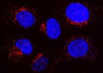
CD36/SR‑B3 in U937 Human Cell Line.
CD36/SR-B3 was detected in immersion fixed U937 human histiocytic lymphoma cell line using Rat Anti-Human CD36/SR-B3 Monoclonal Antibody (Catalog # MAB19551) at 8 µg/mL for 3 hours at room temperature. Cells were stained using the NorthernLights™ 557-conjugated Anti-Rat IgG Secondary Antibody (red; Catalog # NL013) and counterstained with DAPI (blue). Specific staining was localized to cytoplasm. View our protocol for Fluorescent ICC Staining of Non-adherent Cells.Applications for Human CD36/SR-B3 Antibody (255606)
Application
Recommended Usage
CyTOF-ready
Ready to be labeled using established conjugation methods. No BSA or other carrier proteins that could interfere with conjugation.
Flow Cytometry
0.25 µg/106 cells
Sample: HepG2 human hepatocellular carcinoma cell line
Sample: HepG2 human hepatocellular carcinoma cell line
Immunocytochemistry
3-25 µg/mL
Sample: Immersion fixed U937 human histiocytic lymphoma cell line
Sample: Immersion fixed U937 human histiocytic lymphoma cell line
Reviewed Applications
Read 2 reviews rated 4.5 using MAB19551 in the following applications:
Flow Cytometry Panel Builder
Bio-Techne Knows Flow Cytometry
Save time and reduce costly mistakes by quickly finding compatible reagents using the Panel Builder Tool.
Advanced Features
- Spectra Viewer - Custom analysis of spectra from multiple fluorochromes
- Spillover Popups - Visualize the spectra of individual fluorochromes
- Antigen Density Selector - Match fluorochrome brightness with antigen density

Formulation, Preparation, and Storage
Purification
Protein A or G purified from hybridoma culture supernatant
Reconstitution
Reconstitute at 0.5 mg/mL in sterile PBS. For liquid material, refer to CoA for concentration.
Loading...
Formulation
Lyophilized from a 0.2 μm filtered solution in PBS with Trehalose. *Small pack size (SP) is supplied either lyophilized or as a 0.2 µm filtered solution in PBS.
Shipping
Lyophilized product is shipped at ambient temperature. Liquid small pack size (-SP) is shipped with polar packs. Upon receipt, store immediately at the temperature recommended below.
Stability & Storage
Use a manual defrost freezer and avoid repeated freeze-thaw cycles.
- 12 months from date of receipt, -20 to -70 °C as supplied.
- 1 month, 2 to 8 °C under sterile conditions after reconstitution.
- 6 months, -20 to -70 °C under sterile conditions after reconstitution.
Calculators
Background: CD36/SR-B3
References
- Febbraio, M. et al. (2001) J. Clin. Invest. 108:785.
- Khoury, J. et al. (2003) J. Exp. Med. 197:1657.
- Husemann, J. et al. (2002) Glia 40:195.
- Armstrong, L and P. Bornstein (2003) Matrix. Biol. 22:63.
- Febbraio M. et al. (1999) J. Biol. Chem. 274:19055.
Long Name
Scavenger Receptor Class B, Member 3
Alternate Names
CD36, Collagen R, FAT, GPIIIb, GPIV, SCARB3, SR-B3, SRB3, Thrombospondin R
Gene Symbol
CD36
UniProt
Additional CD36/SR-B3 Products
Product Documents for Human CD36/SR-B3 Antibody (255606)
Certificate of Analysis
To download a Certificate of Analysis, please enter a lot or batch number in the search box below.
Note: Certificate of Analysis not available for kit components.
Product Specific Notices for Human CD36/SR-B3 Antibody (255606)
For research use only
Customer Reviews for Human CD36/SR-B3 Antibody (255606) (2)
4.5 out of 5
2 Customer Ratings
Have you used Human CD36/SR-B3 Antibody (255606)?
Submit a review and receive an Amazon gift card!
$25/€18/£15/$25CAN/¥2500 Yen for a review with an image
$10/€7/£6/$10CAN/¥1110 Yen for a review without an image
Submit a review
Customer Images
Showing
1
-
2 of
2 reviews
Showing All
Filter By:
-
Application: Immunocytochemistry/ImmunofluorescenceSample Tested: U937 Human Cell LineSpecies: HumanVerified Customer | Posted 03/03/2022

-
Application: Immunocytochemistry/ImmunofluorescenceSample Tested: Human taste cellsSpecies: HumanVerified Customer | Posted 05/16/2017
There are no reviews that match your criteria.
Protocols
Find general support by application which include: protocols, troubleshooting, illustrated assays, videos and webinars.
- 7-Amino Actinomycin D (7-AAD) Cell Viability Flow Cytometry Protocol
- Appropriate Fixation of IHC/ICC Samples
- Cellular Response to Hypoxia Protocols
- ClariTSA™ Fluorophore Kits
- Detection & Visualization of Antibody Binding
- Extracellular Membrane Flow Cytometry Protocol
- Flow Cytometry Protocol for Cell Surface Markers
- Flow Cytometry Protocol for Staining Membrane Associated Proteins
- Flow Cytometry Staining Protocols
- Flow Cytometry Troubleshooting Guide
- ICC Cell Smear Protocol for Suspension Cells
- ICC Immunocytochemistry Protocol Videos
- ICC for Adherent Cells
- Immunocytochemistry (ICC) Protocol
- Immunocytochemistry Troubleshooting
- Immunofluorescence of Organoids Embedded in Cultrex Basement Membrane Extract
- Immunohistochemistry (IHC) and Immunocytochemistry (ICC) Protocols
- Intracellular Flow Cytometry Protocol Using Alcohol (Methanol)
- Intracellular Flow Cytometry Protocol Using Detergents
- Intracellular Nuclear Staining Flow Cytometry Protocol Using Detergents
- Intracellular Staining Flow Cytometry Protocol Using Alcohol Permeabilization
- Intracellular Staining Flow Cytometry Protocol Using Detergents to Permeabilize Cells
- Preparing Samples for IHC/ICC Experiments
- Preventing Non-Specific Staining (Non-Specific Binding)
- Primary Antibody Selection & Optimization
- Propidium Iodide Cell Viability Flow Cytometry Protocol
- Protocol for Liperfluo
- Protocol for VisUCyte™ HRP Polymer Detection Reagent
- Protocol for the Characterization of Human Th22 Cells
- Protocol for the Characterization of Human Th9 Cells
- Protocol for the Fluorescent ICC Staining of Cell Smears - Graphic
- Protocol for the Fluorescent ICC Staining of Cultured Cells on Coverslips - Graphic
- Protocol for the Preparation and Fluorescent ICC Staining of Cells on Coverslips
- Protocol for the Preparation and Fluorescent ICC Staining of Non-adherent Cells
- Protocol for the Preparation and Fluorescent ICC Staining of Stem Cells on Coverslips
- Protocol for the Preparation of a Cell Smear for Non-adherent Cell ICC - Graphic
- Protocol: Annexin V and PI Staining by Flow Cytometry
- Protocol: Annexin V and PI Staining for Apoptosis by Flow Cytometry
- TUNEL and Active Caspase-3 Detection by IHC/ICC Protocol
- The Importance of IHC/ICC Controls
- Troubleshooting Guide: Fluorokine Flow Cytometry Kits
- View all Protocols, Troubleshooting, Illustrated assays and Webinars


