Human CFTR R Domain Antibody (13-1)
R&D Systems | Catalog # MAB1660

Loading...
Key Product Details
Species Reactivity
Validated:
Human
Cited:
Human, Mouse, Rat, Amphibian, Frog - Xenopus (African Clawed Frog), Hamster, Ovine
Applications
Validated:
Immunohistochemistry, Western Blot, Immunofluorescence, Immunoprecipitation
Cited:
Immunohistochemistry, Immunohistochemistry-Paraffin, Immunohistochemistry-Frozen, Western Blot, Immunocytochemistry, Immunoprecipitation
Label
Unconjugated
Antibody Source
Monoclonal Mouse IgG1 Clone # 13-1
Loading...
Product Specifications
Immunogen
beta -galactosidase-coupled CFTR
Cys590-Lys830 (predicted)
Accession # P13569
Cys590-Lys830 (predicted)
Accession # P13569
Specificity
Detects human CFTR. Specifically recognizes in vitro synthesized CFTR, recombinant CFTR protein, and non-recombinant CFTR protein (1, 2).
Clonality
Monoclonal
Host
Mouse
Isotype
IgG1
Scientific Data Images for Human CFTR R Domain Antibody (13-1)
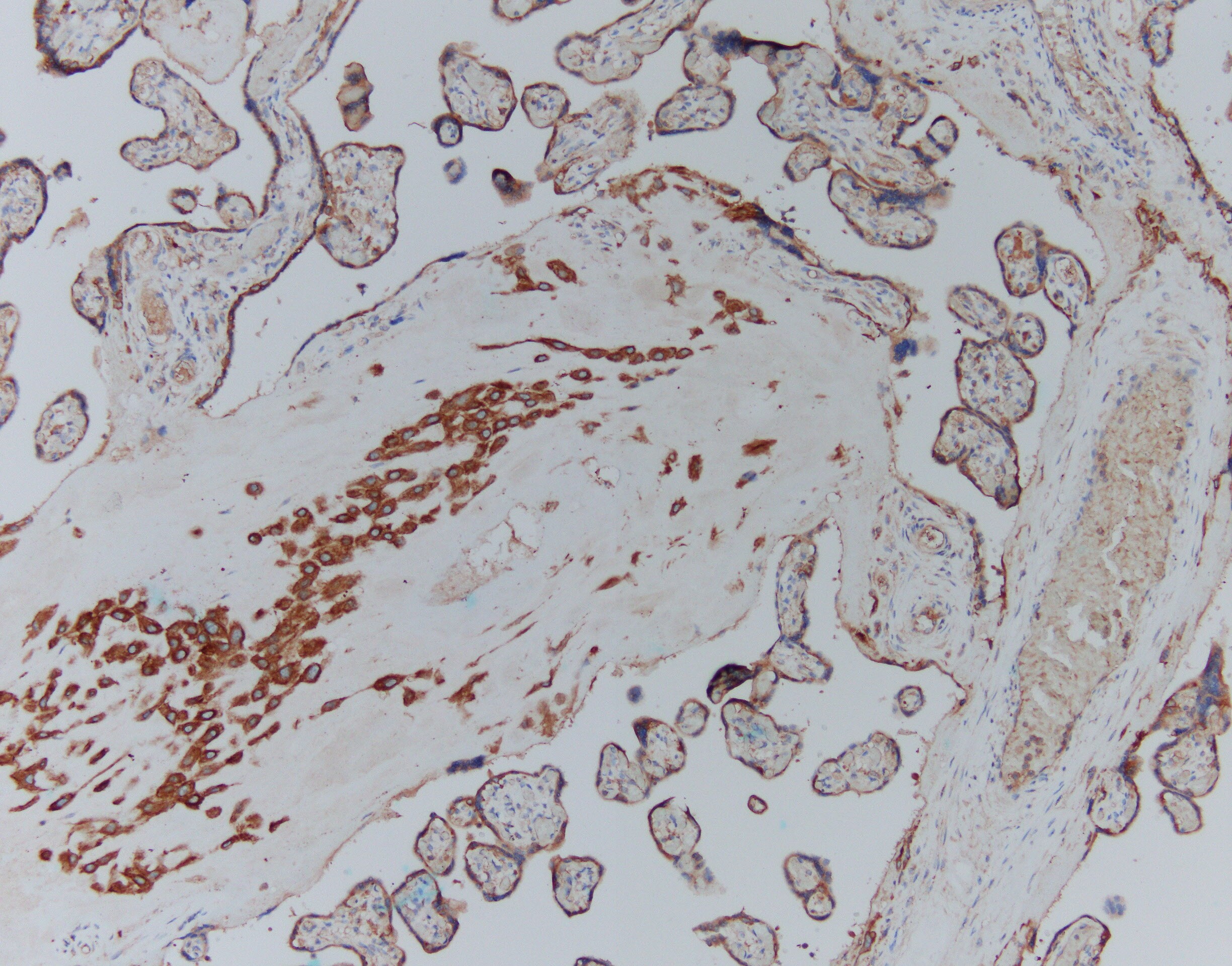
CFTR in Human Placenta.
CFTR was detected in immersion fixed paraffin-embedded sections of human placenta using Mouse Anti-Human CFTR R Domain Monoclonal Antibody (Catalog # MAB1660) at 15 µg/mL overnight at 4 °C. Before incubation with the primary antibody tissue was subjected to heat-induced epitope retrieval using Antigen Retrieval Reagent-Basic (Catalog # CTS013). Tissue was stained using the Anti-Mouse HRP-DAB Cell & Tissue Staining Kit (brown; Catalog # CTS002) and counterstained with hematoxylin (blue). Specific labeling was localized to the plasma membrane and cytoplasm of decidual cells. View our protocol for Chromogenic IHC Staining of Paraffin-embedded Tissue Sections.Applications for Human CFTR R Domain Antibody (13-1)
Application
Recommended Usage
Immunofluorescence
Cheng, S.H. et al. (1990) Cell 63:827.
Marino, C.R. et al. (1991) J. Clin. Invest. 88:712.
Immunohistochemistry
8-25 µg/mL
Sample: Immersion-fixed paraffin-embedded sections of human placenta
Sample: Immersion-fixed paraffin-embedded sections of human placenta
Immunoprecipitation
1-2 µg/106 cells
Sample: T84 human colon carcinoma cell line, see our available Western blot detection antibodies
Sample: T84 human colon carcinoma cell line, see our available Western blot detection antibodies
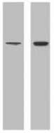
Western Blot
1 µg/mL
Sample: Human CFTR transfected cell line
Sample: Human CFTR transfected cell line
Reviewed Applications
Read 4 reviews rated 4.5 using MAB1660 in the following applications:
Formulation, Preparation, and Storage
Purification
Protein A or G purified from hybridoma culture supernatant
Reconstitution
For liquid material, refer to CoA for concentration.
Formulation
Supplied as a solution in PBS containing BSA.
*Small pack size (SP) is supplied either lyophilized or as a 0.2 µm filtered solution in PBS.
*Small pack size (SP) is supplied either lyophilized or as a 0.2 µm filtered solution in PBS.
Shipping
Lyophilized product is shipped at ambient temperature. Liquid small pack size (-SP) is shipped with polar packs. Upon receipt, store immediately at the temperature recommended below.
Stability & Storage
Use a manual defrost freezer and avoid repeated freeze-thaw cycles.
- 12 months from date of receipt, -20 to -70 °C, as supplied.
- 1 month, 2 to 8 °C under sterile conditions after opening.
- 6 months, -20 to -70 °C under sterile conditions after opening.
Calculators
Background: CFTR
References
- Gregory, R.J. et al. (1990) Nature 347:328.
- Cheng, S.H. et al. (1990) Cell 63:827.
Long Name
Cystic Fibrosis Transmembrane Conductance Regulator
Alternate Names
ABC35, ABCC7CF, ATP-binding cassette sub-family C member 7, ATP-binding cassette transporter sub-family C member 7, cAMP-dependent chloride channel, CFTR/MRP, Channel conductance-controlling ATPase, cystic fibrosis transmembrane conductance regulator, cystic fibrosis transmembrane conductance regulator (ATP-binding cassettesub-family C, member 7), cystic fibrosis transmembrane conductance regulator, ATP-binding cassette(sub-family C, member 7), dJ760C5.1, EC 3.6.3, MRP7EC 3.6.3.49, TNR-CFTR
Entrez Gene IDs
1080 (Human)
Gene Symbol
CFTR
UniProt
Additional CFTR Products
Product Documents for Human CFTR R Domain Antibody (13-1)
Certificate of Analysis
To download a Certificate of Analysis, please enter a lot or batch number in the search box below.
Note: Certificate of Analysis not available for kit components.
Product Specific Notices for Human CFTR R Domain Antibody (13-1)
For research use only
Related Research Areas
Citations for Human CFTR R Domain Antibody (13-1)
 Powered by Bioz
Customer Reviews for Human CFTR R Domain Antibody (13-1) (4)
4.5 out of 5
4 Customer Ratings
Have you used Human CFTR R Domain Antibody (13-1)?
Submit a review and receive an Amazon gift card!
$25/€18/£15/$25CAN/¥2500 Yen for a review with an image
$10/€7/£6/$10CAN/¥1110 Yen for a review without an image
Submit a review
Customer Images
Showing
1
-
4 of
4 reviews
Showing All
Filter By:
-
Application: ImmunohistochemistrySample Tested: Placental tissueSpecies: HumanVerified Customer | Posted 04/14/2025Heat pretreatment in basic buffer Blocking normal horse sera primary Ab MAB1660 x25, 30 min anti-mouse HRP, 30min & DAB

-
Application: Western BlotSample Tested: Cell LysatesSpecies: HumanVerified Customer | Posted 07/18/2021
-
Application: Western BlotSample Tested: See PMID 21811577Species: HumanVerified Customer | Posted 02/10/2015
-
Application: Western BlotSample Tested: See PMID 21952168Species: HumanVerified Customer | Posted 02/10/2015
There are no reviews that match your criteria.
Protocols
Find general support by application which include: protocols, troubleshooting, illustrated assays, videos and webinars.
- Antigen Retrieval Protocol (PIER)
- Antigen Retrieval for Frozen Sections Protocol
- Appropriate Fixation of IHC/ICC Samples
- Cellular Response to Hypoxia Protocols
- Chromogenic IHC Staining of Formalin-Fixed Paraffin-Embedded (FFPE) Tissue Protocol
- Chromogenic Immunohistochemistry Staining of Frozen Tissue
- ClariTSA™ Fluorophore Kits
- Detection & Visualization of Antibody Binding
- Fluorescent IHC Staining of Frozen Tissue Protocol
- Graphic Protocol for Heat-induced Epitope Retrieval
- Graphic Protocol for the Preparation and Fluorescent IHC Staining of Frozen Tissue Sections
- Graphic Protocol for the Preparation and Fluorescent IHC Staining of Paraffin-embedded Tissue Sections
- Graphic Protocol for the Preparation of Gelatin-coated Slides for Histological Tissue Sections
- ICC Cell Smear Protocol for Suspension Cells
- ICC Immunocytochemistry Protocol Videos
- ICC for Adherent Cells
- IHC Sample Preparation (Frozen sections vs Paraffin)
- Immunocytochemistry (ICC) Protocol
- Immunocytochemistry Troubleshooting
- Immunofluorescence of Organoids Embedded in Cultrex Basement Membrane Extract
- Immunofluorescent IHC Staining of Formalin-Fixed Paraffin-Embedded (FFPE) Tissue Protocol
- Immunohistochemistry (IHC) and Immunocytochemistry (ICC) Protocols
- Immunohistochemistry Frozen Troubleshooting
- Immunohistochemistry Paraffin Troubleshooting
- Immunoprecipitation Protocol
- Preparing Samples for IHC/ICC Experiments
- Preventing Non-Specific Staining (Non-Specific Binding)
- Primary Antibody Selection & Optimization
- Protocol for Heat-Induced Epitope Retrieval (HIER)
- Protocol for Making a 4% Formaldehyde Solution in PBS
- Protocol for VisUCyte™ HRP Polymer Detection Reagent
- Protocol for the Fluorescent ICC Staining of Cell Smears - Graphic
- Protocol for the Fluorescent ICC Staining of Cultured Cells on Coverslips - Graphic
- Protocol for the Preparation & Fixation of Cells on Coverslips
- Protocol for the Preparation and Chromogenic IHC Staining of Frozen Tissue Sections
- Protocol for the Preparation and Chromogenic IHC Staining of Frozen Tissue Sections - Graphic
- Protocol for the Preparation and Chromogenic IHC Staining of Paraffin-embedded Tissue Sections
- Protocol for the Preparation and Chromogenic IHC Staining of Paraffin-embedded Tissue Sections - Graphic
- Protocol for the Preparation and Fluorescent ICC Staining of Cells on Coverslips
- Protocol for the Preparation and Fluorescent ICC Staining of Non-adherent Cells
- Protocol for the Preparation and Fluorescent ICC Staining of Stem Cells on Coverslips
- Protocol for the Preparation and Fluorescent IHC Staining of Frozen Tissue Sections
- Protocol for the Preparation and Fluorescent IHC Staining of Paraffin-embedded Tissue Sections
- Protocol for the Preparation of Gelatin-coated Slides for Histological Tissue Sections
- Protocol for the Preparation of a Cell Smear for Non-adherent Cell ICC - Graphic
- R&D Systems Quality Control Western Blot Protocol
- TUNEL and Active Caspase-3 Detection by IHC/ICC Protocol
- The Importance of IHC/ICC Controls
- Troubleshooting Guide: Immunohistochemistry
- Troubleshooting Guide: Western Blot Figures
- Western Blot Conditions
- Western Blot Protocol
- Western Blot Protocol for Cell Lysates
- Western Blot Troubleshooting
- Western Blot Troubleshooting Guide
- View all Protocols, Troubleshooting, Illustrated assays and Webinars
Loading...
Associated Pathways
