Dectin-1, also known as CLEC7A and the beta -glucan receptor, is a 33 kDa type II transmembrane C-type lectin that participates in the innate immune response to fungal pathogens. Although Dectin-1 structurally resembles other CLEC molecules, it binds its ligands in a calcium-independent manner (1, 2). Mature human Dectin-1 consists of a short N-terminal ITAM-containing cytoplasmic tail, a transmembrane segment, and a C-terminal stalk with a carbohydrate recognition domain (CRD) in the extracellular domain (3, 4). Alternate splicing generates one major splice form that lacks the stalk region (3‑5). This isoform is expressed on the surface of monocytes, macrophages, myeloid DC, neutrophils, eosinophils, B cells, and CD4+ T cells (6). The CRD selectively binds beta -glucan polymers, a major component of yeast and mycobacterial cell walls (5‑7). Yeast beta -glucan is accessible to Dectin‑1 only during the process of cell budding. Dectin-1 does not recognize the filamentous form of yeast (8). Dectin-1 mediates the phagocytosis of zymosan particles and intact yeast (8‑10). In the membrane, Dectin-1 colocalizes with TLR2 in the presence of zymosan, and the two receptors cooperate in ligand recognition and the propagation of proinflammatory signaling (9, 11‑13). Dectin-1 also interacts with tetraspanin CD37. This increases its stability on the cell membrane and inhibits ligand-induced signaling (14). Dectin-1 knockout mice show increased susceptibility to pathogenic infection (15‑16). The CRD of human Dectin-1 shares 77%, 60%, and 60% amino acid (aa) sequence identity with that of bovine, mouse and rat Dectin-1, respectively. It shares 29%‑39% aa sequence identity with the CRD of other subgroup members, including CLEC-1, CLEC-2, CLEC9A, CLEC12B, LOX-1, and MICL.
Human Dectin‑1/CLEC7A Antibody
R&D Systems | Catalog # MAB1859
Clone 259931 was used by HLDA to establish CD designation

Key Product Details
Species Reactivity
Validated:
Human
Cited:
Human, Mouse
Applications
Validated:
Blockade of Receptor-ligand Interaction, Flow Cytometry, Immunocytochemistry, CyTOF-ready
Cited:
Immunohistochemistry-Paraffin, Western Blot, Neutralization, Flow Cytometry, Immunocytochemistry, Immunoprecipitation, Bioassay, Blocking, Functional Assay
Label
Unconjugated
Antibody Source
Monoclonal Mouse IgG2B Clone # 259931
Loading...
Product Specifications
Immunogen
Mouse myeloma cell line NS0-derived recombinant human Dectin‑1/CLEC7A
Thr66-Met201
Accession # NP_072092
Thr66-Met201
Accession # NP_072092
Specificity
Detects human Dectin‑1/CLEC7A in direct ELISAs. In directs ELISAs, less than 10% cross-reactivity with recombinant human (rh) DLEC and no cross-reactivity with recombinant mouse Dectin-1 or rhDectin-2 is observed.
Clonality
Monoclonal
Host
Mouse
Isotype
IgG2B
Endotoxin Level
<0.10 EU per 1 μg of the antibody by the LAL method.
Scientific Data Images for Human Dectin‑1/CLEC7A Antibody
Dectin‑1/CLEC7A in Human Dendritic Cells.
Dectin-1/CLEC7A was detected in immersion fixed mature human dendritic cells using Mouse Anti-Human Dectin-1/CLEC7A Monoclonal Antibody (Catalog # MAB1859) at 10 µg/mL for 3 hours at room temperature. Cells were stained using the NorthernLights™ 557-conjugated Anti-Mouse IgG Secondary Antibody (yellow; Catalog # NL007) and counterstained with DAPI (blue). View our protocol for Fluorescent ICC Staining of Non-adherent Cells.Applications for Human Dectin‑1/CLEC7A Antibody
Application
Recommended Usage
Blockade of Receptor-ligand Interaction
In a functional ELISA, 0.1-0.4 µg/mL of this antibody will block 50% of the binding of 75 ng/mL of biotinylated Laminarin to immobilized Recombinant Human Dectin-1/CLEC7A (Catalog # 1859-DC) coated at 250 ng/mL (100 µL/well). At 3 μg/mL, this antibody will block >90% of the binding.
CyTOF-ready
Ready to be labeled using established conjugation methods. No BSA or other carrier proteins that could interfere with conjugation.
Flow Cytometry
2.5 µg/106 cells
Sample: Human whole blood monocytes
Sample: Human whole blood monocytes
Immunocytochemistry
8-25 µg/mL
Sample: Immersion fixed human peripheral blood mononuclear cells and mature human dendritic cells
Sample: Immersion fixed human peripheral blood mononuclear cells and mature human dendritic cells
Reviewed Applications
Read 5 reviews rated 4.8 using MAB1859 in the following applications:
Flow Cytometry Panel Builder
Bio-Techne Knows Flow Cytometry
Save time and reduce costly mistakes by quickly finding compatible reagents using the Panel Builder Tool.
Advanced Features
- Spectra Viewer - Custom analysis of spectra from multiple fluorochromes
- Spillover Popups - Visualize the spectra of individual fluorochromes
- Antigen Density Selector - Match fluorochrome brightness with antigen density

Formulation, Preparation, and Storage
Purification
Protein A or G purified from hybridoma culture supernatant
Reconstitution
Reconstitute at 0.5 mg/mL in sterile PBS. For liquid material, refer to CoA for concentration.
Loading...
Formulation
Lyophilized from a 0.2 μm filtered solution in PBS with Trehalose. *Small pack size (SP) is supplied either lyophilized or as a 0.2 µm filtered solution in PBS.
Shipping
Lyophilized product is shipped at ambient temperature. Liquid small pack size (-SP) is shipped with polar packs. Upon receipt, store immediately at the temperature recommended below.
Stability & Storage
Use a manual defrost freezer and avoid repeated freeze-thaw cycles.
- 12 months from date of receipt, -20 to -70 °C as supplied.
- 1 month, 2 to 8 °C under sterile conditions after reconstitution.
- 6 months, -20 to -70 °C under sterile conditions after reconstitution.
Calculators
Background: Dectin-1/CLEC7A
References
- Kanazawa, N. (2007) J. Dermatol. Sci. 45:77.
- Brown, G.D. (2006) Nat. Rev. Immunol. 6:33.
- Hernanz-Falcon, P. et al. (2001) Immunogenetics 53:288.
- Yokota, K. et al. (2001) Gene 272:51.
- Willment, J.A. et al. (2001) J. Biol. Chem. 276:43818.
- Willment, J.A. et al. (2005) Eur. J. Immunol. 35:1539.
- Palma, A.S. et al. (2006) J. Biol. Chem. 281:5771.
- Gantner, B.N. et al. (2005) EMBO J. 24:1277.
- Gantner, B.N. et al. (2003) J. Exp. Med. 197:1107.
- Kennedy, A.D. et al. (2007) Eur. J. Immunol. 37:467.
- Brown, G.D. et al. (2003) J. Exp. Med. 197:1119.
- Yadav, M. and J.S. Schorey (2006) Blood 108:3168.
- Suram, S. et al. (2006) J. Biol. Chem. 281:5506.
- Meyer-Wentrup, F. et al. (2007) J. Immunol. 178:154.
- Saijo, S. et al. (2007) Nat. Immunol. 8:39.
- Taylor, P.R. et al. (2007) Nat. Immunol. 8:31.
Alternate Names
CD369, CLEC7A, CLECSF12, Dectin1
Gene Symbol
CLEC7A
UniProt
Additional Dectin-1/CLEC7A Products
Product Documents for Human Dectin‑1/CLEC7A Antibody
Certificate of Analysis
To download a Certificate of Analysis, please enter a lot or batch number in the search box below.
Note: Certificate of Analysis not available for kit components.
Product Specific Notices for Human Dectin‑1/CLEC7A Antibody
For research use only
Related Research Areas
Citations for Human Dectin‑1/CLEC7A Antibody
 Powered by Bioz
Customer Reviews for Human Dectin‑1/CLEC7A Antibody (5)
4.8 out of 5
5 Customer Ratings
Have you used Human Dectin‑1/CLEC7A Antibody?
Submit a review and receive an Amazon gift card!
$25/€18/£15/$25CAN/¥2500 Yen for a review with an image
$10/€7/£6/$10CAN/¥1110 Yen for a review without an image
Submit a review
Customer Images
Showing
1
-
5 of
5 reviews
Showing All
Filter By:
-
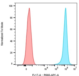
Application: Flow CytometryVerified Customer | Posted 01/16/2024Detection of human Dectin1 on HEK cells overexpressing isoform A of human Dectin1. The cells were treated with 100 nM of the human Dectin1 (catalogue # MAB1859) (BLUE) or mouse IgG2b isotype control antibody (RED) followed by treatments with a secondary goat anti-mouse IgG AF647-conjugated antibody

-
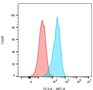
Application: Flow CytometrySample Tested: THP-1 human acute monocytic leukemia cell lineSpecies: HumanVerified Customer | Posted 11/28/2020Dectin1 is detected in THP1 monocytes using 100 nM of the Human Dectin-1/CLEC7A Antibody clone 259931. The 259931 antibody was detected with a secondary goat anti-mouse IgG Fc Alexa Fluor 647 antibody.
-
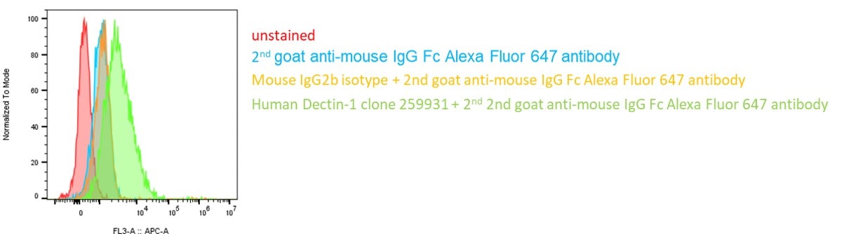
Application: Flow CytometrySample Tested: macrophagesSpecies: HumanVerified Customer | Posted 10/13/2020Detection of human Dectin1 in monocyte-derived cultured macrophages by Flow Cytometry. Human monocytes were cultured in MCSF for 6 days to differentiate into macrophages. After differentiation, the macrophages were stained with the mouse anti-human Dectin1 antibody clone 259931 at 100 nM or the mouse IgG2b isotype control. Binding was detected with a secondary goat a-mouse IgG Fc gamma fragment APC antibody.
-
Application: ImmunofluorescenceSample Tested: See PMID 23514738Species: OtherVerified Customer | Posted 02/12/2015
-
Application: ImmunofluorescenceSample Tested: See PMID 24424029Species: HumanVerified Customer | Posted 02/12/2015
There are no reviews that match your criteria.
Protocols
Find general support by application which include: protocols, troubleshooting, illustrated assays, videos and webinars.
- 7-Amino Actinomycin D (7-AAD) Cell Viability Flow Cytometry Protocol
- Appropriate Fixation of IHC/ICC Samples
- Cellular Response to Hypoxia Protocols
- ClariTSA™ Fluorophore Kits
- Detection & Visualization of Antibody Binding
- Extracellular Membrane Flow Cytometry Protocol
- Flow Cytometry Protocol for Cell Surface Markers
- Flow Cytometry Protocol for Staining Membrane Associated Proteins
- Flow Cytometry Staining Protocols
- Flow Cytometry Troubleshooting Guide
- ICC Cell Smear Protocol for Suspension Cells
- ICC Immunocytochemistry Protocol Videos
- ICC for Adherent Cells
- Immunocytochemistry (ICC) Protocol
- Immunocytochemistry Troubleshooting
- Immunofluorescence of Organoids Embedded in Cultrex Basement Membrane Extract
- Immunohistochemistry (IHC) and Immunocytochemistry (ICC) Protocols
- Intracellular Flow Cytometry Protocol Using Alcohol (Methanol)
- Intracellular Flow Cytometry Protocol Using Detergents
- Intracellular Nuclear Staining Flow Cytometry Protocol Using Detergents
- Intracellular Staining Flow Cytometry Protocol Using Alcohol Permeabilization
- Intracellular Staining Flow Cytometry Protocol Using Detergents to Permeabilize Cells
- Preparing Samples for IHC/ICC Experiments
- Preventing Non-Specific Staining (Non-Specific Binding)
- Primary Antibody Selection & Optimization
- Propidium Iodide Cell Viability Flow Cytometry Protocol
- Protocol for Liperfluo
- Protocol for VisUCyte™ HRP Polymer Detection Reagent
- Protocol for the Characterization of Human Th22 Cells
- Protocol for the Characterization of Human Th9 Cells
- Protocol for the Fluorescent ICC Staining of Cell Smears - Graphic
- Protocol for the Fluorescent ICC Staining of Cultured Cells on Coverslips - Graphic
- Protocol for the Preparation and Fluorescent ICC Staining of Cells on Coverslips
- Protocol for the Preparation and Fluorescent ICC Staining of Non-adherent Cells
- Protocol for the Preparation and Fluorescent ICC Staining of Stem Cells on Coverslips
- Protocol for the Preparation of a Cell Smear for Non-adherent Cell ICC - Graphic
- Protocol: Annexin V and PI Staining by Flow Cytometry
- Protocol: Annexin V and PI Staining for Apoptosis by Flow Cytometry
- TUNEL and Active Caspase-3 Detection by IHC/ICC Protocol
- The Importance of IHC/ICC Controls
- Troubleshooting Guide: Fluorokine Flow Cytometry Kits
- View all Protocols, Troubleshooting, Illustrated assays and Webinars
Loading...
Associated Pathways
