Human Glutathione S-Transferase pi 1/GSTP1 Antibody
Human Glutathione S-Transferase pi 1/GSTP1 Antibody Summary
Met1-Glu210
Accession # AAC51280
Applications
Please Note: Optimal dilutions should be determined by each laboratory for each application. General Protocols are available in the Technical Information section on our website.
Scientific Data
 View Larger
Detection of Human, Mouse, and Rat Glutathione S-Transferase pi 1/GSTP1 by Western Blot. Western blot shows lysates of human placenta tissue, mouse lung tissue, and rat lung tissue. PVDF membrane was probed with 0.25 µg/mL of Mouse Anti-Human Glutathione S-Transferase pi 1/GSTP1 Monoclonal Antibody (Catalog # MAB6455) followed by HRP-conjugated Anti-Mouse IgG Secondary Antibody (Catalog # HAF018). A specific band was detected for Glutathione S-Transferase pi 1/GSTP1 at approximately 25 kDa (as indicated). This experiment was conducted under reducing conditions and using Immunoblot Buffer Group 1.
 View Larger
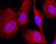
Glutathione S-Transferase pi 1/GSTP1 in HeLa Human Cell Line. Glutathione S-Transferase pi 1/GSTP1 was detected in immersion fixed HeLa human cervical epithelial carcinoma cell line using Mouse Anti-Human Glutathione S-Transferase pi 1/GSTP1 Monoclonal Antibody (Catalog # MAB6455) at 10 µg/mL for 3 hours at room temperature. Cells were stained using the NorthernLights™ 557-conjugated Anti-Mouse IgG Secondary Antibody (red; Catalog # NL007) and counterstained with DAPI (blue). Specific staining was localized to cytoplasm. View our protocol for Fluorescent ICC Staining of Cells on Coverslips.
 View Larger
Glutathione S-Transferase pi 1/GSTP1 in BG01V Human Embryonic Stem Cells. Glutathione S-Transferase pi 1/GSTP1 was detected in immersion fixed BG01V human embryonic stem cells differentiated into hepatocytes using Mouse Anti-Human Glutathione S-Transferase pi 1/GSTP1 Monoclonal Antibody (Catalog # MAB6455) at 10 µg/mL for 3 hours at room temperature. Cells were stained using the NorthernLights™ 557-conjugated Anti-Mouse IgG Secondary Antibody (red; Catalog # NL007) and counterstained with DAPI (blue). Specific staining was localized to cytoplasm. View our protocol for Fluorescent ICC Staining of Stem Cells on Coverslips.
 View Larger
Detection of Human and Rat Glutathione S-Transferase pi 1/GSTP1 by Simple WesternTM. Simple Western lane view shows lysates of human placenta tissue and rat brain tissue, loaded at 0.5 mg/mL. A specific band was detected for Glutathione S-Transferase pi 1/GSTP1 at approximately 26 kDa (as indicated) using 12.5 µg/mL of Mouse Anti-Human Glutathione S-Transferase pi 1/GSTP1 Monoclonal Antibody (Catalog # MAB6455). This experiment was conducted under reducing conditions and using the 12-230 kDa separation system.
Reconstitution Calculator
Preparation and Storage
- 12 months from date of receipt, -20 to -70 °C as supplied.
- 1 month, 2 to 8 °C under sterile conditions after reconstitution.
- 6 months, -20 to -70 °C under sterile conditions after reconstitution.
Background: Glutathione S-Transferase pi 1/GSTP1
Glutathione S-Transferases (GSTs) are members of the phase II detoxification enzyme family that conjugate glutathione to various electrophilic compounds, including metabolites generated by oxidative processes in the body, environmental toxins or carcinogens, and anti-cancer drugs. GSTP1 is a cytosolic protein that belongs to pi class of the GST superfamily. It is crystallized as a homodimer (1), but also exists in solution as an equilibrium mixture of monomer and dimer, depending on the protein concentration (2). Four genetic variants of GSTP1 with different enzymatic activities have been identified, which indicates the particular allelic form expressed in tissues could contribute to variation in catalytic efficiency and biological functions (3, 4). Human GSTP1 is present at elevated levels in many tumor cells, and has unique properties as a cancer marker (5). Genetic polymorphisms and expression patterns of GSTP1 have been associated with a variety of effects on human cancer, anti-cancer drug resistance, and asthma (6). In addition to its role as a drug-metabolizing enzyme, GSTP1 has ligand binding properties and regulates kinase signaling pathways through protein-protein interactions (7).
- Reinemer, P. et al. (1992) J. Mol. Biol. 227:214.
- Huang, Y.C. et al. (2008) J. Biol. Chem. 283:32880.
- Ali-Osman, F. et al. (1997) J. Biol. Chem. 272:10004.
- Hu, X. et al. (1998) Cancer Res. 58:5340.
- Sato, K. et al. (1992) Tohoku J. Exp. Med. 168:97.
- Townsend, D.M. and K.D. Tew (2003) Oncogene 22:7369.
- Adler, V. et al. (1999) EMBO J. 18:1321.
Product Datasheets
Citation for Human Glutathione S-Transferase pi 1/GSTP1 Antibody
R&D Systems personnel manually curate a database that contains references using R&D Systems products. The data collected includes not only links to publications in PubMed, but also provides information about sample types, species, and experimental conditions.
1 Citation: Showing 1 - 1
-
Mining the nucleus accumbens proteome for novel targets of alcohol self-administration in male C57BL/6J mice
Authors: S Faccidomo, KS Swaim, BL Saunders, TS Santanam, SM Taylor, M Kim, GT Reid, VR Eastman, CW Hodge
Psychopharmacology (Berl.), 2018-03-03;0(0):.
Species: Mouse
Sample Types: Tissue Homogenates
Applications: Western Blot
FAQs
No product specific FAQs exist for this product, however you may
View all Antibody FAQsReviews for Human Glutathione S-Transferase pi 1/GSTP1 Antibody
Average Rating: 5 (Based on 1 Review)
Have you used Human Glutathione S-Transferase pi 1/GSTP1 Antibody?
Submit a review and receive an Amazon gift card.
$25/€18/£15/$25CAN/¥75 Yuan/¥2500 Yen for a review with an image
$10/€7/£6/$10 CAD/¥70 Yuan/¥1110 Yen for a review without an image
Filter by:

