Legumain is a lysosomal cysteine protease whose activity is found in several tissues tested (1, 2). Legumain plays a pivotal role in the endosomal/lysosomal degradation system because the Legumain deficiency causes the accumulation of pro cathepsins B, H and L, another group of lysosomal cysteine proteases (3). Over-expression of Legumain in tumors is significant for invasion/metastasis (4). Also known as Asparaginyl Endopeptidase, it specifically cleaves peptide bonds with Asn at the P1 position. Nevertheless, it also cleaves peptide bonds with Asp at the P1 position. Auto-activation of pro Legumain involves both types of the cleavage, which result in the removal of the pro peptides in both C- and N-termini (5). In addition, Legumain activates pro MMP-2 and processes bacterial antigens for MHC class II presentation and pro thymosin alpha to thymosin alpha 1 and thymosin alpha 11, two acidic peptides with immunoregulatory properties (6‑8). Human Legumain is synthesized as a 433 amino acid precursor with a signal peptide (residues 1‑17). The pro enzyme (residues 18‑433) was expressed with an N-terminal His tag. This activity of Legumain can be inhibited by recombinant human (rh) Cystatin C and rhCystatin E/M and recombinant mouse Cystatin C (R&D Systems, Catalog # 1196‑PI, 1286-PI and 1238-PI, respectively).
Human Legumain/Asparaginyl Endopeptidase Antibody
R&D Systems | Catalog # AF2199

Key Product Details
Validated by
Knockout/Knockdown, Biological Validation
Species Reactivity
Validated:
Human
Cited:
Human, Mouse, Fish - Danio rerio (Zebrafish), Gerbil, Transgenic Mouse
Applications
Validated:
Western Blot, Immunoprecipitation
Cited:
Immunohistochemistry, Immunohistochemistry-Paraffin, Immunohistochemistry-Frozen, Western Blot, Immunocytochemistry, Immunoprecipitation, Chromatin Immunoprecipitation (ChIP)
Label
Unconjugated
Antibody Source
Polyclonal Goat IgG
Loading...
Product Specifications
Immunogen
Mouse myeloma cell line NS0-derived recombinant human Legumain/Asparaginyl Endopeptidase
Ile18-Tyr433
Accession # Q99538
Ile18-Tyr433
Accession # Q99538
Specificity
Detects human Legumain/Asparaginyl Endopeptidase in direct ELISAs and Western blots.
Clonality
Polyclonal
Host
Goat
Isotype
IgG
Scientific Data Images for Human Legumain/Asparaginyl Endopeptidase Antibody
Detection of Human Legumain/ Asparaginyl Endopeptidase by Western Blot.
Western blot shows lysates of human kidney tissue, human heart tissue, and human placenta tissue. PVDF membrane was probed with 1 µg/mL of Goat Anti-Human Legumain/Asparaginyl Endopeptidase Antigen Affinity-purified Polyclonal Antibody (Catalog # AF2199) followed by HRP-conjugated Anti-Goat IgG Secondary Antibody (Catalog # HAF109). Specific bands were detected for Pro-Legumain/ Asparaginyl Endopeptidase at approximately 56 kDa and mature Legumain/Asparaginyl Endopeptidase 37 kDa (as indicated). This experiment was conducted under reducing conditions and using Immunoblot Buffer Group 1.
Detection of Human Legumain/Asparaginyl Endopeptidase by Western Blot
The expression of AEP was higher in diffuse type gastric cancer than that in intestinal type gastric cancerA. AEP expression in intestinal type gastric cancer and diffuse type gastric cancer were detected by western blot (Representation: I, intestinal type gastric cancer; D, diffuse type gastric cancer, P=0.032). B. Patients' basic characteristics. Image collected and cropped by CiteAb from the following publication (https://www.oncotarget.com/lookup/doi/10.18632/oncotarget.8879), licensed under a CC-BY license. Not internally tested by R&D Systems.
Detection of Mouse Legumain/Asparaginyl Endopeptidase by Immunocytochemistry/Immunofluorescence
Representative sections showing overall higher expression of legumain (diffuse yellowish fluorescence staining) in the untreated (A) versus the Andosan™-treated (B) intestine.Notably, legumain expression was higher in tumor tissue seen in untreated animals. Scale bars represent 200 μm. Image collected and cropped by CiteAb from the following publication (https://dx.plos.org/10.1371/journal.pone.0167754), licensed under a CC-BY license. Not internally tested by R&D Systems.
Detection of Human Legumain/Asparaginyl Endopeptidase by Immunohistochemistry
AEP and E-cadherin expression were detected both in primary gastric cancer and peritoneal metastatic lociA. The expression of AEP and E-cadherin in primary gastric cancer and metastatic peritoneal loci were firstly investigated by immunohistochemistry. AEP expression were higher and E-cadherin expression was lower in peritoneal metastatic lesions than in primary gastric cancer (magnified 50× in column 1,3; magnified 200× in column 2,4). B. The expression of AEP and E-cadherin in primary gastric cancer and metastatic peritoneal loci were investigated by immunofluorescence assay (magnified 200×). AEP, the secondary antibody of which were labeled with green fluorescence, was more strongly expressed in peritoneal metastatic lesions than in primary gastric cancer. E-cadherin, the secondary antibody of which was labeled with red fluorescence, was expressed in primary gastric cancer and could not be identified in peritoneal metastatic loci. Image collected and cropped by CiteAb from the following publication (https://www.oncotarget.com/lookup/doi/10.18632/oncotarget.8879), licensed under a CC-BY license. Not internally tested by R&D Systems.
Detection of Mouse Legumain/Asparaginyl Endopeptidase by Western Blot
AEP knockdown and overexpressive vectors were constructed and stably transfected gastric cancer cell linesA. Constructing of the AEP knocked-down lentiviral vector, it was verified by western blot that AEP was inhibited corresponding to AEP knockdown. B. Overexpressing AEP lentiviral vector was constructed and AEP indeed increased corresponding to AEP overexpression. C. The proliferative curve in response to AEP overexpression or knockdown as evidenced by the CCK8 method and quantification of proliferative rate. *P<0.05, **P<0.01. (GFP-NC: control of empty vector; AEP-OE: AEP-overexpression; NC: negatively control; AEP-KD1: AEP-knocking down 1; AEP-KD2: AEP-knocking down 2) Image collected and cropped by CiteAb from the following publication (https://www.oncotarget.com/lookup/doi/10.18632/oncotarget.8879), licensed under a CC-BY license. Not internally tested by R&D Systems.
Detection of Human Legumain/Asparaginyl Endopeptidase by Immunocytochemistry/Immunofluorescence
AEP and E-cadherin expression were detected both in primary gastric cancer and peritoneal metastatic lociA. The expression of AEP and E-cadherin in primary gastric cancer and metastatic peritoneal loci were firstly investigated by immunohistochemistry. AEP expression were higher and E-cadherin expression was lower in peritoneal metastatic lesions than in primary gastric cancer (magnified 50× in column 1,3; magnified 200× in column 2,4). B. The expression of AEP and E-cadherin in primary gastric cancer and metastatic peritoneal loci were investigated by immunofluorescence assay (magnified 200×). AEP, the secondary antibody of which were labeled with green fluorescence, was more strongly expressed in peritoneal metastatic lesions than in primary gastric cancer. E-cadherin, the secondary antibody of which was labeled with red fluorescence, was expressed in primary gastric cancer and could not be identified in peritoneal metastatic loci. Image collected and cropped by CiteAb from the following publication (https://www.oncotarget.com/lookup/doi/10.18632/oncotarget.8879), licensed under a CC-BY license. Not internally tested by R&D Systems.
Detection of Mouse Legumain/Asparaginyl Endopeptidase by Immunocytochemistry/Immunofluorescence
Representative sections showing overall higher expression of legumain (diffuse yellowish fluorescence staining) in the untreated (A) versus the Andosan™-treated (B) intestine.Notably, legumain expression was higher in tumor tissue seen in untreated animals. Scale bars represent 200 μm. Image collected and cropped by CiteAb from the following publication (https://dx.plos.org/10.1371/journal.pone.0167754), licensed under a CC-BY license. Not internally tested by R&D Systems.
Detection of Mouse Legumain/Asparaginyl Endopeptidase by Western Blot
AEP knockdown and overexpressive vectors were constructed and stably transfected gastric cancer cell linesA. Constructing of the AEP knocked-down lentiviral vector, it was verified by western blot that AEP was inhibited corresponding to AEP knockdown. B. Overexpressing AEP lentiviral vector was constructed and AEP indeed increased corresponding to AEP overexpression. C. The proliferative curve in response to AEP overexpression or knockdown as evidenced by the CCK8 method and quantification of proliferative rate. *P<0.05, **P<0.01. (GFP-NC: control of empty vector; AEP-OE: AEP-overexpression; NC: negatively control; AEP-KD1: AEP-knocking down 1; AEP-KD2: AEP-knocking down 2) Image collected and cropped by CiteAb from the following publication (https://www.oncotarget.com/lookup/doi/10.18632/oncotarget.8879), licensed under a CC-BY license. Not internally tested by R&D Systems.
Detection of Human Legumain/Asparaginyl Endopeptidase by Western Blot
Legumain, cathepsin B and L expressions in cytosolic and membrane fractions of myotubes.Myotubes were treated with (+) or without (−) 30 µM simvastatin for 48 h before subcellular fractionation was performed. One representative immunoblot of legumain, cathepsin B and L in the cytosolic and membrane fractions is shown. All lanes were loaded with equal amount of total proteins and probed with antibodies as indicated. LAMP-2 and alpha -tubulin are shown as cell compartment controls (n = 3). Image collected and cropped by CiteAb from the following publication (https://pubmed.ncbi.nlm.nih.gov/24416446), licensed under a CC-BY license. Not internally tested by R&D Systems.
Detection of Mouse Human Legumain/Asparaginyl Endopeptidase Antibody by Immunohistochemistry
Representative sections showing overall higher expression of legumain (diffuse yellowish fluorescence staining) in the untreated (A) versus the Andosan™-treated (B) intestine.Notably, legumain expression was higher in tumor tissue seen in untreated animals. Scale bars represent 200 μm. Image collected and cropped by CiteAb from the following publication (https://pubmed.ncbi.nlm.nih.gov/28002446), licensed under a CC-BY license. Not internally tested by R&D Systems.Applications for Human Legumain/Asparaginyl Endopeptidase Antibody
Application
Recommended Usage
Immunoprecipitation
25 µg/mL
Sample: Conditioned cell culture medium spiked with Recombinant Human Legumain (Catalog # 2199‑CY), see our available Western blot detection antibodies
Sample: Conditioned cell culture medium spiked with Recombinant Human Legumain (Catalog # 2199‑CY), see our available Western blot detection antibodies
Western Blot
1 µg/mL
Sample: Human kidney tissue, human heart tissue, and human placenta tissue
Sample: Human kidney tissue, human heart tissue, and human placenta tissue
Reviewed Applications
Read 2 reviews rated 4.5 using AF2199 in the following applications:
Formulation, Preparation, and Storage
Purification
Antigen Affinity-purified
Reconstitution
Reconstitute at 0.2 mg/mL in sterile PBS. For liquid material, refer to CoA for concentration.
Loading...
Formulation
Lyophilized from a 0.2 μm filtered solution in PBS with Trehalose. *Small pack size (SP) is supplied either lyophilized or as a 0.2 µm filtered solution in PBS.
Shipping
Lyophilized product is shipped at ambient temperature. Liquid small pack size (-SP) is shipped with polar packs. Upon receipt, store immediately at the temperature recommended below.
Stability & Storage
Use a manual defrost freezer and avoid repeated freeze-thaw cycles.
- 12 months from date of receipt, -20 to -70 °C as supplied.
- 1 month, 2 to 8 °C under sterile conditions after reconstitution.
- 6 months, -20 to -70 °C under sterile conditions after reconstitution.
Calculators
Background: Legumain/Asparaginyl Endopeptidase
References
- Chen, J.-M. et al. (1997) J. Biol. Chem. 272:8090.
- Tanaka, T. et al. (1996) Cytogenet. Cell Genet. 74:120.
- Shirahama-Noda, K. et al. (2003) J. Biol. Chem. 278:33194.
- Liu, C. et al. (2003) Cancer Res. 63: 2957.
- Li D.N. et al. (2003) J. Biol. Chem. 278:38980.
- Chen, J.M. et al. (2001) Biol. Chem. 382:777.
- Schwarz, G. et al. (2002) Biol. Chem. 383:1813.
- Sarndeses, C.S. et al. (2003) J. Biol. Chem. 278:13286.
Alternate Names
AEP, Asparaginyl Endopeptidase, LGMN
Gene Symbol
LGMN
UniProt
Additional Legumain/Asparaginyl Endopeptidase Products
Product Documents for Human Legumain/Asparaginyl Endopeptidase Antibody
Certificate of Analysis
To download a Certificate of Analysis, please enter a lot or batch number in the search box below.
Note: Certificate of Analysis not available for kit components.
Product Specific Notices for Human Legumain/Asparaginyl Endopeptidase Antibody
For research use only
Related Research Areas
Citations for Human Legumain/Asparaginyl Endopeptidase Antibody
 Powered by Bioz
Customer Reviews for Human Legumain/Asparaginyl Endopeptidase Antibody (2)
4.5 out of 5
2 Customer Ratings
Have you used Human Legumain/Asparaginyl Endopeptidase Antibody?
Submit a review and receive an Amazon gift card!
$25/€18/£15/$25CAN/¥2500 Yen for a review with an image
$10/€7/£6/$10CAN/¥1110 Yen for a review without an image
Submit a review
Customer Images
Showing
1
-
2 of
2 reviews
Showing All
Filter By:
-
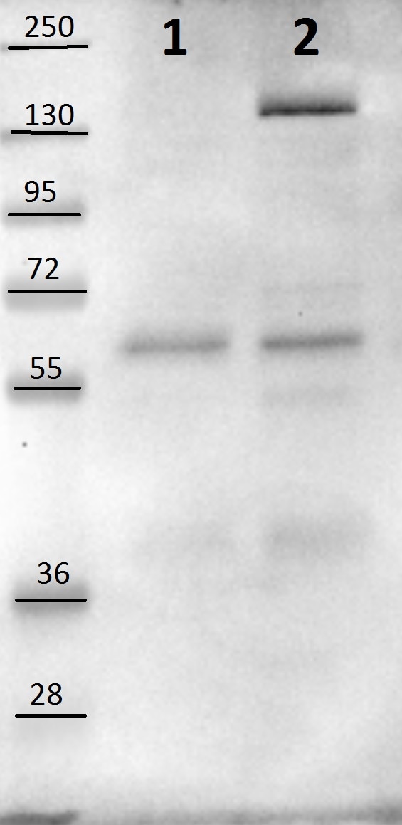
Application: Western BlotSample Tested: HeLa and HEK293 whole cell lysateSpecies: HumanVerified Customer | Posted 12/23/2015Lane 1: HeLa cell lysate; Lane 2: HEK cell lysate

-
Application: Western BlotSample Tested: See PMID 22902879Species: HumanVerified Customer | Posted 01/06/2015
There are no reviews that match your criteria.
Protocols
Find general support by application which include: protocols, troubleshooting, illustrated assays, videos and webinars.
- Cellular Response to Hypoxia Protocols
- Immunoprecipitation Protocol
- R&D Systems Quality Control Western Blot Protocol
- Troubleshooting Guide: Western Blot Figures
- Western Blot Conditions
- Western Blot Protocol
- Western Blot Protocol for Cell Lysates
- Western Blot Troubleshooting
- Western Blot Troubleshooting Guide
- View all Protocols, Troubleshooting, Illustrated assays and Webinars
Loading...