
Lymphotoxin beta Receptor (LT beta R), also known as TNF RIII and TNF R-Related Protein (TNF Rrp) is a member of the TNF receptor superfamily, designated TNFRSF3. Human LT beta R cDNA encodes a 435 amino acid (aa) residue type I membrane protein with a putative 30 aa residue signal peptide, a 193 aa residue extracellular domain and a 171 aa residue cytoplasmic domain. The extracellular domain of LT beta R contains four cysteine-rich motifs characteristic of the TNF receptor superfamily. The cytoplasmic region of LT beta R shares little sequence similarity with other TNF receptor family members, suggesting that different signaling mechanisms may be used. LT beta R is expressed in a variety of tissues including visceral and lymphoid tissues. LT beta R is also expressed by cell lines of monocytic, epithelial, and fibroblastic origins but not by T and B lymphocytes. Human and mouse LT beta R share 76% aa sequence homology. The TNF family ligands that have been shown to bind and activate LT beta R include LIGHT (also a ligand for HVEM) and the heterotrimeric Lymphotoxin LT alpha 1/ beta 2 or LT alpha 2/ beta 1. Depending on the cell type, activation of LT beta R has been shown to induce NF kappa B activation, chemokine production, growth arrest, and apoptosis. In vivo, LT beta R has been shown to play a critical role in controlling cellular immune functions and lymphoid organogenesis.
Human Lymphotoxin beta R/TNFRSF3 Antibody (71319) [PE]
R&D Systems | Catalog # FAB629P

Scientific images may be resized, cropped, or adjusted for brightness or contrast for presentation purposes. These updates are limited to presentation and do not affect the underlying scientific interpretation of the data. Where available, additional source data may be provided upon request.
Key Product Details
Validated by
Species Reactivity
Validated:
Cited:
Applications
Validated:
Cited:
Label
Antibody Source
Product Specifications
Immunogen
Gln31-Met227
Accession # P36941
Specificity
Clonality
Host
Isotype
Scientific Data Images for Human Lymphotoxin beta R/TNFRSF3 Antibody (71319) [PE]
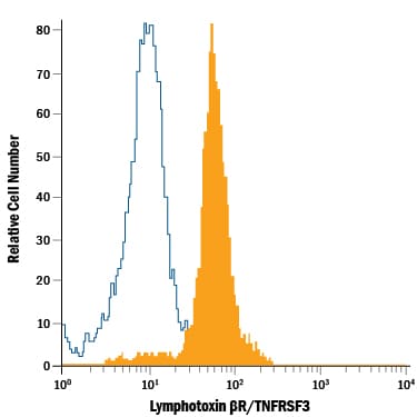
Detection of Lymphotoxin beta R/TNFRSF3 in Human Blood Monocytes by Flow Cytometry.
Human peripheral blood monocytes were stained with Mouse Anti-Human Lymphotoxin beta R/TNFRSF3 PE-conjugated Monoclonal Antibody (Catalog # FAB629P, filled histogram) or isotype control antibody (Catalog # IC002P, open histogram). View our protocol for Staining Membrane-associated Proteins.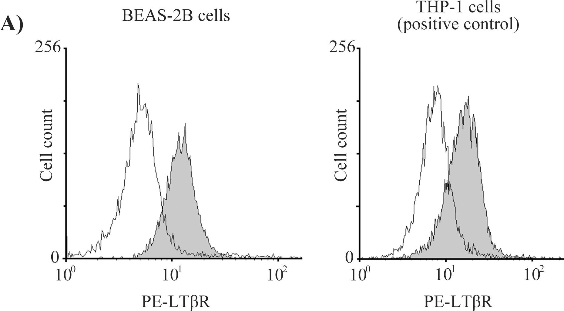
Detection of Human Lymphotoxin beta R/TNFRSF3 by Flow Cytometry
Expression of LT beta R and HVEM receptors on BEAS-2B cells.BEAS-2B cells and THP-1 cells (positive control) were treated with anti-PE labeled LT beta R antibody and anti-FITC labeled HVEM antibody. The fluorescence intensity was measured with a flow cytometer. THP-1 cells, a human monocytic cell line, were used as the positive control because they express both LT beta R and HVEM on their cell surface. The fluorescence intensities of LT beta R (A) and HVEM (B) suggest that BEAS-2B cells express LT beta R but not HVEM. Image collected and cropped by CiteAb from the following publication (https://pubmed.ncbi.nlm.nih.gov/25501580), licensed under a CC-BY license. Not internally tested by R&D Systems.Applications for Human Lymphotoxin beta R/TNFRSF3 Antibody (71319) [PE]
Flow Cytometry
Sample: Human peripheral blood monocytes
Spectra Viewer
Plan Your Experiments
Use our spectra viewer to interactively plan your experiments, assessing multiplexing options. View the excitation and emission spectra for our fluorescent dye range and other commonly used dyes.
Spectra Viewer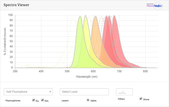
Flow Cytometry Panel Builder
Bio-Techne Knows Flow Cytometry
Save time and reduce costly mistakes by quickly finding compatible reagents using the Panel Builder Tool.
Advanced Features
- Spectra Viewer - Custom analysis of spectra from multiple fluorochromes
- Spillover Popups - Visualize the spectra of individual fluorochromes
- Antigen Density Selector - Match fluorochrome brightness with antigen density

Formulation, Preparation, and Storage
Purification
Formulation
Shipping
Stability & Storage
- 12 months from date of receipt, 2 to 8 °C as supplied.
Background: Lymphotoxin beta R/TNFRSF3
References
- Zhai, Y. et al. (1998) J. Clin. Invest. 102:1142.
- Rennert, P.D. et al. (1998) Immunity 9:71.
- Degli-Esposti, M.A. et al. (1997) J. Immunol 158:1756.
- Mackay, F. et al. (1996) J. Biol. Chem. 271:8618.
- Crowe, P.D. et al. (1994) Science 264:707.
Long Name
Alternate Names
Entrez Gene IDs
Gene Symbol
UniProt
Additional Lymphotoxin beta R/TNFRSF3 Products
Product Documents for Human Lymphotoxin beta R/TNFRSF3 Antibody (71319) [PE]
Certificate of Analysis
To download a Certificate of Analysis, please enter a lot or batch number in the search box below.
Note: Certificate of Analysis not available for kit components.
Product Specific Notices for Human Lymphotoxin beta R/TNFRSF3 Antibody (71319) [PE]
For research use only
Citations for Human Lymphotoxin beta R/TNFRSF3 Antibody (71319) [PE]
 Powered by Bioz
Customer Reviews for Human Lymphotoxin beta R/TNFRSF3 Antibody (71319) [PE]
There are currently no reviews for this product. Be the first to review Human Lymphotoxin beta R/TNFRSF3 Antibody (71319) [PE] and earn rewards!
Have you used Human Lymphotoxin beta R/TNFRSF3 Antibody (71319) [PE]?
Submit a review and receive an Amazon gift card!
$25/€18/£15/$25CAN/¥2500 for a review with an image
$10/€7/£6/$10CAN/¥1110 for a review without an image
Submit a review
Protocols
Find general support by application which include: protocols, troubleshooting, illustrated assays, videos and webinars.
- 7-Amino Actinomycin D (7-AAD) Cell Viability Flow Cytometry Protocol
- Extracellular Membrane Flow Cytometry Protocol
- Flow Cytometry Protocol for Cell Surface Markers
- Flow Cytometry Protocol for Staining Membrane Associated Proteins
- Flow Cytometry Staining Protocols
- Flow Cytometry Troubleshooting Guide
- Intracellular Flow Cytometry Protocol Using Alcohol (Methanol)
- Intracellular Flow Cytometry Protocol Using Detergents
- Intracellular Nuclear Staining Flow Cytometry Protocol Using Detergents
- Intracellular Staining Flow Cytometry Protocol Using Alcohol Permeabilization
- Intracellular Staining Flow Cytometry Protocol Using Detergents to Permeabilize Cells
- Propidium Iodide Cell Viability Flow Cytometry Protocol
- Protocol for Liperfluo
- Protocol for the Characterization of Human Th22 Cells
- Protocol for the Characterization of Human Th9 Cells
- Protocol: Annexin V and PI Staining by Flow Cytometry
- Protocol: Annexin V and PI Staining for Apoptosis by Flow Cytometry
- Troubleshooting Guide: Fluorokine Flow Cytometry Kits
- View all Protocols, Troubleshooting, Illustrated assays and Webinars
Associated Pathways
