
Discontinued Product
MAB20892 has been discontinued.
View all LYVE-1 products.
Key Product Details
Species Reactivity
Validated:
Human
Cited:
Human
Applications
Validated:
Western Blot, Flow Cytometry, CyTOF-ready
Cited:
Immunohistochemistry, Neutralization Control
Label
Unconjugated
Antibody Source
Monoclonal Mouse IgG1 Clone # 537028
Loading...
Product Specifications
Immunogen
Mouse myeloma cell line NS0-derived recombinant human LYVE-1
Ser24-Thr238
Accession # Q9Y5Y7
Ser24-Thr238
Accession # Q9Y5Y7
Specificity
Detects endogenous human LYVE-1 in Western blots.
Clonality
Monoclonal
Host
Mouse
Isotype
IgG1
Scientific Data Images for Human LYVE-1 Antibody (537028)
Detection of Human LYVE‑1 by Western Blot.
Western blot shows lysates of HeLa human cervical epithelial carcinoma cell line, MCF-7 human breast cancer cell line, and 293T human embryonic kidney cell line. PVDF membrane was probed with 1 µg/mL of Mouse Anti-Human LYVE-1 Monoclonal Antibody (Catalog # MAB20892) followed by HRP-conjugated Anti-Mouse IgG Secondary Antibody (Catalog # HAF007). A specific band was detected for LYVE-1 at approximately 70 kDa (as indicated). This experiment was conducted under reducing conditions and using Immunoblot Buffer Group 1.
Detection of LYVE-1 in HUVEC Human Cells by Flow Cytometry.
HUVEC human umbilical vein endothelial cells was stained with Mouse Anti-Human LYVE-1 Monoclonal Antibody (Catalog # MAB20892, filled histogram) or isotype control antibody (Catalog # MAB002, open histogram), followed by Allophycocyanin-conjugated Anti-Mouse IgG Secondary Antibody (Catalog # F0101B).
Detection of LYVE-1 in Human PBMC by Flow Cytometry.
Human PBMC were cultured with 50 ng/ml Recombinant Human M-CSF (Catalog # 216-MC) for 10 days (see Volk-Draper et al (2017) PLoS One 12(6): e0179257), then stained with either (A) Mouse Anti-Human LYVE-1 Monoclonal Antibody (Catalog # MAB20892) or (B) isotype control antibody (Catalog # MAB002), followed by Allophycocyanin-conjugated Anti-Mouse IgG Secondary Antibody (Catalog # F0101B) and Mouse anti-Human CD14 PE-conjugated Monoclonal Antibody (Catalog # FAB3832P). View our protocol for Staining Membrane-associated Proteins.Applications for Human LYVE-1 Antibody (537028)
Application
Recommended Usage
CyTOF-ready
Ready to be labeled using established conjugation methods. No BSA or other carrier proteins that could interfere with conjugation.
Flow Cytometry
0.25 µg/106 cells
Sample: HUVEC human umbilical vein endothelial cells, and M-CSF-treated Human PBMC
Sample: HUVEC human umbilical vein endothelial cells, and M-CSF-treated Human PBMC
Western Blot
1 µg/mL
Sample: HeLa human cervical epithelial carcinoma cell line, MCF-7 human breast cancer cell line, and 293T human embryonic kidney cell line
Sample: HeLa human cervical epithelial carcinoma cell line, MCF-7 human breast cancer cell line, and 293T human embryonic kidney cell line
Reviewed Applications
Read 5 reviews rated 3.8 using MAB20892 in the following applications:
Flow Cytometry Panel Builder
Bio-Techne Knows Flow Cytometry
Save time and reduce costly mistakes by quickly finding compatible reagents using the Panel Builder Tool.
Advanced Features
- Spectra Viewer - Custom analysis of spectra from multiple fluorochromes
- Spillover Popups - Visualize the spectra of individual fluorochromes
- Antigen Density Selector - Match fluorochrome brightness with antigen density

Formulation, Preparation, and Storage
Purification
Protein A or G purified from hybridoma culture supernatant
Reconstitution
Reconstitute at 0.5 mg/mL in sterile PBS. For liquid material, refer to CoA for concentration.
Formulation
Lyophilized from a 0.2 μm filtered solution in PBS with Trehalose. *Small pack size (SP) is supplied either lyophilized or as a 0.2 µm filtered solution in PBS.
Shipping
Lyophilized product is shipped at ambient temperature. Liquid small pack size (-SP) is shipped with polar packs. Upon receipt, store immediately at the temperature recommended below.
Stability & Storage
Use a manual defrost freezer and avoid repeated freeze-thaw cycles.
- 12 months from date of receipt, -20 to -70 °C as supplied.
- 1 month, 2 to 8 °C under sterile conditions after reconstitution.
- 6 months, -20 to -70 °C under sterile conditions after reconstitution.
Calculators
Background: LYVE-1
References
- Knudson, C.B. and W. Knudson (1993) FASEB J. 7:1233.
- Evered, D and J. Whelan (1989) Ciba Found. Symp. 143:1.
- Laurent, T.C. and J.R.F. Fraser (1992) FASEB J. 6:2397.
- Banerji, S. et al. (1999) J. Cell Biol. 144:789.
- Prevo, R. et al. (2001) J. Biol. Chem. 276:19420.
- Jackson, D.J. et al. (2001)Trends Immunol. 22:317.
- Zhou, B. et al. (2000) J. Biol. Chem. 275:37733.
- Achen, M. et al. (1998) Proc. Natl. Acad. Sci. USA 95:548.
- Breiteneder-Gellef, S. et al. (1999) Am. J. Pathol. 154:385.
- Wiggle, J.T. and G. Oliver (1999) Cell 98:769.
Long Name
Lymphatic Vessel Endothelial Hyaluronan Receptor 1
Alternate Names
LYVE1, XLKD1
Gene Symbol
LYVE1
UniProt
Additional LYVE-1 Products
Product Documents for Human LYVE-1 Antibody (537028)
Certificate of Analysis
To download a Certificate of Analysis, please enter a lot or batch number in the search box below.
Note: Certificate of Analysis not available for kit components.
Product Specific Notices for Human LYVE-1 Antibody (537028)
For research use only
Related Research Areas
Citations for Human LYVE-1 Antibody (537028)
 Powered by Bioz
Customer Reviews for Human LYVE-1 Antibody (537028) (5)
3.8 out of 5
5 Customer Ratings
Have you used Human LYVE-1 Antibody (537028)?
Submit a review and receive an Amazon gift card!
$25/€18/£15/$25CAN/¥2500 Yen for a review with an image
$10/€7/£6/$10CAN/¥1110 Yen for a review without an image
Submit a review
Customer Images
Showing
1
-
5 of
5 reviews
Showing All
Filter By:
-
Application: MicroarraysSample Tested: EDTA PlasmaSpecies: HumanVerified Customer | Posted 01/14/2021

-
Application: MicroarraysSample Tested: EDTA PlasmaSpecies: HumanVerified Customer | Posted 11/14/2018
-
Application: MicroarraySample Tested: EDTA PlasmaSpecies: HumanVerified Customer | Posted 11/09/2018
-
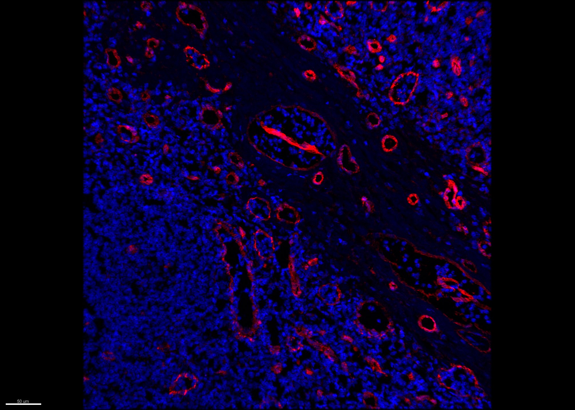
Application: ImmunofluorescenceSample Tested: Human Tonsil tissueSpecies: HumanVerified Customer | Posted 12/13/2017LYVE-1 staining (red) on human tonsil, counterstained with DAPI (blue).Acetone fixed tissue
-
Verified Customer | Posted 04/21/2017Automated staining in Leica Bond. Antigen retrieval pH 6.0 (20 min) Dilution 1:800 Incubation time 30 min Detection-Bond Polymer Refine Detection kit
There are no reviews that match your criteria.
Protocols
Find general support by application which include: protocols, troubleshooting, illustrated assays, videos and webinars.
- 7-Amino Actinomycin D (7-AAD) Cell Viability Flow Cytometry Protocol
- Cellular Response to Hypoxia Protocols
- Extracellular Membrane Flow Cytometry Protocol
- Flow Cytometry Protocol for Cell Surface Markers
- Flow Cytometry Protocol for Staining Membrane Associated Proteins
- Flow Cytometry Staining Protocols
- Flow Cytometry Troubleshooting Guide
- Intracellular Flow Cytometry Protocol Using Alcohol (Methanol)
- Intracellular Flow Cytometry Protocol Using Detergents
- Intracellular Nuclear Staining Flow Cytometry Protocol Using Detergents
- Intracellular Staining Flow Cytometry Protocol Using Alcohol Permeabilization
- Intracellular Staining Flow Cytometry Protocol Using Detergents to Permeabilize Cells
- Propidium Iodide Cell Viability Flow Cytometry Protocol
- Protocol for Liperfluo
- Protocol for the Characterization of Human Th22 Cells
- Protocol for the Characterization of Human Th9 Cells
- Protocol: Annexin V and PI Staining by Flow Cytometry
- Protocol: Annexin V and PI Staining for Apoptosis by Flow Cytometry
- R&D Systems Quality Control Western Blot Protocol
- Troubleshooting Guide: Fluorokine Flow Cytometry Kits
- Troubleshooting Guide: Western Blot Figures
- Western Blot Conditions
- Western Blot Protocol
- Western Blot Protocol for Cell Lysates
- Western Blot Troubleshooting
- Western Blot Troubleshooting Guide
- View all Protocols, Troubleshooting, Illustrated assays and Webinars
Loading...