Human MICA PE-conjugated Antibody Summary
Applications
Please Note: Optimal dilutions should be determined by each laboratory for each application. General Protocols are available in the Technical Information section on our website.
Scientific Data
 View Larger
Detection of MICA in K562 Human Cell Line by Flow Cytometry. K562 human chronic myelogenous leukemia cell line was stained with Mouse Anti-Human MICA PE-conjugated Monoclonal Antibody (Catalog # FAB1300P, filled histogram) or isotype control antibody (Catalog # IC0041P, open histogram). View our protocol for Staining Membrane-associated Proteins.
 View Larger
MICA Specificity is Shown by Flow Cytometry in Knockout Cell Line. MICA knockout K562 human myelogenous leukemia cell line was stained with Mouse Anti-Human MICA PE-conjugated Monoclonal Antibody (Catalog # FAB1300P, filled histogram) or isotype control antibody (Catalog # IC0041P, open histogram). No staining in the MICA knockout K562 cell line was observed. View our protocol for Staining Membrane-associated Proteins.
 View Larger
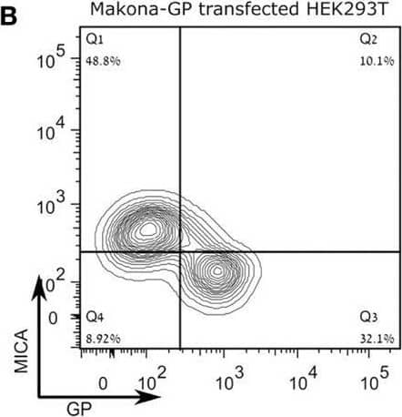
Detection of MICA by Flow Cytometry Expression of the Sudan virus (SUDV)- or Makona-GP results in loss of surface staining of alpha MICA in HEK293T cells and JEG3 cells stably transfected with MICA. The figure shows representative flow cytometry analysis for steric shielding of the MICA antigen by SUDV-GP or Makona-GP. (A) HEK293T cells were transfected, harvested, and stained for SUDV-GP with a biotinylated 3C10 antibody, followed by allophycocyanin (APC)-conjugated streptavidin, and co-stained for MICA surface antigens with phycoerythrin (PE)-conjugated alpha MICA antibody. Results are from one representative experiment of more than 10 performed (B) HEK293T cells were transfected, harvested, and stained for Makona-GP with survivor sera, followed by APC-labeled secondary antibody, and co-stained for MICA surface antigen using PE-conjugated alpha MICA antibody. Results are from one representative experiment of two performed. (C) SUDV-GP-transfected JEG3-MICA cells stained for SUDV-GP and co-stained for MICA surface antigens and NKG2D-Ig ligands. Results are from one representative experiment of more than four performed. (D–F) HEK293T cells were transfected with SUDV-GP or SUDV-GP-GFP, harvested shortly after they were treated with trypsin or left untreated, and finally stained for: (D) HLA-A, B, C, and SUDV-GP, (E) MICA and SUDV-GP, and (F) B7H6. Results are from one representative experiment of more than four performed. Image collected and cropped by CiteAb from the following open publication (https://pubmed.ncbi.nlm.nih.gov/30013549), licensed under a CC-BY license. Not internally tested by R&D Systems.
Reconstitution Calculator
Preparation and Storage
- 12 months from date of receipt, 2 to 8 °C as supplied.
Background: MICA
MICA (MHC class I chain-related gene A) is a transmembrane glycoprotein that functions as a ligand for human NKG2D. A closely related protein, MICB, shares 85% amino acid identity with MICA. These proteins are distantly related to the MHC class I proteins. They possess three extracellular Ig-like domains, but they have no capacity to bind peptide or interact with beta 2-microglobulin. The genes encoding these proteins are found within the Major Histocompatibility Complex on human chromosome 6. The MICA locus is highly polymorphic with more than 50 recognized human alleles. MICA is absent from most cells but is frequently expressed in epithelial tumors and can be induced by bacterial and viral infections. MICA is a ligand for human NKG2D, an activating receptor expressed on NK cells, NKT cells, gamma δ T cells, and CD8+ alpha beta T cells. Recognition of MICA by NKG2D results in the activation of cytolytic activity and/or cytokine production by these effector cells. MICA recognition is involved in tumor surveillance, viral infections, and autoimmune diseases.
- Groh, V. et al. (2001) Nature Immunol. 2:255.
- Stephens, H. (2001) Trends Immunol. 22:378.
- Bauer, S. et al. (1999) Science 285:727.
- Groh, V. et al. (2002) Nature 419:734.
- Steinle, A. et al. (2001) Immunogenetics 53:279.
- Pende, D. et al. (2002) Cancer Res. 62:6178.
- NKG2D and its Ligands (2002) http://www.RnDSystems.com
Product Datasheets
Citations for Human MICA PE-conjugated Antibody
R&D Systems personnel manually curate a database that contains references using R&D Systems products. The data collected includes not only links to publications in PubMed, but also provides information about sample types, species, and experimental conditions.
18
Citations: Showing 1 - 10
Filter your results:
Filter by:
-
Resveratrol promotes MICA/B expression and natural killer cell lysis of breast cancer cells by suppressing c-Myc/miR-17 pathway
Authors: Jie Pan, Jiaying Shen, Wengong Si, Chengyong Du, Danni Chen, Liang Xu et al.
Oncotarget
-
Topoisomerase I Inhibition Radiosensitizing Hepatocellular Carcinoma by RNF144A-mediated DNA-PKcs Ubiquitination and Natural Killer Cell Cytotoxicity
Authors: Chiao-Ling Tsai, Po-Sheng Yang, Feng-Ming Hsu, Ann-Lii Cheng, Wan-Ni Yu, Jason Chia-Hsien Cheng
Journal of Clinical and Translational Hepatology
-
Antitumor effects of NK cells expanded by activation pre?processing of autologous feeder cells before irradiation in colorectal cancer
Authors: Koh, EK;Lee, HR;Son, WC;Park, GY;Bae, J;Park, YS;
Oncology letters
Species: Human
Sample Types: Whole Cells
Applications: Flow Cytometry -
NKG2D receptor activation drives primary graft dysfunction severity and poor lung transplantation outcomes
Authors: DR Calabrese, T Tsao, M Magnen, C Valet, Y Gao, B Mallavia, JJ Tian, EA Aminian, KM Wang, A Shemesh, EB Punzalan, A Sarma, CS Calfee, SA Christenso, CR Langelier, SR Hays, JA Golden, LE Leard, ME Kleinhenz, NA Kolaitis, RJ Shah, A Venado, LL Lanier, JR Greenland, DM Sayah, A Ardehali, J Kukreja, SS Weigt, JA Belperio, JP Singer, MR Looney
JCI Insight, 2022-12-22;0(0):.
Species: Human
Sample Types: Whole Cells
Applications: Flow Cytometry -
Targeting WEE1/AKT restores p53-dependent NK cell activation to induce immune checkpoint blockade responses in 'cold' melanoma
Authors: SS Dinavahi, YC Chen, K Punnath, A Berg, M Herlyn, M Foroutan, ND Huntington, GP Robertson
Cancer Immunology Research, 2022-06-03;0(0):.
Species: Mouse
Sample Types: Whole Cells
Applications: FACS -
Immunomodulatory effect of NEDD8-activating enzyme inhibition in Multiple Myeloma: upregulation of NKG2D ligands and sensitization to Natural Killer cell recognition
Authors: S Petillo, C Capuano, R Molfetta, C Fionda, A Mekhloufi, C Pighi, F Antonangel, A Zingoni, A Soriani, MT Petrucci, R Galandrini, R Paolini, A Santoni, M Cippitelli
Cell Death & Disease, 2021-09-04;12(9):836.
Species: Human
Sample Types: Whole Cells
Applications: Flow Cytometry -
Establishment of HLA class I and MICA/B null HEK-293T panel expressing single MICA alleles to detect anti-MICA antibodies
Authors: JH Jeon, IC Baek, CH Hong, KH Park, H Lee, EJ Oh, TG Kim
Scientific Reports, 2021-08-03;11(1):15716.
Species: Human
Sample Types: Whole Cells
Applications: Flow Cytometry -
Rac1/ROCK-driven membrane dynamics promote natural killer cell cytotoxicity via granzyme-induced necroptosis
Authors: Y Zhu, J Xie, J Shi
Bmc Biology, 2021-07-30;19(1):140.
Species: Human
Sample Types: Whole Cells
Applications: Flow Cytometry -
All-trans retinoic acid enhances cytotoxicity of CIK cells against human lung adenocarcinoma by upregulating MICA and IL-2 secretion
Authors: XY Fan, PY Wang, C Zhang, YL Zhang, Y Fu, C Zhang, QX Li, JN Zhou, BE Shan, DW He
Sci Rep, 2017-11-28;7(1):16481.
Species: Human
Sample Types: Whole Cells
Applications: Flow Cytometry -
Impaired NK cell recognition of vemurafenib-treated melanoma cells is overcome by simultaneous application of histone deacetylase inhibitors
Authors: S López-Cobo, N Pieper, C Campos-Sil, EM García-Cue, HT Reyburn, A Paschen, M Valés-Góme
Oncoimmunology, 2017-11-06;7(2):e1392426.
Species: Human
Sample Types: Whole Cells
Applications: Flow Cytometry -
MICA-Expressing Monocytes Enhance Natural Killer Cell Fc Receptor-Mediated Antitumor Functions
Authors: Amanda R. Campbell, Megan C. Duggan, Lorena P. Suarez-Kelly, Neela Bhave, Kallan S. Opheim, Elizabeth L. McMichael et al.
Cancer Immunology Research
-
Low-dose bortezomib increases the expression of NKG2D and DNAM-1 ligands and enhances induced NK and ?? T cell-mediated lysis in multiple myeloma
Authors: C Niu, H Jin, M Li, S Zhu, L Zhou, F Jin, Y Zhou, D Xu, J Xu, L Zhao, S Hao, W Li, J Cui
Oncotarget, 2017-01-24;8(4):5954-5964.
Species: Human
Sample Types: Whole Cells
Applications: Flow Cytometry -
SEP enhanced the antitumor activity of 5-fluorouracil by up-regulating NKG2D/MICA and reversed immune suppression via inhibiting ROS and caspase-3 in mice
Authors: M Ke, H Wang, Y Zhou, J Li, Y Liu, M Zhang, J Dou, T Xi, B Shen, C Zhou
Oncotarget, 2016-08-02;7(31):49509-49526.
Species: Human
Sample Types: Whole Cells
Applications: Flow Cytometry -
Increase of IFN-gamma and TNF-alpha production in CD107a + NK-92 cells co-cultured with cervical cancer cell lines pre-treated with the HO-1 inhibitor.
Authors: Gomez-Lomeli P, Bravo-Cuellar A, Hernandez-Flores G, Jave-Suarez L, Aguilar-Lemarroy A, Lerma-Diaz J, Dominguez-Rodriguez J, Sanchez-Reyes K, Ortiz-Lazareno P
Cancer Cell Int, 2014-10-01;14(1):100.
Species: Human
Sample Types: Whole Cells
Applications: Flow Cytometry -
A polysaccharide virulence factor of a human fungal pathogen induces neutrophil apoptosis via NK cells.
Authors: Robinet P, Baychelier F, Fontaine T, Picard C, Debre P, Vieillard V, Latge J, Elbim C
J Immunol, 2014-04-30;192(11):5332-42.
Species: Human
Sample Types: Whole Blood
Applications: Flow Cytometry -
HER2/HER3 signaling regulates NK cell-mediated cytotoxicity via MHC class I chain-related molecule A and B expression in human breast cancer cell lines.
Authors: Okita R, Mougiakakos D, Ando T, Mao Y, Sarhan D, Wennerberg E, Seliger B, Lundqvist A, Mimura K, Kiessling R
J. Immunol., 2012-02-01;188(5):2136-45.
Species: Human
Sample Types: Whole Cells
Applications: Flow Cytometry -
The MHC class Ib protein ULBP1 is a nonredundant determinant of leukemia/lymphoma susceptibility to gammadelta T-cell cytotoxicity.
Authors: Lanca T, Correia DV, Moita CF, Raquel H, Neves-Costa A, Ferreira C, Ramalho JS, Barata JT, Moita LF, Gomes AQ, Silva-Santos B
Blood, 2010-01-25;115(12):2407-11.
Species: Human
Sample Types: Whole Cells
Applications: Flow Cytometry -
Expression of WSX1 in tumors sensitizes IL-27 signaling-independent natural killer cell surveillance.
Authors: Dibra D, Cutrera JJ, Xia X, Birkenbach MP, Li S
Cancer Res., 2009-06-23;69(13):5505-13.
Species: Human
Sample Types: Whole Cells
Applications: Flow Cytometry
FAQs
No product specific FAQs exist for this product, however you may
View all Antibody FAQsReviews for Human MICA PE-conjugated Antibody
There are currently no reviews for this product. Be the first to review Human MICA PE-conjugated Antibody and earn rewards!
Have you used Human MICA PE-conjugated Antibody?
Submit a review and receive an Amazon gift card.
$25/€18/£15/$25CAN/¥75 Yuan/¥2500 Yen for a review with an image
$10/€7/£6/$10 CAD/¥70 Yuan/¥1110 Yen for a review without an image
