Human MMR/CD206 Antibody (685645)
R&D Systems | Catalog # MAB25341

Key Product Details
Species Reactivity
Validated:
Human
Cited:
Human, Mouse
Applications
Validated:
Immunohistochemistry, Western Blot, Simple Western
Cited:
Immunohistochemistry, Immunohistochemistry-Paraffin
Label
Unconjugated
Antibody Source
Monoclonal Mouse IgG2B Clone # 685645
Loading...
Product Specifications
Immunogen
Mouse myeloma cell line NS0-derived recombinant human MMR/CD206
Leu19-Lys1383 (Thr399Ala) & (Leu407Phe),
Accession # P22897
Leu19-Lys1383 (Thr399Ala) & (Leu407Phe),
Accession # P22897
Specificity
Detects human MMR/CD206 in direct ELISAs and Western blots. In direct ELISAs, no cross-reactivity
with recombinant mouse (rm) MMR or recombinant human Mrc2 is observed. In
Western blots, approximately 25% cross-reactivity with rmMMR is observed.
Clonality
Monoclonal
Host
Mouse
Isotype
IgG2B
Scientific Data Images for Human MMR/CD206 Antibody (685645)
Detection of Human MMR/CD206 by Western Blot.
Western blot shows lysates of human immature dendritic cells. PVDF Membrane was probed with 1 µg/mL of Mouse Anti-Human MMR/CD206 Monoclonal Antibody (Catalog # MAB25341) followed by HRP-conjugated Anti-Mouse IgG Secondary Antibody (Catalog # HAF007). A specific band was detected for MMR/CD206 at approximately 150 kDa (as indicated). This experiment was conducted under reducing conditions and using Immunoblot Buffer Group 1.
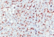
MMR/CD206 in Human Liver.
MMR/CD206 was detected in immersion fixed paraffin-embedded sections of human liver using Mouse Anti-Human MMR/CD206 Monoclonal Antibody (Catalog # MAB25341) at 15 µg/mL overnight at 4 °C. Before incubation with the primary antibody, tissue was subjected to heat-induced epitope retrieval using Antigen Retrieval Reagent-Basic (Catalog # CTS013). Tissue was stained using the Anti-Mouse HRP-DAB Cell & Tissue Staining Kit (brown; Catalog # CTS002) and counterstained with hematoxylin (blue). Specific staining was localized to endothelial cells in sinusoids. View our protocol for Chromogenic IHC Staining of Paraffin-embedded Tissue Sections.
Detection of Human MMR/CD206 by Simple WesternTM.
Simple Western lane view shows lysates of human immature dendritic cells, loaded at 0.2 mg/mL. A specific band was detected for MMR/CD206 at approximately 228 kDa (as indicated) using 20 µg/mL of Mouse Anti-Human MMR/CD206 Monoclonal Antibody (Catalog # MAB25341). This experiment was conducted under reducing conditions and using the 66-440 kDa separation system.Applications for Human MMR/CD206 Antibody (685645)
Application
Recommended Usage
Immunohistochemistry
8-25 µg/mL
Sample: Immersion fixed paraffin-embedded sections of human liver
Sample: Immersion fixed paraffin-embedded sections of human liver
Simple Western
20 µg/mL
Sample: Human immature dendritic cells
Sample: Human immature dendritic cells
Western Blot
1 µg/mL
Sample: Human immature dendritic cells
Sample: Human immature dendritic cells
Reviewed Applications
Read 3 reviews rated 4.3 using MAB25341 in the following applications:
Formulation, Preparation, and Storage
Purification
Protein A or G purified from hybridoma culture supernatant
Reconstitution
Sterile PBS to a final concentration of 0.5 mg/mL. For liquid material, refer to CoA for concentration.
Loading...
Formulation
Lyophilized from a 0.2 μm filtered solution in PBS with Trehalose. *Small pack size (SP) is supplied either lyophilized or as a 0.2 µm filtered solution in PBS.
Shipping
Lyophilized product is shipped at ambient temperature. Liquid small pack size (-SP) is shipped with polar packs. Upon receipt, store immediately at the temperature recommended below.
Stability & Storage
Use a manual defrost freezer and avoid repeated freeze-thaw cycles.
- 12 months from date of receipt, -20 to -70 °C as supplied.
- 1 month, 2 to 8 °C under sterile conditions after reconstitution.
- 6 months, -20 to -70 °C under sterile conditions after reconstitution.
Calculators
Background: MMR/CD206
References
- East, L. and C. Isake (2002) Biochim. Biophys. Acta 1572:364.
- Chieppa, M. et al. (2003) J. Immunol. 171:4552.
- Figdor, C. et al. (2002) Nat. Rev. Immunol. 2:77.
- Taylor, M. et al. (1990) J. Biol. Chem. 265:12156.
- Leteux, C. et al. (2000) J. Exp. Med. 191:1117.
- Martinez-Pomares, L. et al. (2001) Immunobiology 204:527.
- Feinberg, H. et al. (2000) J. Biol. Chem. 275:21539.
Long Name
Macrophage Mannose Receptor
Alternate Names
CD206, CLEC13D, MRC1
Gene Symbol
MRC1
UniProt
Additional MMR/CD206 Products
Product Documents for Human MMR/CD206 Antibody (685645)
Certificate of Analysis
To download a Certificate of Analysis, please enter a lot or batch number in the search box below.
Note: Certificate of Analysis not available for kit components.
Product Specific Notices for Human MMR/CD206 Antibody (685645)
For research use only
Citations for Human MMR/CD206 Antibody (685645)
 Powered by Bioz
Customer Reviews for Human MMR/CD206 Antibody (685645) (3)
4.3 out of 5
3 Customer Ratings
Have you used Human MMR/CD206 Antibody (685645)?
Submit a review and receive an Amazon gift card!
$25/€18/£15/$25CAN/¥2500 Yen for a review with an image
$10/€7/£6/$10CAN/¥1110 Yen for a review without an image
Submit a review
Customer Images
Showing
1
-
3 of
3 reviews
Showing All
Filter By:
-
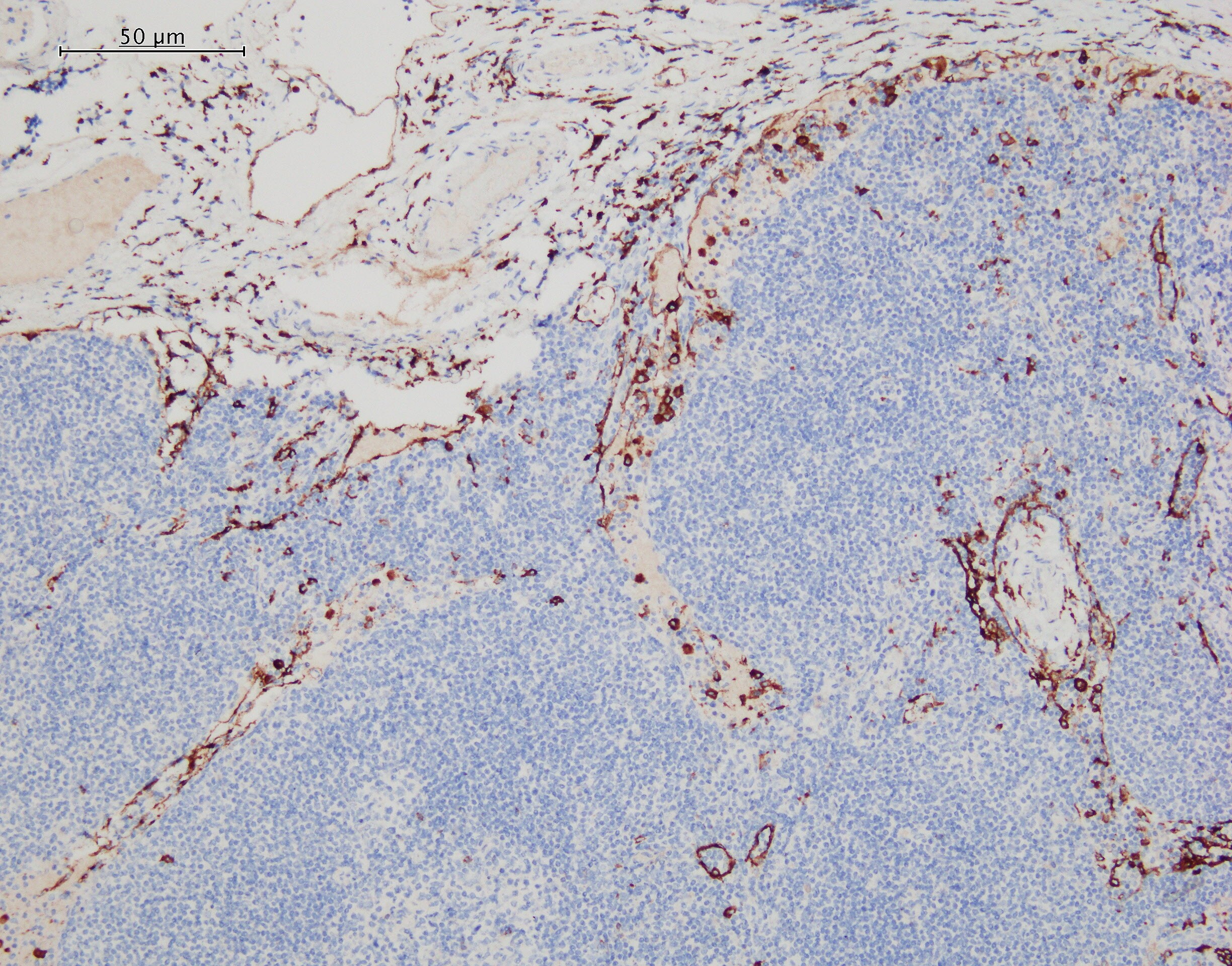
Application: ImmunohistochemistrySample Tested: Liver tissueSpecies: HumanVerified Customer | Posted 11/29/2021

-
Application: ImmunohistochemistrySample Tested: Lymph node tissueSpecies: HumanVerified Customer | Posted 05/07/2021
-
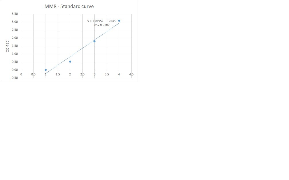
Application: ELISASample Tested: Serum and PlasmaSpecies: HumanVerified Customer | Posted 09/23/2019I used this antibody for developing a sandwich ELISA in combination with pAb (cat.AF2534) and protein (cat.2534 MR). This combination gives good standard curve but unfortunately did not detect any MMR in our samples.
There are no reviews that match your criteria.
Protocols
Find general support by application which include: protocols, troubleshooting, illustrated assays, videos and webinars.
- Antigen Retrieval Protocol (PIER)
- Antigen Retrieval for Frozen Sections Protocol
- Appropriate Fixation of IHC/ICC Samples
- Cellular Response to Hypoxia Protocols
- Chromogenic IHC Staining of Formalin-Fixed Paraffin-Embedded (FFPE) Tissue Protocol
- Chromogenic Immunohistochemistry Staining of Frozen Tissue
- ClariTSA™ Fluorophore Kits
- Detection & Visualization of Antibody Binding
- Fluorescent IHC Staining of Frozen Tissue Protocol
- Graphic Protocol for Heat-induced Epitope Retrieval
- Graphic Protocol for the Preparation and Fluorescent IHC Staining of Frozen Tissue Sections
- Graphic Protocol for the Preparation and Fluorescent IHC Staining of Paraffin-embedded Tissue Sections
- Graphic Protocol for the Preparation of Gelatin-coated Slides for Histological Tissue Sections
- IHC Sample Preparation (Frozen sections vs Paraffin)
- Immunofluorescent IHC Staining of Formalin-Fixed Paraffin-Embedded (FFPE) Tissue Protocol
- Immunohistochemistry (IHC) and Immunocytochemistry (ICC) Protocols
- Immunohistochemistry Frozen Troubleshooting
- Immunohistochemistry Paraffin Troubleshooting
- Preparing Samples for IHC/ICC Experiments
- Preventing Non-Specific Staining (Non-Specific Binding)
- Primary Antibody Selection & Optimization
- Protocol for Heat-Induced Epitope Retrieval (HIER)
- Protocol for Making a 4% Formaldehyde Solution in PBS
- Protocol for VisUCyte™ HRP Polymer Detection Reagent
- Protocol for the Preparation & Fixation of Cells on Coverslips
- Protocol for the Preparation and Chromogenic IHC Staining of Frozen Tissue Sections
- Protocol for the Preparation and Chromogenic IHC Staining of Frozen Tissue Sections - Graphic
- Protocol for the Preparation and Chromogenic IHC Staining of Paraffin-embedded Tissue Sections
- Protocol for the Preparation and Chromogenic IHC Staining of Paraffin-embedded Tissue Sections - Graphic
- Protocol for the Preparation and Fluorescent IHC Staining of Frozen Tissue Sections
- Protocol for the Preparation and Fluorescent IHC Staining of Paraffin-embedded Tissue Sections
- Protocol for the Preparation of Gelatin-coated Slides for Histological Tissue Sections
- R&D Systems Quality Control Western Blot Protocol
- TUNEL and Active Caspase-3 Detection by IHC/ICC Protocol
- The Importance of IHC/ICC Controls
- Troubleshooting Guide: Immunohistochemistry
- Troubleshooting Guide: Western Blot Figures
- Western Blot Conditions
- Western Blot Protocol
- Western Blot Protocol for Cell Lysates
- Western Blot Troubleshooting
- Western Blot Troubleshooting Guide
- View all Protocols, Troubleshooting, Illustrated assays and Webinars
Loading...