
Arginase 1/ARG1 Antibody [Alexa Fluor® 700]
R&D Systems | Catalog # IC5868N

Scientific images may be resized, cropped, or adjusted for brightness or contrast for presentation purposes. These updates are limited to presentation and do not affect the underlying scientific interpretation of the data. Where available, additional source data may be provided upon request.
Key Product Details
Species Reactivity
Applications
Label
Antibody Source
Product Specifications
Immunogen
Accession # P05089
Specificity
Clonality
Host
Isotype
Scientific Data Images for Arginase 1/ARG1 Antibody [Alexa Fluor® 700]
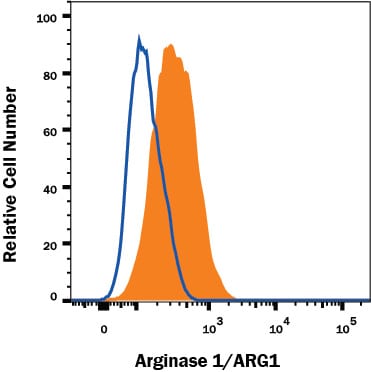
Detection of Arginase 1/ARG1 in HepG2 Human Cell Line by Flow Cytometry.
HepG2 human hepatocellular carcinoma cell line was stained with Sheep Anti-Human/Mouse Arginase 1/ARG1 Alexa Fluor® 700-conjugated Antigen Affinity-purified Polyclonal Antibody (Catalog # IC5868N, filled histogram) or isotype control antibody (Catalog # IC016N, open histogram). To facilitate intracellular staining, cells were fixed with Flow Cytometry Fixation Buffer (Catalog # FC004) and permeabilized with Flow Cytometry Permeabilization/Wash Buffer I (Catalog # FC005). View our protocol for Staining Membrane-associated Proteins.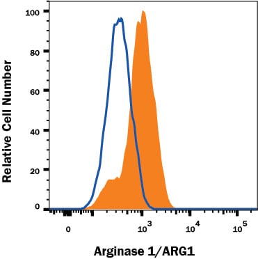
Detection of Arginase 1/ARG1 in Hepa 1-6 Mouse Cell Line by Flow Cytometry.
Hepa 1-6 mouse hepatoma cell line was stained with Sheep Anti-Human/Mouse Arginase 1/ARG1 Alexa Fluor® 700-conjugated Antigen Affinity-purified Polyclonal Antibody (Catalog # IC5868N, filled histogram) or isotype control antibody (Catalog # IC016N, open histogram). To facilitate intracellular staining, cells were fixed with Flow Cytometry Fixation Buffer (Catalog # FC004) and permeabilized with Flow Cytometry Permeabilization/Wash Buffer I (Catalog # FC005). View our protocol for Staining Membrane-associated Proteins.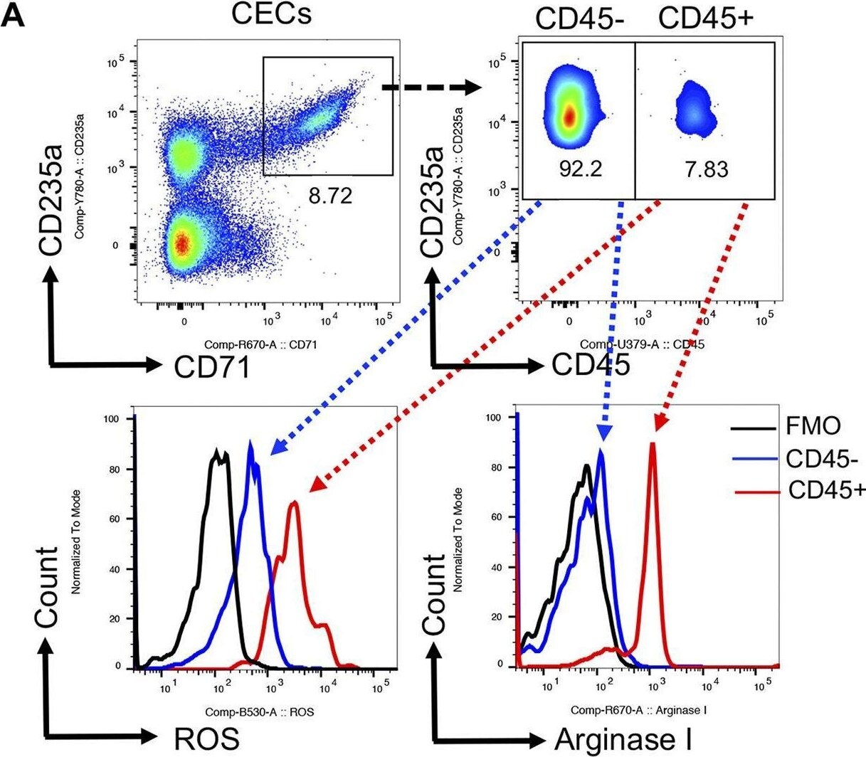
Detection of Arginase 1/ARG1 by Flow Cytometry
CD45+CECs in COVID-19 patients are more potent. (A) Representative flow cytometry plots of the gating strategy for CD45− and CD45+CECs, and the expression of ROS and arginase I in CD45− versus CD45+CECs. (B) Cumulative data of the intensity of ROS expression measured by mean fluorescence intensity (MFI) in CD45− versus CD45+CECs. (C) Cumulative data of the intensity (MFI) of arginase I expression in CD45− versus CD45+CECs. (D) Representative flow cytometry plots, and (E) cumulative data showing % CD8+ T cells expressing GzmB, or (F) CD107a without stimulation (unstim) or stimulated (stim) with anti-CD3/CD28 antibodies in the absence of CECs (−CECs) or presence (+CECs) without and with l-arginine supplementation (2 mM). (G) Cumulative data showing % CD8+ T cells expressing GzmB, or (H) CD107a without stimulation (Unstim) or stimulated (stim) with anti-CD3/CD28 antibodies in the absence of CECs (−CECs) or presence (+CECs). Each dot represents data from a human subject. Image collected and cropped by CiteAb from the following open publication (https://pubmed.ncbi.nlm.nih.gov/35943266), licensed under a CC-BY license. Not internally tested by R&D Systems.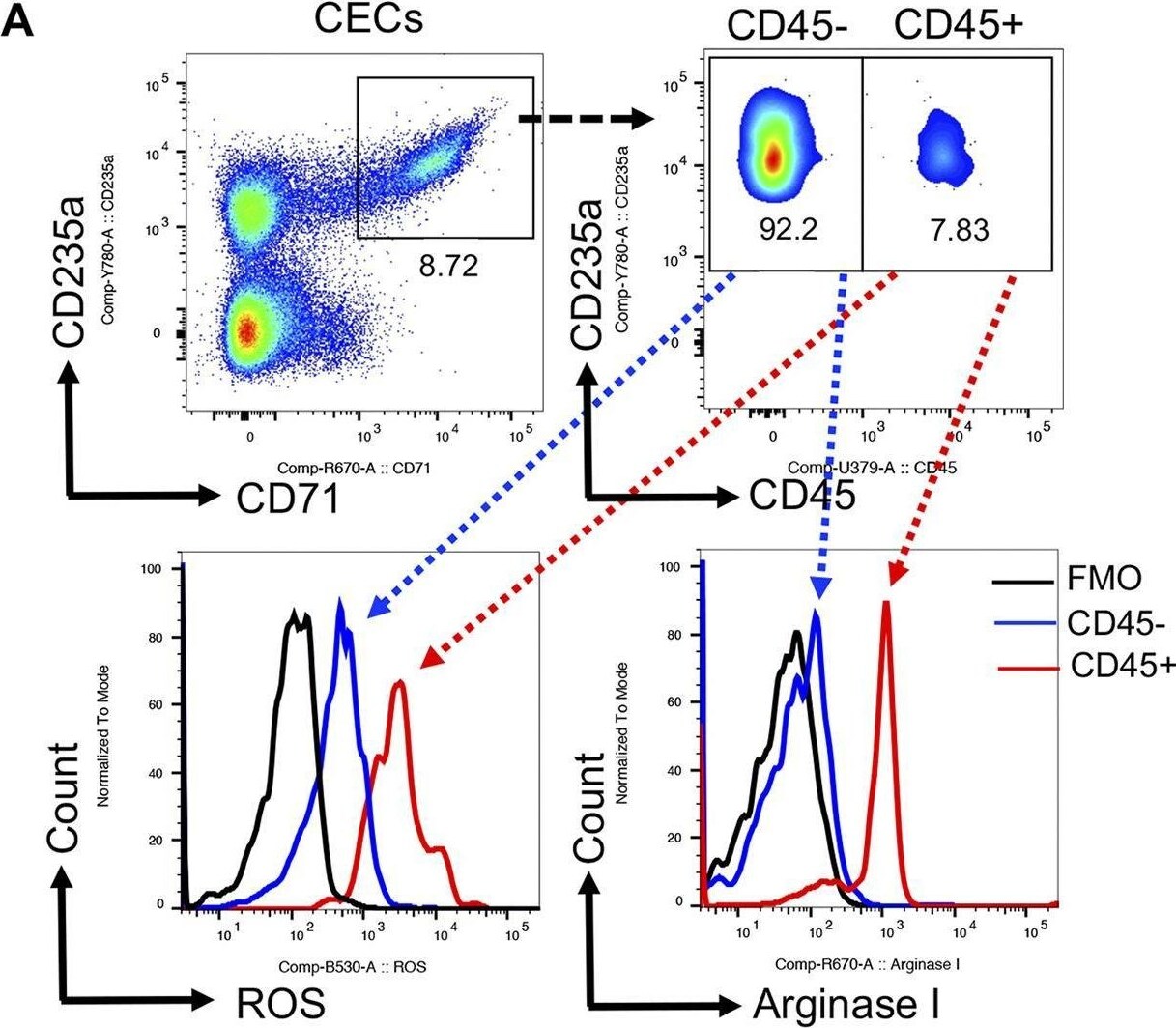
Detection of Arginase 1/ARG1 by Flow Cytometry
CD45+CECs in COVID-19 patients are more potent. (A) Representative flow cytometry plots of the gating strategy for CD45− and CD45+CECs, and the expression of ROS and arginase I in CD45− versus CD45+CECs. (B) Cumulative data of the intensity of ROS expression measured by mean fluorescence intensity (MFI) in CD45− versus CD45+CECs. (C) Cumulative data of the intensity (MFI) of arginase I expression in CD45− versus CD45+CECs. (D) Representative flow cytometry plots, and (E) cumulative data showing % CD8+ T cells expressing GzmB, or (F) CD107a without stimulation (unstim) or stimulated (stim) with anti-CD3/CD28 antibodies in the absence of CECs (−CECs) or presence (+CECs) without and with l-arginine supplementation (2 mM). (G) Cumulative data showing % CD8+ T cells expressing GzmB, or (H) CD107a without stimulation (Unstim) or stimulated (stim) with anti-CD3/CD28 antibodies in the absence of CECs (−CECs) or presence (+CECs). Each dot represents data from a human subject. Image collected and cropped by CiteAb from the following open publication (https://pubmed.ncbi.nlm.nih.gov/35943266), licensed under a CC-BY license. Not internally tested by R&D Systems.Applications for Arginase 1/ARG1 Antibody [Alexa Fluor® 700]
Intracellular Staining by Flow Cytometry
Sample: HepG2 Human Cell Line and Hepa 1-6 Mouse Cell Line
Spectra Viewer
Plan Your Experiments
Use our spectra viewer to interactively plan your experiments, assessing multiplexing options. View the excitation and emission spectra for our fluorescent dye range and other commonly used dyes.
Spectra Viewer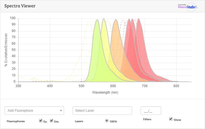
Flow Cytometry Panel Builder
Bio-Techne Knows Flow Cytometry
Save time and reduce costly mistakes by quickly finding compatible reagents using the Panel Builder Tool.
Advanced Features
- Spectra Viewer - Custom analysis of spectra from multiple fluorochromes
- Spillover Popups - Visualize the spectra of individual fluorochromes
- Antigen Density Selector - Match fluorochrome brightness with antigen density

Formulation, Preparation, and Storage
Purification
Formulation
Shipping
Stability & Storage
- 12 months from date of receipt, 2 to 8 °C as supplied.
Background: Arginase 1/ARG1
Long Name
Alternate Names
Gene Symbol
UniProt
Additional Arginase 1/ARG1 Products
Product Documents for Arginase 1/ARG1 Antibody [Alexa Fluor® 700]
Certificate of Analysis
To download a Certificate of Analysis, please enter a lot or batch number in the search box below.
Note: Certificate of Analysis not available for kit components.
Product Specific Notices for Arginase 1/ARG1 Antibody [Alexa Fluor® 700]
This product is provided under an agreement between Life Technologies Corporation and R&D Systems, Inc, and the manufacture, use, sale or import of this product is subject to one or more US patents and corresponding non-US equivalents, owned by Life Technologies Corporation and its affiliates. The purchase of this product conveys to the buyer the non-transferable right to use the purchased amount of the product and components of the product only in research conducted by the buyer (whether the buyer is an academic or for-profit entity). The sale of this product is expressly conditioned on the buyer not using the product or its components (1) in manufacturing; (2) to provide a service, information, or data to an unaffiliated third party for payment; (3) for therapeutic, diagnostic or prophylactic purposes; (4) to resell, sell, or otherwise transfer this product or its components to any third party, or for any other commercial purpose. Life Technologies Corporation will not assert a claim against the buyer of the infringement of the above patents based on the manufacture, use or sale of a commercial product developed in research by the buyer in which this product or its components was employed, provided that neither this product nor any of its components was used in the manufacture of such product. For information on purchasing a license to this product for purposes other than research, contact Life Technologies Corporation, Cell Analysis Business Unit, Business Development, 29851 Willow Creek Road, Eugene, OR 97402, Tel: (541) 465-8300. Fax: (541) 335-0354.
For research use only
Related Research Areas
Customer Reviews for Arginase 1/ARG1 Antibody [Alexa Fluor® 700]
There are currently no reviews for this product. Be the first to review Arginase 1/ARG1 Antibody [Alexa Fluor® 700] and earn rewards!
Have you used Arginase 1/ARG1 Antibody [Alexa Fluor® 700]?
Submit a review and receive an Amazon gift card!
$25/€18/£15/$25CAN/¥2500 for a review with an image
$10/€7/£6/$10CAN/¥1110 for a review without an image
Submit a review
Protocols
Find general support by application which include: protocols, troubleshooting, illustrated assays, videos and webinars.
- 7-Amino Actinomycin D (7-AAD) Cell Viability Flow Cytometry Protocol
- Extracellular Membrane Flow Cytometry Protocol
- Flow Cytometry Protocol for Cell Surface Markers
- Flow Cytometry Protocol for Staining Membrane Associated Proteins
- Flow Cytometry Staining Protocols
- Flow Cytometry Troubleshooting Guide
- Intracellular Flow Cytometry Protocol Using Alcohol (Methanol)
- Intracellular Flow Cytometry Protocol Using Detergents
- Intracellular Nuclear Staining Flow Cytometry Protocol Using Detergents
- Intracellular Staining Flow Cytometry Protocol Using Alcohol Permeabilization
- Intracellular Staining Flow Cytometry Protocol Using Detergents to Permeabilize Cells
- Propidium Iodide Cell Viability Flow Cytometry Protocol
- Protocol for Liperfluo
- Protocol for the Characterization of Human Th22 Cells
- Protocol for the Characterization of Human Th9 Cells
- Protocol: Annexin V and PI Staining by Flow Cytometry
- Protocol: Annexin V and PI Staining for Apoptosis by Flow Cytometry
- Troubleshooting Guide: Fluorokine Flow Cytometry Kits
- View all Protocols, Troubleshooting, Illustrated assays and Webinars