Arginase 1 (ARG1) is a 35‑40 kDa member of the arginase family of enzymes. It is expressed in multiple cell types, including erythrocytes, hepatocytes, neutrophils, smooth muscle and macrophages. ARG1 demonstrates two distinct functions: in the hepatocyte cytoplasm, it catalyzes the conversion of arginine to ornithine and urea, while in multiple cells, it degrades arginine, thus indirectly downregulating NO synthase (NOS) activity by depriving this enzyme of its substrate. Human AGR1 is 322 amino acids (aa) in length. Its enzyme region comprises aa 9‑309 and contains two Mn atoms. ARG1 is modestly active as a monomer, but highly active as a 105 kDa homotrimer. Trimerization is promoted by nitrosylation of Cys303, creating a regulatory feedback loop with NOS. There are two isoform variants, one that shows an eight aa insertion after Gln43, and another that shows a deletion of aa 204‑289. Full-length human ARG1 shares 87% aa identity with mouse and rat ARG1.
Human/Mouse Arginase 1/ARG1 Fluorescein‑conjugated Antibody
R&D Systems | Catalog # IC5868F

Key Product Details
Validated by
Species Reactivity
Validated:
Cited:
Applications
Validated:
Cited:
Label
Antibody Source
Product Specifications
Immunogen
Met1-Lys322
Accession # P05089
Specificity
Clonality
Host
Isotype
Scientific Data Images for Human/Mouse Arginase 1/ARG1 Fluorescein‑conjugated Antibody
Detection of Arginase 1/ARG1 in HepG2 Human Cell Line by Flow Cytometry.
HepG2 human hepatocellular carcinoma cell line was stained with Sheep Anti-Human/Mouse Arginase 1/ARG1 Fluorescein-conjugated Antigen Affinity-purified Polyclonal Antibody (Catalog # IC5868F, filled histogram) or isotype control antibody (Catalog # IC016F, open histogram). To facilitate intracellular staining, cells were fixed with Flow Cytometry Fixation Buffer (Catalog # FC004) and permeabilized with Flow Cytometry Permeabilization/Wash Buffer I (Catalog # FC005). View our protocol for Staining Intracellular Molecules.
Detection of Arginase 1/ARG1 in Hepa 1‑6 Mouse Cell Line by Flow Cytometry.
Hepa 1-6 mouse hepatoma cell line was stained with Sheep Anti-Human/Mouse Arginase 1/ARG1 Fluorescein-conjugated Antigen Affinity-purified Polyclonal Antibody (Catalog # IC5868F, filled histogram) or isotype control antibody (Catalog # IC016F, open histogram). To facilitate intracellular staining, cells were fixed with Flow Cytometry Fixation Buffer (Catalog # FC004) and permeabilized with Flow Cytometry Permeabilization/Wash Buffer I (Catalog # FC005). View our protocol for Staining Intracellular Molecules.
Detection of Human Arginase 1/ARG1/liver Arginase by Flow Cytometry
Within PBMCs population, GSC-derived exosomes promote an immunosuppressive phenotype in monocytes and stimulate the production of arginase-1 and IL-10 by Mo-MDSCs.Unstimulated PBMCs were incubated in absence (CTRL) or presence (GSC-EXO) of GSC-derived exosomes. Cells were surface stained with anti-CD14, anti CD33, anti CD11b and HLA-DR and then stained to detect intracellular level of IL-10 and arginase-1 by flow cytometry. (A) Gating strategy: physical parameters, i.e. forward scatter (FSC) and side scatter (SSC), were used to select monocytes (gate R3, left panel). Monocytes were recognized evaluating the expression of CD11b/CD33 (gate R4, middle panel) and CD14/HLA-DR (gate R5, right panel). (B) Representative FACS histograms of the intracellular staining of IL-10 and arginase-1 and of HLA-DR staining of CD14+/CD11b/CD33+ cells are shown. (C) The percentage of cells expressing IL-10 and arginase-1 and the MFI ratio of HLA-DR expression are shown (n = 6); bars, SD;*, significantly different from the control; P<0.05. Image collected and cropped by CiteAb from the following publication (https://dx.plos.org/10.1371/journal.pone.0169932), licensed under a CC-BY license. Not internally tested by R&D Systems.
Detection of Human Arginase 1/ARG1/liver Arginase by Flow Cytometry
Ultracentrifugated (UC) GSC-derived exosomes promote an immunosuppressive phenotype in monocytes similarly to ExoQuick (EQ) purified GSC- or GBM-derived exosomes.(A) CFSE-labelled PBMCs isolated from healthy donors (left) or CD14 negatively-sorted PBMCs (CD14-) (right) were pre-treated for 24 hours without (white column, CTRL) or with EQ or UC isolated GSC-derived exosomes (black column, EQ GSC-EXO; grey column, UC GSC-EXO, respectively) and stimulated for 4 days with anti-CD3 and anti-CD28. Histograms show a significant difference in the percentage of proliferating CD3+ (n = 4); bars, SD;*; from the control; P<0.05. (B-D) Induction of a Mo-MDSC phenotype on monocytes. Unstimulated PBMCs were incubated in the absence (CTRL) or presence of GSC-derived exosomes purified by EQ (black column, EQ EXO-GSC) or UC(grey column, UC EXO-GSC). Cells were surface stained with (B) anti-CD14, anti CD33, anti CD11b and (C) HLA-DR and then stained to detect intracellular level of IL-10 and arginase-1 by flow cytometry. (B) Gating strategy: physical parameters, i.e. forward scatter (FSC) and side scatter (SSC), were used to select monocytes (gate R1, left panel). Monocytes were recognized by evaluating the expression of CD11b/CD33 (gate R2, middle panel) and CD14/HLA-DR (gate R3, right panel). (C) Representative FACS histograms of the intracellular staining of IL-10 and arginase-1 and of HLA-DR staining of CD14+/CD11b/CD33+ cells are shown. (D) The percentage of cells expressing IL-10 and arginase-1 and the MFI ratio of HLA-DR expression are shown (n = 3); bars, SD;*, significantly different from the control; *, P<0.05; **,P<0.01. (E) PBMCs were stimulated with anti-CD3 and anti-CD28 in the absence (white column, CTRL) or presence (black column, GBM-EXO) of exosomes isolated from plasma of glioblastoma patients (GBM) by either EQ or UC and used at 1:10 dilution. Healthy donor plasma-derived exosomes were used as a control (striped column, EXO-HEALTHY). Proliferation of CD3+ was measu
Detection of Human Arginase 1/ARG1/liver Arginase by Flow Cytometry
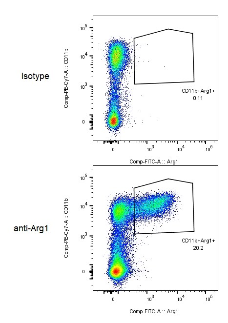
Arginase 1 expression in different subsets of circulating MDSC in patients with PC. Flow cytometric evaluation of ARG1 expression in CD33, CD11b, CD15 and CD14 in whole blood is shown in the representative dot plots. Gates were set based on negative controls. Numbers represent the percentages from the original populations gated. P (number) above each FACS plot indicates the population gated which was analysed. The gate was first set on HLA-DR negative against side scatter as shown in the top dot plot. Next, the CD11b+ & CD33+ (right) and CD14+ & CD15+ (left) subsets were identified. The expression of ARG1 in each subset was then determined as shown in the bottom dot plots. Image collected and cropped by CiteAb from the following publication (https://pubmed.ncbi.nlm.nih.gov/24741628), licensed under a CC-BY license. Not internally tested by R&D Systems.Applications for Human/Mouse Arginase 1/ARG1 Fluorescein‑conjugated Antibody
Intracellular Staining by Flow Cytometry
Sample: HepG2 human hepatocellular carcinoma cell line and Hepa 1‑6 mouse hepatoma cell line fixed with Flow Cytometry Fixation Buffer (Catalog # FC004) and permeabilized with Flow Cytometry Permeabilization/Wash Buffer I (Catalog # FC005)
Reviewed Applications
Read 1 review rated 5 using IC5868F in the following applications:
Spectra Viewer
Plan Your Experiments
Use our spectra viewer to interactively plan your experiments, assessing multiplexing options. View the excitation and emission spectra for our fluorescent dye range and other commonly used dyes.
Spectra Viewer
Flow Cytometry Panel Builder
Bio-Techne Knows Flow Cytometry
Save time and reduce costly mistakes by quickly finding compatible reagents using the Panel Builder Tool.
Advanced Features
- Spectra Viewer - Custom analysis of spectra from multiple fluorochromes
- Spillover Popups - Visualize the spectra of individual fluorochromes
- Antigen Density Selector - Match fluorochrome brightness with antigen density

Formulation, Preparation, and Storage
Purification
Formulation
Shipping
Stability & Storage
- 12 months from date of receipt, 2 to 8 °C as supplied.
Background: Arginase 1/ARG1
Long Name
Alternate Names
Gene Symbol
UniProt
Additional Arginase 1/ARG1 Products
Product Documents for Human/Mouse Arginase 1/ARG1 Fluorescein‑conjugated Antibody
Certificate of Analysis
To download a Certificate of Analysis, please enter a lot or batch number in the search box below.
Note: Certificate of Analysis not available for kit components.
Product Specific Notices for Human/Mouse Arginase 1/ARG1 Fluorescein‑conjugated Antibody
For research use only
Related Research Areas
Citations for Human/Mouse Arginase 1/ARG1 Fluorescein‑conjugated Antibody
 Powered by Bioz
Customer Reviews for Human/Mouse Arginase 1/ARG1 Fluorescein‑conjugated Antibody (1)
Have you used Human/Mouse Arginase 1/ARG1 Fluorescein‑conjugated Antibody?
Submit a review and receive an Amazon gift card!
$25/€18/£15/$25CAN/¥2500 Yen for a review with an image
$10/€7/£6/$10CAN/¥1110 Yen for a review without an image
Submit a review
Customer Images
-
Application: Flow CytometrySample Tested: MonocytesSpecies: MouseVerified Customer | Posted 08/18/2017

There are no reviews that match your criteria.
Protocols
Find general support by application which include: protocols, troubleshooting, illustrated assays, videos and webinars.
- 7-Amino Actinomycin D (7-AAD) Cell Viability Flow Cytometry Protocol
- Extracellular Membrane Flow Cytometry Protocol
- Flow Cytometry Protocol for Cell Surface Markers
- Flow Cytometry Protocol for Staining Membrane Associated Proteins
- Flow Cytometry Staining Protocols
- Flow Cytometry Troubleshooting Guide
- Intracellular Flow Cytometry Protocol Using Alcohol (Methanol)
- Intracellular Flow Cytometry Protocol Using Detergents
- Intracellular Nuclear Staining Flow Cytometry Protocol Using Detergents
- Intracellular Staining Flow Cytometry Protocol Using Alcohol Permeabilization
- Intracellular Staining Flow Cytometry Protocol Using Detergents to Permeabilize Cells
- Propidium Iodide Cell Viability Flow Cytometry Protocol
- Protocol for Liperfluo
- Protocol for the Characterization of Human Th22 Cells
- Protocol for the Characterization of Human Th9 Cells
- Protocol: Annexin V and PI Staining by Flow Cytometry
- Protocol: Annexin V and PI Staining for Apoptosis by Flow Cytometry
- Troubleshooting Guide: Fluorokine Flow Cytometry Kits
- View all Protocols, Troubleshooting, Illustrated assays and Webinars