Arginase 1 (ARG1) is a 35‑40 kDa member of the arginase family of enzymes. It is expressed in multiple cell types, including erythrocytes, hepatocytes, neutrophils, smooth muscle and macrophages. ARG1 demonstrates two distinct functions: in the hepatocyte cytoplasm, it catalyzes the conversion of arginine to ornithine and urea, while in multiple cells, it degrades arginine, thus indirectly downregulating NO synthase (NOS) activity by depriving this enzyme of its substrate. Human AGR1 is 322 amino acids (aa) in length. Its enzyme region comprises aa 9‑309 and contains two Mn atoms. ARG1 is modestly active as a monomer, but highly active as a 105 kDa homotrimer. Trimerization is promoted by nitrosylation of Cys303, creating a regulatory feedback loop with NOS. There are two isoform variants, one that shows an eight aa insertion after Gln43, and another that shows a deletion of aa 204‑289. Full-length human ARG1 shares 87% aa identity with mouse and rat ARG1.
Human/Mouse Arginase 1/ARG1 APC‑conjugated Antibody
R&D Systems | Catalog # IC5868A

Key Product Details
Validated by
Species Reactivity
Validated:
Cited:
Applications
Validated:
Cited:
Label
Antibody Source
Product Specifications
Immunogen
Met1-Lys322
Accession # P05089
Specificity
Clonality
Host
Isotype
Scientific Data Images for Human/Mouse Arginase 1/ARG1 APC‑conjugated Antibody
Detection of Arginase 1/ARG1 in HepG2 Human Cell Line by Flow Cytometry.
HepG2 human hepatocellular carcinoma cell line was stained with Sheep Anti-Human/Mouse Arginase 1/ARG1 APC-conjugated Antigen Affinity-purified Polyclonal Antibody (Catalog # IC5868A, filled histogram) or isotype control antibody (Catalog # IC016A, open histogram). To facilitate intracellular staining, cells were fixed with Flow Cytometry Fixation Buffer (Catalog # FC004) and permeabilized with Flow Cytometry Permeabilization/Wash Buffer I (Catalog # FC005). View our protocol for Staining Intracellular Molecules.
Detection of Arginase 1/ARG1 in Hepa 1-6 Mouse Cell Line by Flow Cytometry.
Hepa 1-6 mouse hepatoma cell line was stained with Sheep Anti-Human/Mouse Arginase 1/ARG1 APC-conjugated Antigen Affinity-purified Polyclonal Antibody (Catalog # IC5868A, filled histogram) or isotype control antibody (Catalog # IC016A, open histogram). To facilitate intracellular staining, cells were fixed with Flow Cytometry Fixation Buffer (Catalog # FC004) and permeabilized with Flow Cytometry Permeabilization/Wash Buffer I (Catalog # FC005). View our protocol for Staining Intracellular Molecules.
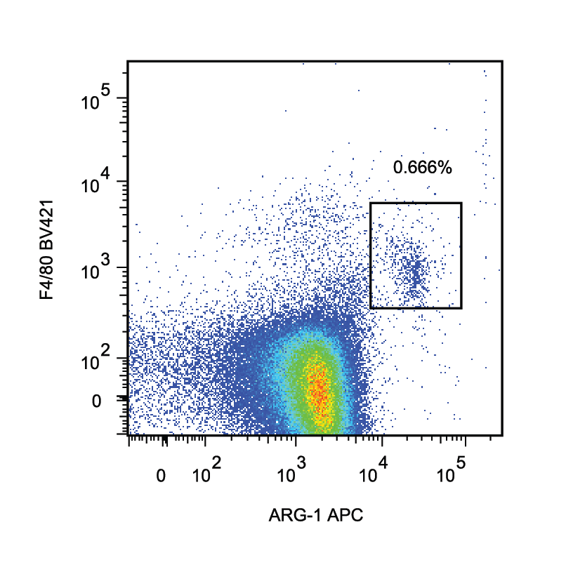
Detection of Mouse Arginase 1/ARG1/liver Arginase by Flow Cytometry
ILC2 transfers to apoE−/− mice increase peritoneal B1 cells, eosinophils and AAMs. Flow cytometry gating strategies of B1 cells identified as CD19+B220lowCD11b+IgM+ (a), eosinophils as CD45+SiglecF+CD11b+ cells (d) and AAMs as CD45+CD11b+F480+Arg1+ cells (i) of apoE−/− mice for both control and ILC2 groups respectively as indicated. Percentages and numbers of peritoneal B1 cells (b, c), eosinophils (e, f) and AAMs (j, k). CD11c+ expression from gated eosinophils and respective cell numbers of activated eosinophils identified as CD45+SiglecF+CD11b+CD11c+cells (g, h). Mann Whitney U test, **P < 0.01, ***P < 0.001, ****P < 0.0001. Each data point represents one mouse Image collected and cropped by CiteAb from the following publication (https://pubmed.ncbi.nlm.nih.gov/31823769), licensed under a CC-BY license. Not internally tested by R&D Systems.
Detection of Mouse Arginase 1/ARG1/liver Arginase by Flow Cytometry
Flow cytometry and qRT/PCR analyses of immune cell infiltrates. (A) Flow cytometry gating strategy for macrophages (CD11b+CD45hiLy6G-), microglia (CD11b+CD45loLy6G-), and neutrophils (CD11b+CD45hiLy6G+). (B) Macrophages or microglia were further characterized as alternatively activated (M2) CD206+Ym1+Arg1+ or (C) classically activated (M1) CD86+MHCII+iNOS+. (D) Flow cytometry gating for T cell subsets: Th1 = CD4+IFN-gamma +, Th17 = CD4+IL17+, Treg = CD4+CD25+FOXP3. (E, F, G) Flow cytometry of cells isolated from pooled brain and spinal cord showed that the only significant difference in total numbers of T cell subtypes isolated from the CNS at 21 dpi was a decrease in the ratio of IFN-gamma + to IL-17+ cells in the astroglial CXCL10 knockout mice (n = 3 mice/group, P = 0.0063). (H, I) No significant differences between astroglial CXCL10 knockout and control mice in total numbers of macrophages, microglia, and neutrophils (H) or M1 and M2 subtypes of macrophages and microglia (I) isolated from the CNS at 21 dpi (n = 3). (J) qRT/PCR of spinal cord tissue isolated at 14 dpi normalized to the housekeeping gene HSP90; CXCR3 expression was significantly upregulated in astroglial CXCL10 knockout mice compared to controls (P = 0.0157). IFN-gamma, FOXP3, ROR gamma t, iNOS, and arginase-1 mRNA levels were not significantly different between astroglial CXCL10 knockout and control mice at 14 dpi (n = 6 mice/group). Vertical bars = SEMs. Image collected and cropped by CiteAb from the following publication (https://pubmed.ncbi.nlm.nih.gov/24924222), licensed under a CC-BY license. Not internally tested by R&D Systems.
Detection of Mouse Arginase 1/ARG1/liver Arginase by Flow Cytometry
Tumor permissive microenvironment in Brca1 MT mammary glands.aS100a9/S100a8 mRNA expression in the subpopulations of luminal and stromal cells of WT 4-month mammary gland (WTV4MG) and MT 4-months-old virgin mammary gland (MTV4MG) (n = 3 mice). b Protein level of S100a9 in both WT (B477) and MT (G600) mammary epithelial cell lines and tumor tissues by Western blots (n = 3 individual experiment-up and n = 3 mice-down). c Co-staining of S100a9 (red) and CK18 (green) with antibodies on WTV4MG, MTV4MG, WTV6MG, and MTV6MG tissues (n = 3 pairs in each group, Scale bar: 20 μM). d The S100a9 and Arg1 positive cell populations by FACS analysis from the blood and mammary tissues of both WT and MT mice at 4-month and 6-month, respectively (FACS gating strategies see in Supplementary Fig. 8c, n = 3 mice/ group). e Co-staining with S100a9 (red) and CD206 (green) antibodies (left panel) and co-staining with S100a9 (red) and CK18 (green) antibodies (right panel) on tumor-adjacent tissues by IF (40X confocal microscope, Scale bar: 20 μM.) (n = 3 mice and 3 individual experiment). f Secreted S100a9 proteins (left) from both tumor cell and MDSC cells in tumor-adjacent mammary gland (n = 3 mice) and present in the supernatant of cultured cancer cells (right) (n = 3 individual experiment, Scale bar: 10 μM). g Protein levels of S100a9, TGF-beta, and Il-10 in mammary gland tissues of both WT and Brca1 MT mice at 4-month (n = 3 mice). h Protein levels of S100a9, TGF-beta, and IL-10 in mammary tissues of both WT and Brca1 MT mice at 6-month (n = 3 mice). The data are expressed as means ± SD (a) and P values determined by unpaired two-tailed Student’s t test. The experiments were independently repeated three times with similar results (a, b). Source data are provided as a Source data file. Image collected and cropped by CiteAb from the following publication (https://pubmed.ncbi.nlm.nih.gov/35304461), licensed under a CC-BY license. Not internally tested by R&D Systems.Applications for Human/Mouse Arginase 1/ARG1 APC‑conjugated Antibody
Intracellular Staining by Flow Cytometry
Sample: HepG2 human hepatocellular carcinoma cell line and Hepa 1-6 mouse hepatoma cell line fixed with Flow Cytometry Fixation Buffer (Catalog # FC004) and permeabilized with Flow Cytometry Permeabilization/Wash Buffer I (Catalog # FC005).
Reviewed Applications
Read 1 review rated 4 using IC5868A in the following applications:
Spectra Viewer
Plan Your Experiments
Use our spectra viewer to interactively plan your experiments, assessing multiplexing options. View the excitation and emission spectra for our fluorescent dye range and other commonly used dyes.
Spectra Viewer
Flow Cytometry Panel Builder
Bio-Techne Knows Flow Cytometry
Save time and reduce costly mistakes by quickly finding compatible reagents using the Panel Builder Tool.
Advanced Features
- Spectra Viewer - Custom analysis of spectra from multiple fluorochromes
- Spillover Popups - Visualize the spectra of individual fluorochromes
- Antigen Density Selector - Match fluorochrome brightness with antigen density

Formulation, Preparation, and Storage
Purification
Formulation
Shipping
Stability & Storage
- 12 months from date of receipt, 2 to 8 °C as supplied.
Background: Arginase 1/ARG1
Long Name
Alternate Names
Gene Symbol
UniProt
Additional Arginase 1/ARG1 Products
Product Documents for Human/Mouse Arginase 1/ARG1 APC‑conjugated Antibody
Certificate of Analysis
To download a Certificate of Analysis, please enter a lot or batch number in the search box below.
Note: Certificate of Analysis not available for kit components.
Product Specific Notices for Human/Mouse Arginase 1/ARG1 APC‑conjugated Antibody
For research use only
Related Research Areas
Citations for Human/Mouse Arginase 1/ARG1 APC‑conjugated Antibody
 Powered by Bioz
Customer Reviews for Human/Mouse Arginase 1/ARG1 APC‑conjugated Antibody (1)
Have you used Human/Mouse Arginase 1/ARG1 APC‑conjugated Antibody?
Submit a review and receive an Amazon gift card!
$25/€18/£15/$25CAN/¥2500 Yen for a review with an image
$10/€7/£6/$10CAN/¥1110 Yen for a review without an image
Submit a review
Customer Images
-
Application: Flow CytometrySample Tested: Mouse splenocytesSpecies: MouseVerified Customer | Posted 09/07/2016

There are no reviews that match your criteria.
Protocols
Find general support by application which include: protocols, troubleshooting, illustrated assays, videos and webinars.
- 7-Amino Actinomycin D (7-AAD) Cell Viability Flow Cytometry Protocol
- Extracellular Membrane Flow Cytometry Protocol
- Flow Cytometry Protocol for Cell Surface Markers
- Flow Cytometry Protocol for Staining Membrane Associated Proteins
- Flow Cytometry Staining Protocols
- Flow Cytometry Troubleshooting Guide
- Intracellular Flow Cytometry Protocol Using Alcohol (Methanol)
- Intracellular Flow Cytometry Protocol Using Detergents
- Intracellular Nuclear Staining Flow Cytometry Protocol Using Detergents
- Intracellular Staining Flow Cytometry Protocol Using Alcohol Permeabilization
- Intracellular Staining Flow Cytometry Protocol Using Detergents to Permeabilize Cells
- Propidium Iodide Cell Viability Flow Cytometry Protocol
- Protocol for Liperfluo
- Protocol for the Characterization of Human Th22 Cells
- Protocol for the Characterization of Human Th9 Cells
- Protocol: Annexin V and PI Staining by Flow Cytometry
- Protocol: Annexin V and PI Staining for Apoptosis by Flow Cytometry
- Troubleshooting Guide: Fluorokine Flow Cytometry Kits
- View all Protocols, Troubleshooting, Illustrated assays and Webinars