Arginase 1 (ARG1) is a 35‑40 kDa member of the arginase family of enzymes. It is expressed in multiple cell types, including erythrocytes, hepatocytes, neutrophils, smooth muscle and macrophages. ARG1 demonstrates two distinct functions: in the hepatocyte cytoplasm, it catalyzes the conversion of arginine to ornithine and urea, while in multiple cells, it degrades arginine, thus indirectly downregulating NO synthase (NOS) activity by depriving this enzyme of its substrate. Human AGR1 is 322 amino acids (aa) in length. Its enzyme region comprises aa 9‑309 and contains two Mn atoms. ARG1 is modestly active as a monomer, but highly active as a 105 kDa homotrimer. Trimerization is promoted by nitrosylation of Cys303, creating a regulatory feedback loop with NOS. There are two isoform variants, one that shows an eight aa insertion after Gln43, and another that shows a deletion of aa 204‑289. Full-length human ARG1 shares 87% aa identity with mouse and rat ARG1.
Human/Mouse/Rat Arginase 1/ARG1 Antibody
R&D Systems | Catalog # AF5868

Key Product Details
Validated by
Biological Validation
Species Reactivity
Validated:
Human, Mouse, Rat
Cited:
Human, Mouse, Rat, Transgenic Mouse, Xenograft
Applications
Validated:
Western Blot, Simple Western, Immunoprecipitation
Cited:
Immunohistochemistry, Western Blot, Flow Cytometry, ELISA Development (Capture), CyTOF-reported
Label
Unconjugated
Antibody Source
Polyclonal Sheep IgG
Loading...
Product Specifications
Immunogen
E. coli-derived recombinant human Arginase 1/ARG1
Met1-Lys322
Accession # P05089
Met1-Lys322
Accession # P05089
Specificity
Detects human, mouse, and rat Arginase 1/ARG1 in direct ELISAs and Western blots.
Clonality
Polyclonal
Host
Sheep
Isotype
IgG
Scientific Data Images for Human/Mouse/Rat Arginase 1/ARG1 Antibody
Detection of Human and Rat Arginase 1/ARG1 by Western Blot.
Western blot shows lysates of human liver tissue and rat liver tissue. PVDF membrane was probed with 0.25 µg/mL of Sheep Anti-Human/Mouse/Rat Arginase 1/ARG1 Antigen Affinity-purified Polyclonal Antibody (Catalog # AF5868) followed by HRP-conjugated Anti-Sheep IgG Secondary Antibody (Catalog # HAF016). A specific band was detected for Arginase 1/ARG1 at approximately 41 kDa (as indicated). This experiment was conducted under reducing conditions and using Immunoblot Buffer Group 8.
Detection of Human, Mouse, and Rat Arginase 1/ARG1 by Simple WesternTM.
Simple Western lane view shows lysates of human liver tissue, mouse liver tissue, and rat liver tissue, loaded at 0.2 mg/mL. A specific band was detected for Arginase 1/ARG1 at approximately 41 kDa (as indicated) using 2.5 µg/mL of Sheep Anti-Human/Mouse/Rat Arginase 1/ARG1 Antigen Affinity-purified Polyclonal Antibody (Catalog # AF5868) followed by 1:50 dilution of HRP-conjugated Anti-Sheep IgG Secondary Antibody (Catalog # HAF016). This experiment was conducted under reducing conditions and using the 12-230 kDa separation system.
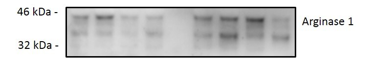
Detection of Mouse Arginase 1/ARG1/liver Arginase by Western Blot
Sepsis induces polarization of M2-like macrophages.(a–e) Lung tissue and peritoneal cells from C57BL/6 J (a,c,d) and BALB/c (b,e) mice undergoing CLP and antibiotic treatment were harvested at the indicated time points. (a) mRNA expression of Cebpb (C/EBP beta ), Arg1 (arginase-1), Mrc1 (MR) and Igf1 in the total lungs were determined by RT-qPCR at indicated times after CLP (n≥4 mice per group). (b) Lungs obtained from six mice, either naive or CLP (day 10 after CLP), were pooled from two independent samples and CD11b+ cells were isolated. mRNA expression of Arg1 (arginase-1), Mrc1 (MR), Rentla (encoding Fizz1) in isolated CD11b+ cells were determined by RT-qPCR. (c) IGF-1, CCL17 and CCL22 concentrations in the lungs were determined by ELISA at indicated times after CLP (n≥3 mice per group). (d) Representative FACS plots and frequency of peritoneal CD206+F4/80+ macrophages at indicated times after CLP (n≥3 mice per group). (e) Representative western blot of Arg1 (Arginase-1) expression in the peritoneal cells at day 10 after CLP (n=9 for naive group and n=5 for CLP group). ND, not detected. *P<0.05, **P<0.01 and ***P<0.001 (one-way ANOVA result with Dunnett posthoc tests in a,c, two-tailed unpaired Student's t-test in b,d). Data are from one (b) and representative of two (a,c–e) independent experiments (mean±s.e.m. in a–d). Image collected and cropped by CiteAb from the following publication (https://pubmed.ncbi.nlm.nih.gov/28374774), licensed under a CC-BY license. Not internally tested by R&D Systems.Applications for Human/Mouse/Rat Arginase 1/ARG1 Antibody
Application
Recommended Usage
Immunoprecipitation
25 µg/mL
Sample: Cell lysates spiked with Recombinant Human Arginase 1/ARG1 (Catalog # 5868-AR), see our available Western blot detection antibodies
Sample: Cell lysates spiked with Recombinant Human Arginase 1/ARG1 (Catalog # 5868-AR), see our available Western blot detection antibodies
Simple Western
2.5 µg/mL
Sample: Human liver tissue, mouse liver tissue, and rat liver tissue
Sample: Human liver tissue, mouse liver tissue, and rat liver tissue
Western Blot
0.25 µg/mL
Sample: Human liver tissue and rat liver tissue
Sample: Human liver tissue and rat liver tissue
Reviewed Applications
Read 2 reviews rated 3.5 using AF5868 in the following applications:
Formulation, Preparation, and Storage
Purification
Antigen Affinity-purified
Reconstitution
Reconstitute at 0.2 mg/mL in sterile PBS. For liquid material, refer to CoA for concentration.
Loading...
Formulation
Lyophilized from a 0.2 μm filtered solution in PBS with Trehalose. *Small pack size (SP) is supplied either lyophilized or as a 0.2 µm filtered solution in PBS.
Shipping
Lyophilized product is shipped at ambient temperature. Liquid small pack size (-SP) is shipped with polar packs. Upon receipt, store immediately at the temperature recommended below.
Stability & Storage
Use a manual defrost freezer and avoid repeated freeze-thaw cycles.
- 12 months from date of receipt, -20 to -70 °C as supplied.
- 1 month, 2 to 8 °C under sterile conditions after reconstitution.
- 6 months, -20 to -70 °C under sterile conditions after reconstitution.
Calculators
Background: Arginase 1/ARG1
Long Name
Liver-Type Arginase
Alternate Names
AI, ARG1, Arginase-1, Liver Arginase, PGIF, Type I Arginase
Gene Symbol
ARG1
UniProt
Additional Arginase 1/ARG1 Products
Product Documents for Human/Mouse/Rat Arginase 1/ARG1 Antibody
Certificate of Analysis
To download a Certificate of Analysis, please enter a lot or batch number in the search box below.
Note: Certificate of Analysis not available for kit components.
Product Specific Notices for Human/Mouse/Rat Arginase 1/ARG1 Antibody
For research use only
Related Research Areas
Citations for Human/Mouse/Rat Arginase 1/ARG1 Antibody
 Powered by Bioz
Customer Reviews for Human/Mouse/Rat Arginase 1/ARG1 Antibody (2)
3.5 out of 5
2 Customer Ratings
Have you used Human/Mouse/Rat Arginase 1/ARG1 Antibody?
Submit a review and receive an Amazon gift card!
$25/€18/£15/$25CAN/¥2500 Yen for a review with an image
$10/€7/£6/$10CAN/¥1110 Yen for a review without an image
Submit a review
Customer Images
Showing
1
-
2 of
2 reviews
Showing All
Filter By:
-
Application: Western BlotSample Tested: endothelial cellsSpecies: MouseVerified Customer | Posted 02/17/2020

-
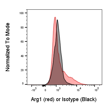
Application: Flow CytometrySample Tested: Mature myeloid dendritic cellsSpecies: MouseVerified Customer | Posted 08/24/2017
There are no reviews that match your criteria.
Protocols
Find general support by application which include: protocols, troubleshooting, illustrated assays, videos and webinars.
- Cellular Response to Hypoxia Protocols
- Immunoprecipitation Protocol
- R&D Systems Quality Control Western Blot Protocol
- Troubleshooting Guide: Western Blot Figures
- Western Blot Conditions
- Western Blot Protocol
- Western Blot Protocol for Cell Lysates
- Western Blot Troubleshooting
- Western Blot Troubleshooting Guide
- View all Protocols, Troubleshooting, Illustrated assays and Webinars
Loading...