
Key Product Details
Validated by
Biological Validation
Species Reactivity
Validated:
Human, Mouse
Cited:
Mouse, Transgenic Mouse
Applications
Validated:
Western Blot
Cited:
Immunohistochemistry-Frozen, Western Blot
Label
Unconjugated
Antibody Source
Monoclonal Rat IgG1 Clone # 91508
Loading...
Product Specifications
Immunogen
E. coli-derived recombinant mouse BID
Met1-Asp195
Accession # P70444
Met1-Asp195
Accession # P70444
Specificity
Detects human and mouse BID in Western blots. Detects full-length mouse BID and the 15 kDa carboxyl-terminal fragment (tBID) generated by cleavage with Caspase-8. The antibody also detects human BID. However, in Western blots of recombinant human and mouse BID, the antibody is most sensitive for mouse BID.
Clonality
Monoclonal
Host
Rat
Isotype
IgG1
Scientific Data Images for BID Antibody (91508)
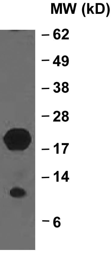
Detection of Human and Mouse BID by Western Blot.
Western blot shows lysates of Recombinant Mouse BID and Recombinant Human BID untreated (-) or treated (+) with Caspase-8. PVDF membrane was probed with 1 µg/mL Rat Anti-Human/Mouse BID Monoclonal Antibody (Catalog # MAB860) followed by HRP-conjugated Anti-Rat IgG Secondary Antibody (Catalog # HAF005). Specific bands for BID were detected at approximately 12 kDa and 20 kDa (as indicated). This experiment was conducted under reducing conditions and using Immunoblot Buffer Group 4.
Detection of Human and Mouse BID by Western Blot.
Western blot shows lysates of SVEC4-10 mouse vascular endothelial cell line and Jurkat human acute T cell leukemia cell line. PVDF membrane was probed with 1 µg/mL of Rat Anti-Human/Mouse BID Monoclonal Antibody (Catalog # MAB860) followed by HRP-conjugated Anti-Rat IgG Secondary Antibody (Catalog # HAF005). A specific band was detected for BID at approximately 20 kDa (as indicated). This experiment was conducted under reducing conditions and using Immunoblot Buffer Group 1.
Detection of BID by Western Blot
Vector control S. Typhimurium does not activate apoptotic backup pathways in vitro.(A–E) Bone marrow-derived macrophages (BMMs) were infected with indicated SPI2-induced S. Typhimurium strains. (A) Western blot analysis of whole cell lysates at 4 hpi. Results from one experiment. (B) Lactate dehydrogenase (LDH) release at 1–8 hpi. Results representative of three independent experiments. Data are represented as mean ± SD of three technical replicates. Data performed at the same time as Figure 1E, graphed separately for visualization. (C) LDH release at 1–8 hpi. Results from one experiment. Data are represented as mean ± SD of three technical replicates. (D–E) Immunofluorescence and brightfield at 4 hpi. Cells were stained with PI, cleaved caspase-3/7, and Hoechst. Representative image from two (brightfield, PI) or one (cleaved caspase-3/7) independent experiments. Data performed at the same time as Figure 1F. (D) 60 x magnification, scale bar 20 µm. (E) 20 x stitched image, scale bar 500 µm.Figure 1—figure supplement 1—source data 1.Western blot images for Figure 1—figure supplement 1A.Figure 1—figure supplement 1—source data 2.Data for Figure 1—figure supplement 1B–C.Western blot images for Figure 1—figure supplement 1A.Data for Figure 1—figure supplement 1B–C. Image collected and cropped by CiteAb from the following open publication (https://pubmed.ncbi.nlm.nih.gov/38055781), licensed under a CC-BY license. Not internally tested by R&D Systems.Applications for BID Antibody (91508)
Application
Recommended Usage
Western Blot
1 µg/mL
Sample: Recombinant Mouse BID and Recombinant Human BID untreated or treated with Caspase-8, SVEC4‑10 mouse vascular endothelial cell line, and Jurkat human acute T cell leukemia cell line
Sample: Recombinant Mouse BID and Recombinant Human BID untreated or treated with Caspase-8, SVEC4‑10 mouse vascular endothelial cell line, and Jurkat human acute T cell leukemia cell line
Reviewed Applications
Read 3 reviews rated 4.3 using MAB860 in the following applications:
Formulation, Preparation, and Storage
Purification
Protein A or G purified from hybridoma culture supernatant
Reconstitution
Reconstitute at 0.5 mg/mL in sterile PBS. For liquid material, refer to CoA for concentration.
Loading...
Formulation
Lyophilized from a 0.2 μm filtered solution in PBS with Trehalose. *Small pack size (SP) is supplied either lyophilized or as a 0.2 µm filtered solution in PBS.
Shipping
Lyophilized product is shipped at ambient temperature. Liquid small pack size (-SP) is shipped with polar packs. Upon receipt, store immediately at the temperature recommended below.
Stability & Storage
Use a manual defrost freezer and avoid repeated freeze-thaw cycles.
- 12 months from date of receipt, -20 to -70 °C as supplied.
- 1 month, 2 to 8 °C under sterile conditions after reconstitution.
- 6 months, -20 to -70 °C under sterile conditions after reconstitution.
Calculators
Background: BID
Long Name
BH3 Interacting Domain Death Agonist
Alternate Names
apoptic death agonist, BH3 interacting domain death agonist, BH3-interacting domain death agonist, BID isoform ES(1b), BID isoform L(2), BID isoform Si6, desmocollin type 4, FP497, Human BID coding sequence, MGC15319, MGC42355, p22 BID, tbid
Gene Symbol
BID
UniProt
Additional BID Products
Product Documents for BID Antibody (91508)
Certificate of Analysis
To download a Certificate of Analysis, please enter a lot or batch number in the search box below.
Note: Certificate of Analysis not available for kit components.
Product Specific Notices for BID Antibody (91508)
For research use only
Related Research Areas
Citations for BID Antibody (91508)
 Powered by Bioz
Customer Reviews for BID Antibody (91508) (3)
4.3 out of 5
3 Customer Ratings
Have you used BID Antibody (91508)?
Submit a review and receive an Amazon gift card!
$25/€18/£15/$25CAN/¥2500 Yen for a review with an image
$10/€7/£6/$10CAN/¥1110 Yen for a review without an image
Submit a review
Customer Images

Showing
1
-
3 of
3 reviews
Showing All
Filter By:
-
Application: Western BlotSample Tested: Colon tissue and Tissue LysatesSpecies: MouseVerified Customer | Posted 01/29/2022Could only detect the pro form in my samples.

-
Application: Western BlotSample Tested: fibroblastsSpecies: MouseVerified Customer | Posted 11/17/2021
-
Application: Western BlotSample Tested: Mouse embryonic fibroblasts treated with TNFa (50 ng/ml) and cycloheximide (5 ug/ml) for 8 hoursSpecies: MurineVerified Customer | Posted 05/19/2017
There are no reviews that match your criteria.
Protocols
Find general support by application which include: protocols, troubleshooting, illustrated assays, videos and webinars.
- Cellular Response to Hypoxia Protocols
- R&D Systems Quality Control Western Blot Protocol
- Troubleshooting Guide: Western Blot Figures
- Western Blot Conditions
- Western Blot Protocol
- Western Blot Protocol for Cell Lysates
- Western Blot Troubleshooting
- Western Blot Troubleshooting Guide
- View all Protocols, Troubleshooting, Illustrated assays and Webinars
Loading...
Associated Pathways
