CELSR2 (Cadherin EGF LAG seven-pass G-type receptor 2; also cadherin family member 10/CDHF10, Flamingo1 and EGFL2) is a 300-330 kDa member of the LN‑7TM subfamily, GPCR 2 family of proteins. It is expressed on neurons, breast epithelium, Sertoli cells and germ cells, and through homophilic interactions, serves as either an adhesion or guidance molecule. Mature human CELSR2 is 2892 amino acids in length (aa 32-2923). It is a highly complex 7-transmembrane protein that contains a 2349 aa extended N-terminal extracellular region (aa 32-2380) plus a 310 aa C-terminal cytoplasmic domain. The N-terminal region contains nine consecutive cadherin domains (aa 182-1146) followed by a mixture of seven EGF-like and three laminin-like domains. There is a proteolytic cleavage site between Met2356-Thr2357 that generates a 250 kDa soluble fragment and a (mature) 60-65 kDa transmembrane segment that may reside on the cell membrane. Over aa 51‑231, human CELSR2 shares 93% aa identity with mouse CELSR2.

Discontinued Product
AF6739 has been discontinued.
View all CELSR2 products.
Key Product Details
Species Reactivity
Validated:
Human, Mouse
Cited:
Mouse
Applications
Validated:
Immunohistochemistry, Western Blot, Flow Cytometry, CyTOF-ready
Cited:
Western Blot
Label
Unconjugated
Antibody Source
Polyclonal Goat IgG
Loading...
Product Specifications
Immunogen
E. coli-derived recombinant human CELSR2
Cys51-Phe231
Accession # Q9HCU4
Cys51-Phe231
Accession # Q9HCU4
Specificity
Detects human CELSR2 in direct ELISAs and Western blots. In direct ELISAs, less than 5% cross-reactivity with recombinant human (rh) CELSR1 and rhCELSR3 is observed.
Clonality
Polyclonal
Host
Goat
Isotype
IgG
Scientific Data Images for CELSR2 Antibody
Detection of CELSR2 by Western Blot.
Western blot shows lysates of HEK293 human embryonic kidney cell line either mock transfected or transfected with human CELSR2. PVDF Membrane was probed with 1 µg/mL of Goat Anti-Human CELSR2 Antigen Affinity-purified Polyclonal Antibody (Catalog # AF6739) followed by HRP-conjugated Anti-Goat IgG Secondary Antibody (Catalog # HAF019). A specific band was detected for CELSR2 at approximately 240 kDa (as indicated). This experiment was conducted under reducing conditions and using Immunoblot Buffer Group 8.
Detection of CELSR2 in SH‑SY5Y Human Cell Line by Flow Cytometry.
SH-SY5Y human neuroblastoma cell line was stained with Goat Anti-Human CELSR2 Antigen Affinity-purified Polyclonal Antibody (Catalog # AF6739, filled histogram) or isotype control antibody (Catalog # AB-108-C, open histogram), followed by Allophycocyanin-conjugated Anti-Goat IgG Secondary Antibody (Catalog # F0108).
Detection of CELSR2 in bEnd.3 Mouse Cell Line by Flow Cytometry.
bEnd.3 mouse endothelioma cell line was stained with Goat Anti-Human CELSR2 Antigen Affinity-purified Polyclonal Antibody (Catalog # AF6739, filled histogram) or isotype control antibody (Catalog # AB-108-C, open histogram), followed by Allophycocyanin-conjugated Anti-Goat IgG Secondary Antibody (Catalog # F0108).
CELSR2 in Human Breast.
CELSR2 was detected in immersion fixed paraffin-embedded sections of human breast using Goat Anti-Human CELSR2 Antigen Affinity-purified Polyclonal Antibody (Catalog # AF6739) at 10 µg/mL overnight at 4 °C. Tissue was stained using the Anti-Goat HRP-DAB Cell & Tissue Staining Kit (brown; Catalog # CTS008) and counterstained with hematoxylin (blue). Lower panel shows a lack of labeling when primary antibodies are omitted and tissue is stained only with secondary antibody followed by incubation with detection reagents. Specific staining was localized to ductal epithelium. View our protocol for Chromogenic IHC Staining of Paraffin-embedded Tissue Sections.Applications for CELSR2 Antibody
Application
Recommended Usage
CyTOF-ready
Ready to be labeled using established conjugation methods. No BSA or other carrier proteins that could interfere with conjugation.
Flow Cytometry
2.5 µg/106 cells
Sample: SH‑SY5Y human neuroblastoma cell line and bEnd.3 mouse endothelioma cell line
Sample: SH‑SY5Y human neuroblastoma cell line and bEnd.3 mouse endothelioma cell line
Immunohistochemistry
5-15 µg/mL
Sample: Immersion fixed paraffin-embedded sections of human breast
Sample: Immersion fixed paraffin-embedded sections of human breast
Western Blot
1 µg/mL
Sample: HEK293 human embryonic kidney cell line transfected with human CELSR2
Sample: HEK293 human embryonic kidney cell line transfected with human CELSR2
Reviewed Applications
Read 1 review rated 4 using AF6739 in the following applications:
Flow Cytometry Panel Builder
Bio-Techne Knows Flow Cytometry
Save time and reduce costly mistakes by quickly finding compatible reagents using the Panel Builder Tool.
Advanced Features
- Spectra Viewer - Custom analysis of spectra from multiple fluorochromes
- Spillover Popups - Visualize the spectra of individual fluorochromes
- Antigen Density Selector - Match fluorochrome brightness with antigen density

Formulation, Preparation, and Storage
Purification
Antigen Affinity-purified
Reconstitution
Sterile PBS to a final concentration of 0.2 mg/mL. For liquid material, refer to CoA for concentration.
Formulation
Lyophilized from a 0.2 μm filtered solution in PBS with Trehalose. *Small pack size (SP) is supplied either lyophilized or as a 0.2 µm filtered solution in PBS.
Shipping
Lyophilized product is shipped at ambient temperature. Liquid small pack size (-SP) is shipped with polar packs. Upon receipt, store immediately at the temperature recommended below.
Stability & Storage
Use a manual defrost freezer and avoid repeated freeze-thaw cycles.
- 12 months from date of receipt, -20 to -70 °C as supplied.
- 1 month, 2 to 8 °C under sterile conditions after reconstitution.
- 6 months, -20 to -70 °C under sterile conditions after reconstitution.
Calculators
Background: CELSR2
Long Name
Cadherin, EGF LAG Seven-pass G-type Receptor 2
Alternate Names
CDHF10, EGFL2, Flamingo1, MEGF3
Gene Symbol
CELSR2
UniProt
Additional CELSR2 Products
Product Documents for CELSR2 Antibody
Certificate of Analysis
To download a Certificate of Analysis, please enter a lot or batch number in the search box below.
Note: Certificate of Analysis not available for kit components.
Product Specific Notices for CELSR2 Antibody
For research use only
Related Research Areas
Citations for CELSR2 Antibody
 Powered by Bioz
Customer Reviews for CELSR2 Antibody (1)
4 out of 5
1 Customer Rating
Have you used CELSR2 Antibody?
Submit a review and receive an Amazon gift card!
$25/€18/£15/$25CAN/¥2500 Yen for a review with an image
$10/€7/£6/$10CAN/¥1110 Yen for a review without an image
Submit a review
Customer Images
Showing
1
-
1 of
1 review
Showing All
Filter By:
-
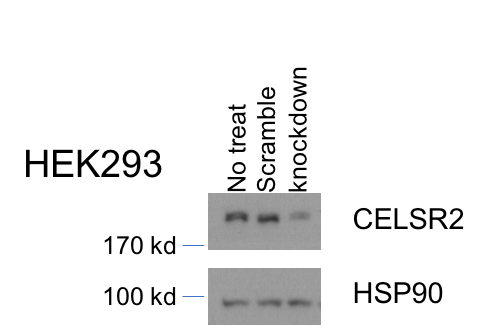
Application: Western BlotSample Tested: Cell LysatesSpecies: HumanVerified Customer | Posted 05/03/2017

There are no reviews that match your criteria.
Protocols
Find general support by application which include: protocols, troubleshooting, illustrated assays, videos and webinars.
- 7-Amino Actinomycin D (7-AAD) Cell Viability Flow Cytometry Protocol
- Antigen Retrieval Protocol (PIER)
- Antigen Retrieval for Frozen Sections Protocol
- Appropriate Fixation of IHC/ICC Samples
- Cellular Response to Hypoxia Protocols
- Chromogenic IHC Staining of Formalin-Fixed Paraffin-Embedded (FFPE) Tissue Protocol
- Chromogenic Immunohistochemistry Staining of Frozen Tissue
- ClariTSA™ Fluorophore Kits
- Detection & Visualization of Antibody Binding
- Extracellular Membrane Flow Cytometry Protocol
- Flow Cytometry Protocol for Cell Surface Markers
- Flow Cytometry Protocol for Staining Membrane Associated Proteins
- Flow Cytometry Staining Protocols
- Flow Cytometry Troubleshooting Guide
- Fluorescent IHC Staining of Frozen Tissue Protocol
- Graphic Protocol for Heat-induced Epitope Retrieval
- Graphic Protocol for the Preparation and Fluorescent IHC Staining of Frozen Tissue Sections
- Graphic Protocol for the Preparation and Fluorescent IHC Staining of Paraffin-embedded Tissue Sections
- Graphic Protocol for the Preparation of Gelatin-coated Slides for Histological Tissue Sections
- IHC Sample Preparation (Frozen sections vs Paraffin)
- Immunofluorescent IHC Staining of Formalin-Fixed Paraffin-Embedded (FFPE) Tissue Protocol
- Immunohistochemistry (IHC) and Immunocytochemistry (ICC) Protocols
- Immunohistochemistry Frozen Troubleshooting
- Immunohistochemistry Paraffin Troubleshooting
- Intracellular Flow Cytometry Protocol Using Alcohol (Methanol)
- Intracellular Flow Cytometry Protocol Using Detergents
- Intracellular Nuclear Staining Flow Cytometry Protocol Using Detergents
- Intracellular Staining Flow Cytometry Protocol Using Alcohol Permeabilization
- Intracellular Staining Flow Cytometry Protocol Using Detergents to Permeabilize Cells
- Preparing Samples for IHC/ICC Experiments
- Preventing Non-Specific Staining (Non-Specific Binding)
- Primary Antibody Selection & Optimization
- Propidium Iodide Cell Viability Flow Cytometry Protocol
- Protocol for Heat-Induced Epitope Retrieval (HIER)
- Protocol for Liperfluo
- Protocol for Making a 4% Formaldehyde Solution in PBS
- Protocol for VisUCyte™ HRP Polymer Detection Reagent
- Protocol for the Characterization of Human Th22 Cells
- Protocol for the Characterization of Human Th9 Cells
- Protocol for the Preparation & Fixation of Cells on Coverslips
- Protocol for the Preparation and Chromogenic IHC Staining of Frozen Tissue Sections
- Protocol for the Preparation and Chromogenic IHC Staining of Frozen Tissue Sections - Graphic
- Protocol for the Preparation and Chromogenic IHC Staining of Paraffin-embedded Tissue Sections
- Protocol for the Preparation and Chromogenic IHC Staining of Paraffin-embedded Tissue Sections - Graphic
- Protocol for the Preparation and Fluorescent IHC Staining of Frozen Tissue Sections
- Protocol for the Preparation and Fluorescent IHC Staining of Paraffin-embedded Tissue Sections
- Protocol for the Preparation of Gelatin-coated Slides for Histological Tissue Sections
- Protocol: Annexin V and PI Staining by Flow Cytometry
- Protocol: Annexin V and PI Staining for Apoptosis by Flow Cytometry
- R&D Systems Quality Control Western Blot Protocol
- TUNEL and Active Caspase-3 Detection by IHC/ICC Protocol
- The Importance of IHC/ICC Controls
- Troubleshooting Guide: Fluorokine Flow Cytometry Kits
- Troubleshooting Guide: Immunohistochemistry
- Troubleshooting Guide: Western Blot Figures
- Western Blot Conditions
- Western Blot Protocol
- Western Blot Protocol for Cell Lysates
- Western Blot Troubleshooting
- Western Blot Troubleshooting Guide
- View all Protocols, Troubleshooting, Illustrated assays and Webinars
Loading...