
Key Product Details
Species Reactivity
Validated:
Human, Mouse
Cited:
Human, Mouse, Transgenic Mouse
Applications
Validated:
Immunohistochemistry
Cited:
Immunohistochemistry, Immunohistochemistry-Paraffin, Immunohistochemistry-Frozen, Western Blot, Immunocytochemistry, Bioassay
Label
Unconjugated
Antibody Source
Polyclonal Goat IgG
Loading...
Product Specifications
Immunogen
E. coli-derived recombinant human Desmin
Val261-Leu470
Accession # P17661
Val261-Leu470
Accession # P17661
Specificity
Detects human and mouse Desmin.
Clonality
Polyclonal
Host
Goat
Isotype
IgG
Scientific Data Images for Desmin Antibody
Desmin in Human Heart.
Desmin was detected in immersion fixed paraffin-embedded sections of human heart using 15 µg/mL Human Desmin Antigen Affinity-purified Polyclonal Antibody (Catalog # AF3844) overnight at 4 °C. Tissue was stained with the Anti-Goat HRP-DAB Cell & Tissue Staining Kit (brown; Catalog # CTS008) and counterstained with hematoxylin (blue). Specific labeling was localized to the sarcoplasm of cardiomyocytes. View our protocol for Chromogenic IHC Staining of Paraffin-embedded Tissue Sections.
Desmin in Mouse Skeletal Muscle.
Desmin was detected in immersion fixed frozen sections of mouse skeletal muscle using Human Desmin Antigen Affinity-purified Polyclonal Antibody (Catalog # AF3844) at 10 µg/mL overnight at 4 °C. Tissue was stained using the NorthernLights™ 557-conjugated Anti-Goat IgG Secondary Antibody (red; Catalog # NL001) and counterstained with DAPI (blue). Specific staining was localized to z-lines. View our protocol for Fluorescent IHC Staining of Frozen Tissue Sections.
Detection of Human Desmin by Immunocytochemistry/Immunofluorescence
Overview of Multi-dimensional Microscopic Molecular Profiling (MMMP).The overall MMMP approach is depicted using an example tissue section from normal human duodenum (sample #1.9.7). (a) Slides were subjected to repeated cycles of staining and imaging with fluorescent primary antibodies and DAPI. At the end of each cycle, fluorescent signal was removed by a chemical bleaching process, and slides were again imaged, before proceeding to the next round of this iterative procedure. After the final antibody stain (#15 Sma), slides were analyzed with a series of histochemical stains. (b) A set of tiling images spanning each tissue section was initially generated by the microscope system. The tiling images were then computationally ‘stitched’ together to produce a single image per staining cycle for each sample. (c) Image registration was performed to align images from the same tissue section across cycles. Mean intensities of the DAPI signal from all immuno-fluorescence images are shown from before (Unregistered) and after (Registered) the image registration procedure was completed. (d) Following registration, signal intensities from the relevant channels for each image (columns) in the MMMP series were extracted for each pixel (rows) within the tissue section and compiled into a large data matrix of in situ molecular profiles. Image collected and cropped by CiteAb from the following publication (https://dx.plos.org/10.1371/journal.pone.0128975), licensed under a CC-BY license. Not internally tested by R&D Systems.
Detection of Human Desmin by Immunocytochemistry/Immunofluorescence
In vitro and in vivo differentiation of GMP-transitioned RiPSC lines.(A) Directed in vitro differentiation of three GMP-transitioned RiPSC lines into three germ layers followed by FACS analysis (endoderm) and immunocytochemistry (ectoderm, mesoderm). Scale bar = 150 μm. (B) Comparison of differentiation potential into neuroectoderm (PAX6) and mesoderm (DESMIN) between non-GMP (research-grade) and GMP-grade lines (BJ, HUF1). Comparisons are based on counting analysis of cells that were positive for the specific differentiation markers. N = 200–300. (C) Hematoxylin and eosin staining of teratomas derived from GMP-transitioned lines showing ectoderm (neural rosettes, epidermis), mesoderm (cartilage) and endoderm (gut-like endothelium). Scale bar = 200 μm. Image collected and cropped by CiteAb from the following publication (https://pubmed.ncbi.nlm.nih.gov/24718618), licensed under a CC-BY license. Not internally tested by R&D Systems.
Immunofluorescent Staining of iPSC-derived Human Intestinal Organoids.
iPSC-derived human intestinal organoids were generated following the steps detailed in the human intestinal organoid culture protocol. Human intestinal organoids were stained using a Rat Anti-Human/Mouse/Rat Vimentin Monoclonal Antibody (Catalog # MAB2105; green) and a Goat Anti-Human/Mouse Desmin Antigen Affinity-purified Polyclonal Antibody (Catalog # AF3844; red) to visualize myofibroblast cells and counterstained with DAPI (Catalog # 5748; blue).Applications for Desmin Antibody
Application
Recommended Usage
Immunohistochemistry
5-15 µg/mL
Sample: Immersion fixed paraffin-embedded sections of human heart and immersion fixed frozen sections of mouse skeletal muscle
Sample: Immersion fixed paraffin-embedded sections of human heart and immersion fixed frozen sections of mouse skeletal muscle
Reviewed Applications
Read 2 reviews rated 4.5 using AF3844 in the following applications:
Formulation, Preparation, and Storage
Purification
Antigen Affinity-purified
Reconstitution
Reconstitute at 0.2 mg/mL in sterile PBS. For liquid material, refer to CoA for concentration.
Loading...
Formulation
Lyophilized from a 0.2 μm filtered solution in PBS with Trehalose. *Small pack size (SP) is supplied either lyophilized or as a 0.2 µm filtered solution in PBS.
Shipping
Lyophilized product is shipped at ambient temperature. Liquid small pack size (-SP) is shipped with polar packs. Upon receipt, store immediately at the temperature recommended below.
Stability & Storage
Use a manual defrost freezer and avoid repeated freeze-thaw cycles.
- 12 months from date of receipt, -20 to -70 °C as supplied.
- 1 month, 2 to 8 °C under sterile conditions after reconstitution.
- 6 months, -20 to -70 °C under sterile conditions after reconstitution.
Calculators
Background: Desmin
Alternate Names
CMD1I, DES
Gene Symbol
DES
UniProt
Additional Desmin Products
Product Documents for Desmin Antibody
Certificate of Analysis
To download a Certificate of Analysis, please enter a lot or batch number in the search box below.
Note: Certificate of Analysis not available for kit components.
Product Specific Notices for Desmin Antibody
For research use only
Related Research Areas
Citations for Desmin Antibody
 Powered by Bioz
Customer Reviews for Desmin Antibody (2)
4.5 out of 5
2 Customer Ratings
Have you used Desmin Antibody?
Submit a review and receive an Amazon gift card!
$25/€18/£15/$25CAN/¥2500 Yen for a review with an image
$10/€7/£6/$10CAN/¥1110 Yen for a review without an image
Submit a review
Customer Images
Showing
1
-
2 of
2 reviews
Showing All
Filter By:
-
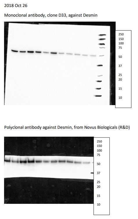
Application: Western BlotSample Tested: Heart tissueSpecies: CatVerified Customer | Posted 10/27/20181:1000 in 1%milk/TBST overnight at 4C; washed 10 min for 3 times and incubate with mouse-anti-goat-HRP monoclonal antibody for 1 hour; washed for another 5 min 3 times. Developed with ECL.

-
Application: Immunohistochemistry-ParaffinSample Tested: See PMID 24200904Species: HumanVerified Customer | Posted 01/08/2015
There are no reviews that match your criteria.
Protocols
Find general support by application which include: protocols, troubleshooting, illustrated assays, videos and webinars.
- Antigen Retrieval Protocol (PIER)
- Antigen Retrieval for Frozen Sections Protocol
- Appropriate Fixation of IHC/ICC Samples
- Cellular Response to Hypoxia Protocols
- Chromogenic IHC Staining of Formalin-Fixed Paraffin-Embedded (FFPE) Tissue Protocol
- Chromogenic Immunohistochemistry Staining of Frozen Tissue
- ClariTSA™ Fluorophore Kits
- Detection & Visualization of Antibody Binding
- Fluorescent IHC Staining of Frozen Tissue Protocol
- Graphic Protocol for Heat-induced Epitope Retrieval
- Graphic Protocol for the Preparation and Fluorescent IHC Staining of Frozen Tissue Sections
- Graphic Protocol for the Preparation and Fluorescent IHC Staining of Paraffin-embedded Tissue Sections
- Graphic Protocol for the Preparation of Gelatin-coated Slides for Histological Tissue Sections
- IHC Sample Preparation (Frozen sections vs Paraffin)
- Immunofluorescent IHC Staining of Formalin-Fixed Paraffin-Embedded (FFPE) Tissue Protocol
- Immunohistochemistry (IHC) and Immunocytochemistry (ICC) Protocols
- Immunohistochemistry Frozen Troubleshooting
- Immunohistochemistry Paraffin Troubleshooting
- Preparing Samples for IHC/ICC Experiments
- Preventing Non-Specific Staining (Non-Specific Binding)
- Primary Antibody Selection & Optimization
- Protocol for Heat-Induced Epitope Retrieval (HIER)
- Protocol for Making a 4% Formaldehyde Solution in PBS
- Protocol for VisUCyte™ HRP Polymer Detection Reagent
- Protocol for the Preparation & Fixation of Cells on Coverslips
- Protocol for the Preparation and Chromogenic IHC Staining of Frozen Tissue Sections
- Protocol for the Preparation and Chromogenic IHC Staining of Frozen Tissue Sections - Graphic
- Protocol for the Preparation and Chromogenic IHC Staining of Paraffin-embedded Tissue Sections
- Protocol for the Preparation and Chromogenic IHC Staining of Paraffin-embedded Tissue Sections - Graphic
- Protocol for the Preparation and Fluorescent IHC Staining of Frozen Tissue Sections
- Protocol for the Preparation and Fluorescent IHC Staining of Paraffin-embedded Tissue Sections
- Protocol for the Preparation of Gelatin-coated Slides for Histological Tissue Sections
- TUNEL and Active Caspase-3 Detection by IHC/ICC Protocol
- The Importance of IHC/ICC Controls
- Troubleshooting Guide: Immunohistochemistry
- View all Protocols, Troubleshooting, Illustrated assays and Webinars
Loading...
Associated Pathways
